
Chapter Outline PowerPoint PPT Presentation
1 / 59
Title: Chapter Outline
1
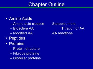
Chapter Outline
- Amino Acids
- Amino acid classes Stereoisomers
- Bioactive AA Titration of AA
- Modified AA AA reactions
- Peptides
- Proteins
- Protein structure
- Fibrous proteins
- Globular proteins
2
5.1 Amino Acid Definition
- An alpha amino acid is a carboxylic acid with an
amino group on the carbon alpha to the carboxylic
acid . - The alpha carbon also has an R group side chain
except for glycine which has two Hs.
3
Definition, cont.
- If the R group is not H, the AA can exist in two
enantiomeric forms (nonsuperimposable mirror
image) forms.)
4
Amino Acids
- General form 1. an amino acid (AA) 2. two AA
linked to form the peptide bond.
L-form
5
Amino Acids-2
- Only the L form of amino acids is commonly found
in proteins. - Depending on the nature of the R group, AA are
classified into four groups. - nonpolar
- polar
- acidic
- basic
6
AA with nonpolar side chains-1
7
AA with nonpolar side chains-2
8
AA with polar side chains-1
9
AA with polar side chains-2
10
AA acidic and basic
11
Amino Acid Titration
- At physiological pH, the carboxyl group of the AA
is negatively charged and the amino group is
positively charged. - Amino acids without charged side chains are
zwitterions and have no net charge.
H3N-CHR-COO-. - A titration curve shows how the amine and
carboxyl groups react with hydrogen ion.
12
Amino Acid Titration-2
- At low pH a nonacidic/nonbasic amino acid is
protonated and has the structure below. - H3NCHRCOOH
- The charge behavior of acidic and basic AAs is
more complex.
13
Titration of Alanine
1
14
Isoelectric point
- The isoelectric point (pI) for an AA occurs when
there is no net charge. - For a neutral AA, the pI is calculated using the
equation pK1 pK2/2 - Eg. alanine 2.34 9.7 / 2 6.0
- For acidic or basic AAs, the pI is the average of
the two pKa values bracketing the isoelectric
structure.
15
Isoelectric point-2
- In general the pI is the average of the two pKas
bracketing the isoelectric structure. Eg.
glutamic acid, pI 3.2
pK39.9
pK24.3
pK12.2
16
5.2 Peptides
- Peptide a polymer of about 2-100 AAs linked by
the peptide(amide) bond. As the amino group and
the carboxyl group link, water is lost.
17
Peptides-2
- A peptide is written with the N-terminal end to
the left and the C-terminal end to the right. - H2N-Tyr-Ala-Cys-Gly-COOH
- Name Tyrosylalanylcysteinylglycine
- The peptide bond is rigid and planar due to the
resonance contribution shown right.
18
Peptides-3
- The peptide bond angles force specific
conformations of proteins and, on extended
chains, successive R groups are on opposite sides.
19
Physiologically Interesting Peptides
Common name carnosine found in muscle tissue
20
Physiologically Interesting Peptides
Glutathione the reduced form reduces oxidizing
agents by dimerizing to form the disulfide bond
with release of 2 H.
21
Physiologically Interesting Peptides
Tyr-Gly-Gly-Phe-Leu
C-terminal AA
N-terminal AA
22
Physiologically Interesting Peptides
Oxytocin Induces labor and aids in forcing
milk from the mammary glands.
Vassopressin has a Phe at position 3 instead of
Ile and an Arg at position 8 instead of a Leu.
Its role is in regulating blood pressure.
23
Protein Function
- Catalysis
- 2. Structure
- 3. Movement
- 4. Defense
- 5. Regulation
- 6. Transport
- 7. Storage
- 8. Stress Response
24
Proteins by Shape-1
- Fibrous proteins exist as long stranded
molecules Eg. Silk, collagen, wool. A collagen
segment in space-filling mode illustrates this
point.
Red spheres represent oxygen, grey carbon, and
blue nitrogen
25
Proteins by Shape-2
- Globular proteins have somewhat spherical shapes.
Most enzymes are globular. Eg. myoglobin,
hemoglobin. Myoglobin in space-filling mode is
the chosen example.
26
Proteins by Composition
- Simple
- Contain only amino acids
- Conjugated
- simple protein (apoprotein)
- prostetic group (nonprotein)
- glycoproteins
- lipoproteins
- metaloproteins
- etc.
27
Four Levels of Protein Structure
- Primary, 1o
- the amino acid sequence
- Secondary, 2o
- 3-D arrangement of backbone atoms in space
- Tertiary, 3o
- 3-D arrangement of all the atoms in space
- Quaternary, 4o
- 3-D arrangement of subunit chains
28
Determining Primary Structure
- 1. Hydrolyze protein with hot 6M HCl.
- Identify AA and of each.
- Usually done by chromatography
- 2. Identify the N-term and C-term AAs
- C-term via carboxypeptidase
- N-term via Sangers Reagent, DNFB
- 2,4-dinitrofluorobenzene
- Often step 2 can be skipped today.
29
Det. Primary Structure 2
- 3. Selectively fragment large proteins into
smaller ones. - Eg. Tripsin cleave to leave Arg or Lys as C-term
AA - Eg. Chymotrypsin cleave to leave Tyr or Trp or
Phe as C-term AA - Eg. Cyanogen bromide cleaves at internal Met
leaving Met as C-term homoserine lactone
30
Det. Primary Structure 3
- 4. Determine AA sequence of peptides with AA
sequencer using Edmans reagent - phenyl isothiocyanate which reacts with the
N-term AA - See the next slide
31
Det. Primary Structure 3b
protein
Edmans reagent
Phenylthiohydantoin (PTH) derivative of N-term AA
32
Det. Primary Structure 4
- 5. Reassemble peptide fragments from step 3 to
give protein. - An example follows on the next slide.
33
Det. Primary Structure 4b
- A twelve AA peptide was hydrolyzed.
- Trypsin hydrolysis
- Leu-Ser-Tyr-Gly-Ile-Arg
- Thr-Ala-Met-Phe-Val-Lys
- Chymotrypsin hydrolysis
- Val-Lys-Leu-Ser-Tyr
- Gly-Ile-Arg
- Thr-Ala-Met-Phe
- Deduce the AA sequence
Lys is internal!
34
Det. Primary Structure 4c
Keeping in mind the N-term AA and overlaping the
sequences properly gives
- Tr
Leu-Ser-Tyr-Gly-Ile-Arg - Ct
Gly-Ile-Arg - Ct
Val-Lys-Leu-Ser-Tyr - Tr Thr-Ala-Met-Phe-Val-Lys
- Ct Thr-Ala-Met-Phe
- The complete sequence is
- Thr-Ala-Met-Phe-Val-Lys-Leu-Ser-Tyr-Gly-Ile-Ar
g
35
Secondary Structure
- The two very important secondary structures of
proteins are - a-helix
- b-pleated sheet
- Both depend on hydrogen bonding between the amide
H and the carbonyl O further down the chain or on
a parallel chain.
36
a Helix Peptide w Hbonds
First six CO to N hydrogen bonds shown
37
b Sheet stick form Protein G
H bonds in dotted red-blue
Chain segment 1
Seg 2
Seg 3
Chain 1
Seg 4
38
B Sheet Lewis Structure
Antiparallel sheet
Parallel sheet
39
Supersecondary Structure
- Reverse turns in a protein chain allow helices
and sheets to align side-by-side - Common AA found at turns are
- glycine small size allows a turn
- proline geometry favors a turn
40
Supersecondary Structure 2
Combinations of a helix and b sheet.
41
Tertiary Structure
- The configuration of all the atoms in the protein
chain - side chains
- prosthetic groups
- helical and pleated sheet regions
42
Tertiary Structure 2
- Protein folding attractions
- 1. Noncovalent forces
- a. Inter and intrachain H bonding
- b. Hydrophobic interactions
- c. Electrostatic attractions
- to - ionic attraction
- d. Complexation with metal ions
- e. Ion-dipole
- 2. Covalent disulfide bridges
43
Tertiary interactions diag.
metal coordn
44
Domains
- Domains are common structural units within the
protein that bind an ion or small molecule.
45
Quaternary Structure-1
- Quaternary structure is the result of noncovalent
interactions between two or more protein chains. - Oligomers are multisubunit proteins with all or
some identical subunits. - The subunits are called protomers.
- two subunits are called dimers
- four subunits are called tetramers
46
Quaternary Structure-2
- If a change in structure on one chain causes
changes in structure at another site, the protein
is said to be allosteric. - Many enzymes exhibit allosteric control features.
- Hemoglobin is a classic example of an allosteric
protein.
47
Denaturation
- -loss of protein structure, 2o? 4o, but not 1o.
- 1. Strong acid or base
- 2. Organic solvents
- 3. Detergents
- 4. Reducing agents
- 5. Salt concentrations
- 6. Heavy metal ions
- 7. Temperature changes
- 8. Mechanical stress
48
Denaturation-2
- Denaturing destroys the physiological function of
the protein. - Function may be restored if the correct
conditions for the protein function are restored. - But! Cooling a hardboiled egg does not restore
protein function!!
49
Fibrous Proteins
- Fibrous proteins have a high concentration of
a-helix or b-sheet. Most are structural
proteins. - Examples include
- a-keratin
- collagen
- silk fibroin
50
Globular Proteins
- Usually bind substrates within a hydrophobic
cleft in the structure. - Myoglobin and hemoglobin are typical examples of
globular proteins. - Both are hemoproteins and each is involved in
oxygen metabolism.
51
Myoglobin 2o and 3o aspects
- Globular myoglobin has 153 AA arranged in eight
a-helical regions labeled A-H. - The prosthetic heme group is necessary for its
function, oxygen storage in mammalian muscle
tissue. - His E7 and F8 are important for locating the heme
group within the protein and for binding oxygen. - A representation of myoglobin follows with the
helical regions shown as ribbons.
52
Myoglobin 2o and 3o aspects
53
The Heme Group
N of His F8 binds to fifth site on the iron.
His E7 acts as a gate for oxygen.
54
Binding Site for Heme
- Lower His bonds covalently to iron(II)
- Oxygen coordinates to sixth site on iron and the
upper His acts as a gate for the oxygen.
55
Hemoglobin
- A tetrameric protein
- two a-chains (141 AA)
- two b-chains (146 AA)
- four heme units, one in each chain
- Oxygen binds to heme in hemoglobin cooperatively
as one O2 is bound, it becomes easier for the
next to bind. - Lengthy segments of the a and b chains homologous
to myoglobin.
56
Hemoglobin ribbons hemes
- Each chain is in ribbon form and color coded.
- The heme groups are in space filling form
57
Oxygen Binding Curves
- Oxygen bonds differently to hemoglobin and
myoglobin. - Myoglobin shows normal behavior while hemoglobin
shows cooperative behavior. Each oxygen added to
a heme makes addition of the next one easier. - The myoglobin curve is hyperbolic.
- The hemoglobin curve is sigmoidal.
58
Oxygen Binding Curves-2
59
The Bohr Effect (H and Hb)
- Lungs
- pH higher than in actively metabolizing tissue.
(Low H). Hb binds oxygen and releases H. - Muscle at Work
- pH lower (H product of metabolism). Hb releases
oxygen and binds H.
Recommended
