References: - PowerPoint PPT Presentation
1 / 1
Title: References:
1
- Introduction
- Diffusion tensor imaging (DTI) has been applied
to a great variety of neurological diseases and
brain injuries. One of the problems that still
faces this technology originates from the fact
that tensors are difficult to visualize and the
various scalar quantities that can be derived
from the tensors eigenvalues and eigenvectors,
like fractional anisotropy (FA), mean diffusivity
(MD) or the linear, planar and spherical
coefficient (see fig. 1), may or may not be
appropriate for a given application. Here we
extend DTI into the temporal domain by comparing
such measures within sequences of scans obtained
from subjects who suffered a concussion. In such
scans we expect to find differences in the
structural integrity of the white matter during
the recovery period. We also propose an analysis
technique that can lead to a better diagnosis and
classification of the severity of mild traumatic
brain injuries (MTBI).
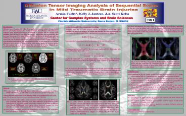
For closer investigation, the vector fields
corresponding to the dominating eigenvalue for
the three scans in regions with LIgt0.2 are shown
in fig. 3 (right). Vectors plotted on top of each
other in red, green and blue show clear
differences in the region where the colors appear
in fig. 2, indicating a reorganization of the
white matter integrity over the time span of two
weeks.
- Conclusions
- Reorganization in the white matter is found in
sequences of DTI scans taken within 24h of a
concussion and about one and two weeks later - The lattice index is a compelling measure for the
comparison of the white matter integrity as is
allows for a comparison of tensor properties in
regions (not single voxels) therefore increasing
the S/N ratio - Color component encoding is a powerful tool to
identify brain regions where the white matter
integrity in a sequence of scans differs and may
be a powerful diagnosic technique for detecting
MTBI and recovery from MTBI.
- Methods
- DTI scans from College football players who
suffered a concussion were taken within 24h of
the injury with follow up scans about one and two
weeks later - Eigenvalues and eigenvectors of the diffusion
tensor were calculated for each voxel using FSL
1 and the three scans were co-registered using
TBSS 2 - The lattice index together with a color component
encoding scheme (see below) was used to identify
regions where the scans show differences in the
integrity of the white matter. In such a scheme
the scans in the sequence are encoded by
different colors thereby highlighting brain
regions where structural changes over time take
place as colored whereas areas that do not change
appear gray - In these regions additional analysis was
performed by investigating the vector field
that corresponds to the dominating eigenvalue.
References 1 S.M. Smith et al. (2004) Advances
in functional and structural MR image analysis
and implementation in FSL. Neuroimage
23208-219 2 S.M. Smith et al. (2006)
Tract-based spatial statistics Voxelwise
analysis of multi-subject diffusion data.
Neuroimage 311487-1505 3 C. Pierpaoli, P.J.
Basser (1996) Toward a quantitative assessment of
diffusion anisotropy. Magnetic Resonance in
Medicine 36893-906
Acknowledgement Work supported by NINDS grant
48299 (JASK).


