
3- mammals : - PowerPoint PPT Presentation
1 / 46
Title:
3- mammals :
Description:
Title: GENERAL AND COMPARATIVE ANIMAL PHYSIOLOGY Biology 556 Author: Biology Last modified by: siwini Created Date: 8/27/2003 8:54:14 PM Document presentation format – PowerPoint PPT presentation
Number of Views:102
Avg rating:3.0/5.0
Title: 3- mammals :
1
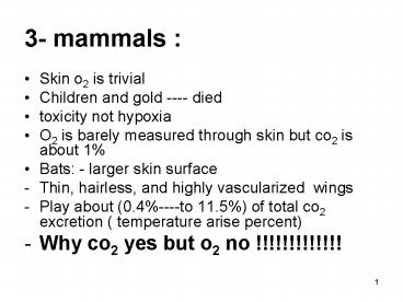
3- mammals
- Skin o2 is trivial
- Children and gold ---- died
- toxicity not hypoxia
- O2 is barely measured through skin but co2 is
about 1 - Bats - larger skin surface
- Thin, hairless, and highly vascularized wings
- Play about (0.4----to 11.5) of total co2
excretion ( temperature arise percent) - Why co2 yes but o2 no !!!!!!!!!!!!!
2
Mammals lungs
- The lung is founded in amphibians as divided sac
but - Frog lung 1 cubic cm of lung tissue 20
squarecm of gas-exchange surface - Mouse lung 1 cubic cm of lung tissue 800
square cm of gas-exchange tissue - Surface area of human lung 100 square m size
of tennis court! - Large surface area essential for high rate of
oxygen uptake required for high metabolic rate of
endothermic organisms
3
- Membrane that separates the air in the lungs from
the blood is thin 2micrometers thick (thickness
of page 50 micrometers - Large surface ( tennis court) 100m2 thin
membrane very high rate of gaze exchange
4
Lung volume
- In mammals about 5 of body weight
5
(No Transcript)
6
(No Transcript)
7
inhalation and expiration
- Volume of air taken in single breath is termed
tidal volume - A person at rest has a tidal volume 500 cubic
cm - Dead space already present in lungs (150 cubic
cm) - Therefore, only about 350 Cubic cm of fresh air
reach the lungs - Dead space space already occupied with air in
passageways, resulting in less volume for
incoming air
8
inhalation and expiration
- Lungs never completely devoid of air
- For a human, 1000 cubic cm of air left in lungs
after exhalation thus impossible for person to
fill lungs with fresh air - In respiration at rest, a person may have about
1650 cubic cm of air in the lungs when inhalation
begins - If 350 cubic cm reach the lungs, and mixed with
the 1650 already there, then renewal of air is
only about 1 in 5 (20) Result Alveolar gas
remains constant 15 oxygen 5 carbon dioxide
9
Tidal Ventilation
- Inhalation
- diaphragm, intercostals contract
- negative pressure
- Exhalation
- muscles relax
- elastic recoil pushes air out
10
- Mechanical work of breathing
- Movement of air in and out of the lungs requires
work how much? - During rest (human) 1.2 of total resting
oxygen consumption - During exercise (human) increases 3
11
(No Transcript)
12
Respiratory Membrane
Figure 22.9b
13
Respiratory Membrane
Figure 22.9c ,d
14
Physical Properties of the Lungs
- Ventilation occurs as a result of pressure
differences induced by changes in lung volume. - Physical properties that affect lung function
- Compliance.
- Elasticity.
- Surface tension.
15
Compliance
- Distensibility (stretchability)
- Ease with which the lungs can expand.
- Change in lung volume per change in
transpulmonary pressure. - 100 x more distensible than a balloon.
- Compliance is reduced by factors that produce
resistance to distension.
16
Elasticity
- Tendency to return to initial size after
distension. - High content of elastin proteins.
- Very elastic and resist distension.
- Recoil ability.
- Elastic tension increases during inspiration and
is reduced by recoil during expiration.
17
Surface Tension
- Force exerted by fluid in alveoli to resist
distension. - Lungs secrete and absorb fluid, leaving a very
thin film of fluid. - This film of fluid causes surface tension.
- Fluid absorption is driven (osmosis) by Na
active transport. - Fluid secretion is driven by the active transport
of Cl- out of the alveolar epithelial cells. - H20 molecules at the surface are attracted to
other H20 molecules by attractive forces. - Force is directed inward, raising pressure in
alveoli.
18
(Silverthorn, Fig. 17-12)
13
19
14
20
Surfactant
- Phospholipid produced by alveolar type II cells.
- Lowers surface tension.
- Reduces attractive forces of hydrogen bonding by
becoming interspersed between H20 molecules. - Surface tension in alveoli is reduced.
- As alveoli radius decreases, surfactants ability
to lower surface tension increases.
Insert fig. 16.12
21
Respiratory Distress Syndrome (RDS)
- Leading cause of death and illness in infants,
especially premature infants - 2 surfactant production pathways
- One develops 22-24 weeks
- The other develops at 35 weeks (very soon to
birth) - If type II alveolar cells do not produce enough
surfactant - Lungs collapse easily
- Hard to inflate strains diaphragm
22
Respiratory control centers
1- Medullary respiratory center
2- Pons respiratory center
(Sherwood, Fig. 13-33)
23
III. Gas exchange in air
- 4. Regulation of breathing
- Two major respiratory centers in the brain stem
- Medullary respiratory center
- Controls inspiration and expiration
- Consists of dorsal respiratory group (DRG) and
ventral respiratory group (VRG) - DRG contain mostly inspiratory neurons
(I-neurons) - VRG contain expiratory neurons (E-neurons) and I
neurons (greater than normal ventilation) - Rhythmic breathing produced by pacemaker neurons
(rostral ventromedial medulla?)
24
III. Gas exchange in air
- 2) Pons respiratory center
- Influences output from medullary respiratory
center - Pneumotaxic neurons switch off I-neurons
(limits duration of inspiration) - Apneustic neurons prevent I neurons from being
switched off - Pneumotaxic dominant over apneustic, allowing for
smooth breathing
25
III. Gas exchange in air
- Control of ventilation by PO2, PCO2 and H
- Achieved via chemoreceptors (2 types)
- Peripheral- located in the carotid bodies and
aortic bodies - Central- located on the ventral surface of the
medulla - Controls breathing via nerve fibers to the
respiratory control centers
26
Peripheral chemoreceptors
(Sherwood, Fig. 13-35)
27
III. Gas exchange in air
- Peripheral chemoreceptors
- Sense changes in arterial O2, CO2 and H
- ?PCO2 ? chemoreceptor ?sensory neurons ?
- respiratory control ctr ?motor neurons ?
respiratory muscle ??ventilation (CO2 blown off)
??PCO2 - ?H (keto or lactic acids) ? chemoreceptor ? resp
control ctr ? ?ventilation ? ?PCO2 ??H
28
III. Gas exchange in air
- Control of respiration in mammals is regulated by
changes in PCO2 (not PO2) - Peripheral O2 chemoreceptors do not contribute in
regulating normal ventilation unless arterial PO2
falls below 60 mm Hg - Peripheral O2, CO2 and H chemoreceptors are
weakly responsive and play a minor role in
controlling respiration
29
III. Gas exchange in air
- Central chemoreceptors
- Most important regulator of ventilation
- Do not monitor changes in PCO2 directly
- Respond to changes in CO2-induced production of
H in cerebrospinal fluid (brain interstitial
fluid) - Blood-brain barrier allows the diffusion of CO2
but is impermeable to H
30
Central chemoreceptor
(Silverthorn, Fig. 17-31)
31
Control of Breathing in Humans
- The main breathing control centers
- Are located in two regions of the brain, the
medulla oblongata and the pons
4
32
(No Transcript)
33
Regulation of respiration
34
Hering Breuer reflex
- ?? Mediated by vagus nerve
- ?? Hering-Breuer Reflex. Slowly adapting stretch
- receptors (SARs) in bronchial airways send
- sensory information to medulla respiratory
- centers through vagus.
- ?? If vagus is severed on both sides, lungs will
- inflate maximally and use IRV
- ?? Hering-Breuer reflex is important in adults
- during exercise when tidal volume is increased
35
Central chemoreceptors
- ?? Change in PaCO2 alters CSF pH
- ?? Increase PaCO2 will decrease CSF pH
- ?? Decrease PaCO2 will increase CSF pH
- ?? Decreased pH (Increased H) in CSF
- ?? Located on the ventral surface of medulla,
- bathed by Cerebrospinal fluid
- ?? CSF CO2 combines with water to form
- carbonic acid which dissociates to form
- hydrogen ions and bicarbonate.
36
Central chemoreceptors
- ?? The CSF H diffuse into brain tissue to
- stimulate medullary chemoreceptors.
- ?? Increased arterial H may also stimulate
- central chemoreceptors slightly, but it
- does not diffuse into CSF as easily as CO2.
- ?? Stimulates receptors to increase
- ventilation
37
(No Transcript)
38
Peripheral chemoreceptors
- ?? Located in carotid bodies at bifurcation of
- common carotid
- ?? Carotid body afferents in glossopharyngeal
- nerve.
- ?? Neural impulses from the carotid body
- increase as PaO2 falls below about 60
- mmHg
- ?? Also responds to pH
39
Peripheral chemoreceptors
- ?? Aortic bodies, afferents in vagus nerve.
- ?? Respond to PaCO2 and PO2 but not pH
40
(No Transcript)
41
(No Transcript)
42
(No Transcript)
43
Pneumotaxic center
- Located in the upper pons
- Turns off inspiratory activity
- Controls tidal volume and respiratory rate
- Normal breathing can persist without this
- center
44
Dorsal respiratory group
- Inspiration
- ?? Controls basic rhythm of breathing
- ?? Oscillations in activity are due to multiple
- inputs /- pacemaker cells
- ?? Crescendo of activity leads to inspiration
- and decreases in expiration
45
Dorsal respiratory group
- ?? Input from IXth and Xth nerves that
- terminate in nucleus of the solitary tract
- (NTS)
- ?? Output to inspiratory muscles
46
Ventral respiratory group
- ?? Expiration
- ?? Inactive in normal, quiet breathing
- ?? Inspiration (DRG) is active, and expiration
- is passive without need for VRG output to
- expiratory muscles
- ?? Increases activity with exercise
Recommended
CrystalGraphics Presentations


