
Digestive System I - PowerPoint PPT Presentation
Title:
Digestive System I
Description:
Lecture 21 Digestive System I Digestive System Anatomy Digestive tract Alimentary tract or canal Gastrointestinal (GI) tract Accessory organs Primarily glands Liver ... – PowerPoint PPT presentation
Number of Views:636
Avg rating:3.0/5.0
Title: Digestive System I
1
Lecture 21
- Digestive System I
2
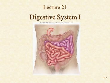
Digestive System Anatomy
- Digestive tract
- Alimentary tract or canal
- Gastrointestinal (GI) tract
- Accessory organs
- Primarily glands
- Liver, gallbladder, pancreas, salivary glands
- Regions
- Mouth or oral cavity
- Pharynx
- Esophagus
- Stomach
- Small intestine
- Large intestine
- Anus
Fig. 26.1
3
Functions
- Ingestion Introduction of food into mouth
- Mastication Chewing
- Propulsion
- Peristalsis Moves material through digestive
tract - Mass movements Moves material through large
intestine
Fig. 26.2
4
Functions
- Segmentation Segmental contraction that occurs
in small intestine - Secretion Lubricate, liquefy, digest
- Digestion Mechanical and chemical
- Absorption Movement from tract into circulation
or lymph - Elimination Waste products removed from body
Fig. 26.2
5
Oral Cavity
- Mouth or oral cavity
- Lips (labia)
- Orbicularis oris
- Cheeks
- Buccinator
- Palate Oral cavity roof
- Hard and soft
- Palatine tonsils
- Tongue
- Involved in speech, taste, mastication,
swallowing - Skeletal muscles
Upper lip
Hard palate
Soft palate
Uvula
Palatine tonsil
Tongue
Salivary duct orifices
Sublingual
Teeth
Submandibular
Lower lip
Fig. 26.3
6
Salivary Glands
- Produce saliva
- Prevents bacterial infection
- Lubrication
- Contains salivary amylase
- Breaks down starch
- Three pairs
- Parotid Largest
- Submandibular
- Sublingual Smallest
Fig. 26.4
7
Pharynx and Esophagus
- Pharynx
- Food passes through the oropharynx and
laryngopharynx
Internal nares
Opening of auditory tube
Nasopharynx
Oropharynx
Pharynx
Laryngopharynx
Esophagus
Trachea
Fig. 25.2
8
Review Question
- Food moves along the esophagus by
- Peristalsis
- Gravity alone
- Mass movement
- Force of swallowing
- Contraction of ribs
9
Pharynx and Esophagus
- Esophagus
- Transports food from pharynx to stomach
- Passes through esophageal hiatus (opening) of
diaphragm and ends at stomach - Hiatal hernia
- Sphincters
- Circular muscles
- Upper
- Lower
Oral cavity
Pharynx
Esophagus
Liver
Stomach
Fig. 26.1
10
Stomach Anatomy
Fundus
Fig. 26.12
Esophagus
Longi- tudinal layer (outer)
Cardia
Pyloric orifice
Pyloric sphincter
Three layers of smooth muscle
Circular layer (middle)
Oblique layer (inner)
Duodenum
Pylorus
Body
Gastric folds
- Openings
- Gastroesophageal to esophagus
- Pyloric to duodenum
- Parts
- Cardia
- Fundus
- Body
- Pyloric
11
Stomach Histology
- Layers
- Three layers of muscles
- Outer longitudinal
- Middle circular
- Inner oblique
Fig. 26.13
12
Stomach Histology
Fig. 26.12
- Rugae Folds in stomach when empty
- Gastric pits Openings for gastric glands
- Contain cells
- Mucous cells Mucus along surface and in pits
- Parietal cells Hydrochloric acid
- Chief cells Pepsinogen
Fig. 26.13
13
Points to Remember
- Digestive system consists of digestive tract and
accessory organs (primarily glands) - Functions include mechanical and chemical
breakdown of food, absorption of nutrients and
elimination of wastes - Mechanical and chemical breakdown start with oral
cavity - Food transported through pharynx and esophagus to
rest of digestive tract - Stomach
- Mixes food
- Protein digestion
- Limited absorption (aspirin)
14
Questions?
Recommended
CrystalGraphics Presentations


