
1' dia - PowerPoint PPT Presentation
1 / 7
Title:
1' dia
Description:
LM micrographs of striated muscle. Low power EM micrograph. High power EM micrograph ... Arrangement of the myosin molecules within the filament (250-350 ... – PowerPoint PPT presentation
Number of Views:28
Avg rating:3.0/5.0
Title: 1' dia
1
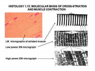
HISTOLOGY 1.12 MOLECULAR BASIS OF
CROSS-STRIATION AND MUSCLE CONTRACTION
LM micrographs of striated muscle Low power EM
micrograph High power EM micrograph
2
Structure of the myofibrils
Sarcomer 2,2-2,5 mm
Myofilaments Thick (myosin) Thin (actin)
3
Myofilaments 1. The thin filament
6-9 nm
F-actin (made up by several G-actins)
Further proteins involved
4
Myofilaments 2. The thick filament
Building block the myosin molecule
L-chains
H-chains
Heads
Arrangement of the myosin molecules within the
filament (250-350 molecule/filament)
Bipolar structure
Myosin heads
12- 15 nm
5
The sliding filament mechanism of the
contraction
6
Cytoskeletal proteins within the sarcomer
7
The sarcoplasmic reticulum and the transverse
tubular system (T-system)
Sarcoplasmic reticulum Longitudinal
tubules Terminal cisterns
triads
Triad T-tubule two adjacent terminal cisterns
of the sarcoplasmic reticulum
Recommended
CrystalGraphics Presentations


