
Female Reproductive Anatomy and Physiology - PowerPoint PPT Presentation
1 / 21
Title:
Female Reproductive Anatomy and Physiology
Description:
Endocrine function estrogen, progesterone. Exocrine function eggs ... Fundus. Body and. Cervix. Pap test. Cervical cancer is caused by a virus. Vagina ... – PowerPoint PPT presentation
Number of Views:45
Avg rating:3.0/5.0
Title: Female Reproductive Anatomy and Physiology
1
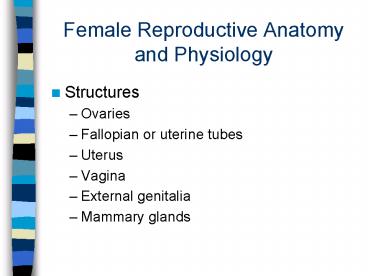
Female Reproductive Anatomy and Physiology
- Structures
- Ovaries
- Fallopian or uterine tubes
- Uterus
- Vagina
- External genitalia
- Mammary glands
2
The Female Reproductive
- System
3
Ovaries
- Female gonads - in pairs
- Produce ova and female hormones
- Endocrine function estrogen, progesterone
- Exocrine function eggs
- Have follicles in various stages of development
4
Oogenesis the process of egg formation
- In utero about 5, 000,000
- Born with about 2,000,000
- By puberty only about 400,000
- By age 50,none left
- They left through the excretory system, dried up
and went away
5
Follicles and corpus luteum
- Primary follicle
- Layers of cells around oocyte
- Secondary follicle
- Fluid filled space develops (antrum)
- Graafian follicle
- Mature follicle with the chosen oocyte for
ovulation - Corpus luteum
- Once oocyte has left ovary, follicle becomes this
endocrine tissue(estrogen and progesterone)
6
Continued
- Corpus luteum secretes
- Addition ovulation stops
- Uterine wall thickens, mammaries develop
- Corpus albicans
7
Ovulation
- FSH and LH cause rupturing of mature follicle
(Graafian, chosen one) - Secondary oocyte formed. Others degenerate after
releasing progesterone and estrogen. - Rupture site heals, leaving scar on exterior.
8
continued
- Twins
- Ordinarily one egg per month
- Fertility drugs
- Genetically predisposed to generating more than
one egg - Fraternal twins
9
Fallopian Tubes
- Receives secondary oocyte
- Tubes are not directly connected to ovaries
- Superior end opens into cavity
- Inferior end opens into uterus
- Infundibulum fringed with fimbriae which have
cilia to draw in oocyte
10
Fertilization and Implantation
- Carried to Tube by peristaltic contractions and
cilia because oocyte cannot move on its own - Fertilization will occur here usually
- Then travel down to uterus to become implanted in
the wall
11
Ectopic pregnancy
- Implantation in fallopian tube
- Terminated because of
- lack of nourishment,
- no space to develop
- Chance of rupturing and endangering mothers life
12
Uterus
- Has two linings
- Endometrium tissue
- Myometrium thick muscle
- Has three major parts
- Fundus
- Body and
- Cervix
- Pap test. Cervical cancer is caused by a virus
13
Vagina
- Removal of menstrual tissue and blood
- Semen is deposited
- Birth canal for baby
14
Mammary glands
- Modified sweat glands
- Produce milk for newborns
15
- Disclaimer
- Any numbers shown in the next few slides are
averages. Most women function in these areas
however, some do not. Some months stress or
other factors could cause a change.
16
Time line
- Day one to 14 estrogen phase. FSH
- Days 14 to 28 progesterone phase
- Progesterone
- Prepares uterus for egg implantation
- Maintains pregnancy
- Prevents further ovulation during pregnancy
- Made by adrenals, ovaries and placenta
17
Dysmenorrhea (Greek for painful menstruation.
- Primary dysmenorrhea most common
- Secondary dysmenorrhea
- Uterus is a muscle, contracts and relaxes
- Strong contractions cause blood supply to be
temporarily cut off - Caused by excessive prostaglandins
18
Maternal events of pregnancy
- Some signs of pregnancy
- Missed period
- 5th or 6th week prominent superficial veins
- 16th begin to show
- 17th and 20th feel movement
- 26th actually see movement
19
Both eggs and sperm
- Are packages for chromosomes
- Cells divide 2-4-8-16, etc.
- Cells are the same for several months,
differentiation has not occurred - Stem cells
- Males and females derive from same embryological
tissue
20
More similarities
- Come from the same place
- Develop at the same rate
- Both produce hormones
- Hormones will control primary and secondary sex
characteristics
21
Differences
- Ovaries
- Produce hormone estogen
- Produce sex cell cells, eggs
- Testes
- Produce hormone testosterone
- Produce sex cells, sperm
Recommended
CrystalGraphics Presentations


