
Histomonas meleagridis - PowerPoint PPT Presentation
1 / 40
Title:
Histomonas meleagridis
Description:
Histomonas meleagridis Cosmopolitan parasite of Birds in the order Galiformes. Causes a severe and often fatal disease called histomoniasis, blackhead in turkeys. – PowerPoint PPT presentation
Number of Views:1903
Avg rating:3.0/5.0
Title: Histomonas meleagridis
1
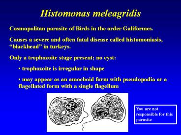
Histomonas meleagridis
- Cosmopolitan parasite of Birds in the order
Galiformes. - Causes a severe and often fatal disease called
histomoniasis, blackhead in turkeys. - Only a trophozoite stage present no cyst
- trophozoite is irregular in shape
- may appear as an amoeboid form with pseudopodia
or a flagellated form with a single flagellum
You are not responsible for this parasite
2
(No Transcript)
3
(No Transcript)
4
(No Transcript)
5
Histomonas meleagridis Life Cycle
Transmission is within the egg of the cecal
nematode of chickens and turkeys (Heterakis
gallinarum)
You are not responsible for this parasite
6
Histomonas meleagridis Life Cycle
Transmission is within the egg of the cecal
nematode of chickens and turkeys (Heterakis
gallinarum) - trophozoites from the cecum of an
infected bird are ingested by the nematode and
invade the eggs
You are not responsible for this parasite
7
Histomonas meleagridis Life Cycle
- Transmission is within the egg of the cecal
nematode of chickens and turkeys (Heterakis
gallinarum) - trophozoites from the cecum of an infected bird
are ingested by the nematode and invade the eggs - - infected eggs of the nematode are released onto
the soil where they are eaten by young birds
during pecking activities
You are not responsible for this parasite
8
Histomonas meleagridis Life Cycle
Transmission is within the egg of the cecal
nematode of chickens and turkeys (Heterakis
gallinarum) - trophozoites from the cecum of an
infected bird are ingested by the nematode and
invade the eggs - infected eggs of the nematode
are released onto the soil where they are eaten
by young birds during pecking activities - as
nematode eggs hatch in small intestine,
Histomonas trophozoites are released to invade
cecum.
You are not responsible for this parasite
9
(No Transcript)
10
Histomonas meleagridis pathology
Habitat of trophozoites Cecum Pathology
You are not responsible for this parasite
11
Histomonas meleagridis pathology
Habitat of trophozoites Cecum Pathology Young
turkeys are more susceptible to the infection
than are chickens. Mortality can reach 100 in
young turkeys - millions of dollars worth of
turkeys are lost to this parasite.
You are not responsible for this parasite
12
Look at Mr. Pro Diver!!!
13
Hello, The VISIBVILITY IS GREAT!!!!
14
Steering Wheel
Matts hose and his bubbles
This is Matt!! Holding a steering wheel of a
sunken boat!! Melissa took the picture from too
far away. Sorry Matt
15
(No Transcript)
16
Amoebic Meningitis
17
Naegleria fowleri
- Free-living in freshwater and soil including
thermal pools are bacteriophagous. - They have even been isolated from bottled mineral
water in Mexico.
18
(No Transcript)
19
Naegleria fowleri Life Cycle
20
Naegleria fowleri Pathology
- After entering the nose and nasal cavities, the
trophozoites migrate along the olfactory nerves,
through the cribriform plate, and into the
cranium.
21
Naegleria fowleri Pathology
- After entering the nose and nasal cavities, the
trophozoites migrate along the olfactory nerves,
through the cribriform plate, and into the
cranium. - Ameboid trophozoites multiply rapidly by binary
fission in the brain and cause rapid brain tissue
destruction.
22
Naegleria fowleri Pathology
- After entering the nose and nasal cavities, the
trophozoites migrate along the olfactory nerves,
through the cribriform plate, and into the
cranium. - Ameboid trophozoites multiply rapidly by binary
fission in the brain and cause rapid brain tissue
destruction. - Symptoms include a headache, fever, neck
rigidity, and mental confusion followed by coma
and death.
23
Naegleria fowleri Pathology
- After entering the nose and nasal cavities, the
trophozoites migrate along the olfactory nerves,
through the cribriform plate, and into the
cranium. - Ameboid trophozoites multiply rapidly by binary
fission in the brain and cause rapid brain tissue
destruction. - Symptoms include a headache, fever, neck
rigidity, and mental confusion followed by coma
and death. - Death usually occurs from brain destruction.
24
Trophozoites are clustered around small vessels
near the brain surface
Primary Amebic Meningoencephalits (PAM)
25
(No Transcript)
26
Figure 1. A) Computed tomographic scan note the
right fronto-basal collection (arrow) with a
midline shift right to left. B) Brain histology
three large clusters of amebic vegetative forms
are seen (H-E stain, x 250). Inset Positive
indirect immunofluorescent analysis on tissue
section with anti Naegleria fowleri serum.
27
Naegleria in Oklahoma
- Two boys, ages 7 and 9, in Tulsa, Oklahoma, died
from rare parasite Saturday August 5, 2005 from
infection with Naegleria fowleri.
28
Naegleria in Oklahoma
- Two boys, ages 7 and 9, in Tulsa, Oklahoma, die
from rare parasite Saturday August 5, 2005 from
infection with Naegleria fowleri. - The two boys were not related, but both came to
their doctors with symptoms of fever,
hallucinations, and headaches, and despite
medical care neither was able to survive the
deadly infection.
29
Naegleria in Oklahoma
- Two boys, ages 7 and 9, in Tulsa, Oklahoma, die
from rare parasite Saturday August 5, 2005 from
infection with Naegleria fowleri. - The two boys were not related, but both came to
their doctors with symptoms of fever,
hallucinations, and headaches, and despite
medical care neither was able to survive the
deadly infection. - Of the 200 known cases of Naegleria infection in
the past 40 years, only two people have survived.
Only 24 infections were documented in the U.S.
between 1989 and 2000.
30
Acanthamoeba spp.
At least 5 species of Acanthamoeba have been
identified in human tissues, this is one of the
most common amebas in soil and freshwater. Tropho
zoites occur only as amoeboid forms
31
Life Cycle Stages
Free-living trophozoites and cysts occur in both
the soil and freshwater.
32
Acanthamoeba spp.
These species cause 2 pathological effects
1) Over 100 cases of granulomatous amebic
meningoencephalitis caused by Acanthamoeba have
been documented.
33
(No Transcript)
34
2) Incriminated in a number of cases of
inflammation and opacity of the cornea.
35
Most of these ocular infections were in contact
lens wearers who used home-made saline.
36
Symptoms
- Foreign body sensation, severe ocular pain,
photophobia and blurred vision. - Often pain is more severe than signs in early
course of the disease.
37
Pathology
- Usually unilateral diffuse punctate
epitheliopathy, dendritic epithelial lesion which
may gradually progress to stromal infection
associated with ring infiltrate formation.
38
Pathology
- Usually unilateral diffuse punctate
epitheliopathy, dendritic epithelial lesion which
may gradually progress to stromal infection
associated with ring infiltrate formation. - Enlarged corneal nerve (keratoneuritis) is
pathognomonic of the infection.
39
Pathology
- Usually unilateral diffuse punctate
epitheliopathy, dendritic epithelial lesion which
may gradually progress to stromal infection
associated with ring infiltrate formation. - Enlarged corneal nerve (keratoneuritis) is
pathognomonic of the infection. - Scleritis may be found in advanced cases.
40
Acanthamoeba spp.
- Management
- Early diagnosis a prognostic factor of a
successful outcome. - Topical anti-amoeba agents.
- Penetrating keratoplasty in a severe progressive
keratitis.
Recommended
CrystalGraphics Presentations


