
Urinary Tract Infection UTI: - PowerPoint PPT Presentation
1 / 10
Title:
Urinary Tract Infection UTI:
Description:
Percutaneous Antegrade Nephrostoureterolithotomy. Laparoscopic Ureterolithotomy ... Treatment: antihypertensives, percutaneous balloon ... – PowerPoint PPT presentation
Number of Views:939
Avg rating:3.0/5.0
Title: Urinary Tract Infection UTI:
1
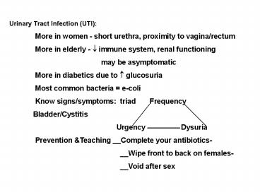
Urinary Tract Infection (UTI) More in women -
short urethra, proximity to vagina/rectum More
in elderly - ? immune system, renal functioning
may be asymptomatic More in diabetics
due to ? glucosuria Most common bacteria
e-coli Know signs/symptoms triad
Frequency Bladder/Cystitis
Urgency Dysuria Prevention
Teaching __Complete your antibiotics-
__Wipe front to back on females-
__Void
after sex
2
PYELONEPHRITIS Infection of kidney - renal
pelvis, calices ? abscess Acute or
Chronic Can be result of poor
hygiene/ sex practices Can lead to septic
shock if untreated Flank pain, fever,
chills, N V, malaise IV
antibiotics,Urinary Antiseptic,Labs,bedrest
3
RENAL ABSCESS Collection of fluid - bacterial
invasion Same s/s as pyelonephritis but not
relieved with antibiotics (Abx) May
need to surgically or percutaneously
drain RENAL TUBERCULOSIS Most common extra
pulmonary site for TB Hematogenous spread Scar
Tissue impaired renal function Treatment with
anti- tubercular therapy
4
GLOMERULONEPRITIS (Acute Nephritic
Syndrome) Also classified Infectious/Inflam.
Etiology Beta Hemolytic
Strep A Antigen- Antibody reaction Damage to
capillary membrane - allows protein/RBCs to pass
through Chronic is a leading cause of ESRD -
End Stage Renal Disease S S
Edema, abdominal/flank pain, oliguria occasiona
lly misdiagnosed as CHF Cardinal Signs
proteinuria, hematuria,? BUN and Creatinine ?
GFR - noted in 24h urine test, ? serum albumin,
hyperkalemia, uremia Care Match fluid
intake with output, Bedrest, Diruetics ,
? BP, Diet - ? protein,? CHO, ? K, ?
Na
5
NEPHROTIC SYNDROME ? Glomerular permeability
massive Proteinuria, edema hypo-
albuminemia Etiology Immune process -
agents, diseases S S
Proteinuria and Hypo albuminemia lt 3 ? albumin
edema due to ? oncotic pressure RAA system
activated ? BP Seizures May progress to
Uremia - ESRD (dialysis/transplant)
Care Steroids, Cytotoxic drugs Diet -
depends on GFR - if OK then give proteins if ?
then restrict proteins. Restrict Na Meds -
Diuretics - Lasix ? (Foley) - monitor K Teach
to monitor weight indicator of fluid retention
or diuresis
6
Polycystic Kidney Disease 100 of Nephrons are
involved 50 of people with disease develop ESRD
by age 50 Pathology Fluid filled cysts in
epithelial cells of the nephron Glomerular/Tubul
ar membranes damaged Cystic Kidney - enlarges
2-3x ? BP due to renal ischemia - RAA system
activated May also see cysts in liver and
vascular system Can lead to
aneurysms Assessment Family history Pain -
sharp or intermittent when cyst
ruptures Hematuria, Dysuria if
infection Protruding abdomen Aneurysms -
severe headache
7
Polycystic Kidney Disease Treatment
Hydration, Antihypertensives/Genetic Counseling
Pain Watch NSAIDs as
can effect renal function ASA
compounds ?? risk for bleeding
Infection Lipid soluble abx so
will penetrate cyst walls -
Bactrim or Cipro --- ? Creatinine as these
abx are Nephrotoxic
Bowels Enlarged kidney can
put pressure on intestines ? peristalsis
Weight Monitor daily
8
Extroacorporal Shock Wave Lithotripsy Sound
/ laser wave energies to break stone into
fragments Conscious Sedation Local
anesthetic agent - occasionally immersed in
water Monitor cardiac rhythm - waves
synchronized with EKG Strain urine after
procedure May be bruising on flank -
occasionally ureteral stent placed prior
procedure May have foley cath - if clots
obstruct then milk tubing
Do not irrigate unless ordered Surgery
Endoscopic - ureteroscopy stent placement
Percutaneous Antegrade Nephrostoureterolithotomy
Laparoscopic Ureterolithotomy Open
procedures - Nephrolithotomy - ? risk for
infection
9
NEPHROSCLEROSIS Blood vessels thicken - stenosis
so ? renal blood flow Occurs with HBP,
Atherosclerosis, Diabetes Can lead to
ESRD Demographics seen in African
Americans Treatment Control BP, meds (ACE
inhibitors, Diuretics - watch electrolyte
imbalances), diet RENOVASCULAR DISEASE (Renal
Artery Stenosis - RAS) Sudden onset of
hypertension Seen on Renal Arteriogram Risk for
acute renal failure from nephrotoxic drugs
Aminoglycosides - gentamycin or
Cephalosporins Radiopaque contrast
media Treatment antihypertensives, percutaneous
balloon angioplasty or surgical bypass to
restore flow
10
DIABETIC NEPHROPATHY Leading cause of ESRD -
microvascular complication
of diabetes Assessment
persistent albuminuria retinal changes often
correlate (Retinopathy) insulin remains in
system longer as kidney metabolism slows so
need less false sense of improving
diabetes Treatment Avoid fluid volume
deficit Risk for acute renal failure with
Nephrotoxic drugs or contrast media Diabetic
management
Recommended
CrystalGraphics Presentations


