
Optometry department. - PowerPoint PPT Presentation
Title:
Optometry department.
Description:
... Antidepressant drugs. (medication used to relieve depression). - Antispasmodic drugs. ... the pupils to constrict. 6-In senility when the pupils become small ... – PowerPoint PPT presentation
Number of Views:1703
Avg rating:3.0/5.0
Title: Optometry department.
1
??? ???? ?????? ??????
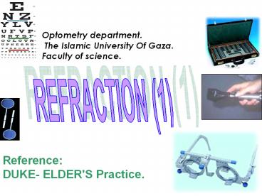
Optometry department. The Islamic University Of
Gaza. Faculty of science.
REFRACTION (1)
Reference DUKE- ELDER'S Practice. .
2
CONTENTS
- CLINICAL IMPORTANCE OF REFRACTION
- VISUAL ACUITY ( V . A.(Testing
- Testing V.A in adults
- Testing V.A in preverbal children
- Testing V.A in verbal children
- THE TESTING OF NEAR VISION
- OBJECTIVE METHODS OF REFRACTION
- RETINOSCOPY
- TRIAL FRAME (T.F.)
- TRIAL LENSES
- The power of the neutralizing lens and the
meridian of astigmatism. - The value of spherocylindrical (s-c) combination
in retinoscopy
3
CONTENTS
- REFRACTIVE STATES OF THE EYE.
- The calculation of the final refraction.
- DIFFICULTIES IN RETINOSCOPY.
- Advantages of cycloplegia.
- Disadvantage (side effects) of cycloplegia.
- SUBJECTIVE VERIFICATION OF REFRACTION.
- ASTIGMATIC FAN.
- THE ELEMINATION OF ACCOMMODATION DURING
SUBJECTIVE REFRACTION. - STAENOPIC SLIT.
- BINOCULAR CORRECTION.
- THE ORDERING OF SPECTACLE FOR DISTANCE.
4
CONTENTS
- THE RANGE AND AMPLETUDE OF ACCOMMODATION.
- FATIGUE OF ACCOMMODATION.
- SYMPTOMS OF FAILURE OF ACCOMMODATION.
- INCREASED ACCOMMODATION.
- DIMINISHED ACCOMMODATION
- Convergence.
5
- Review of geometrical optics
- Light travels through space in straight lines. If
a ray of light meets a body in its passage
through space one of three things may happen - It may be absorbed.
- It may be reflected.
- It may be transmitted. (refracted)
- When the light transmits from one medium into
another medium of different density, it is called
to be refracted. - Principles of vergence
- As applied to light rays, the term vergence
describes the direction of a ray as it passes
between some luminous point to a lens. - Vergence is the reciprocal of the distance from
the lens to the point of convergence of the
light. - Light rays that moving away from each other are
termed divergent. - Light rays that are moving toward each other are
termed convergent. - Parallel light rays have zero vergence.
6
Divergent rays convergent rays
parallel rays
Divergent rays
convergent rays
parallel rays
7
Postpone
- Visual thresholds
- Visual thresholds can be classified into three
groups - 1-Light discrimination ( which include
brightness sensitivity, brightness
discrimination, brightness contrast, and color
discrimination). - 2-Spatial discrimination (which include visual
acuity, distance discrimination, and movement
discrimination). - 3-Temporal discrimination This refers to
perception of transient visual phenomena like
flickering lights.
8
vergence
- -Light rays emanating from a point source of
light are divergent. - -Convergent light rays do not usually occur in
nature but are the result of the action of an
optical system (e.g., a lens). - -Light rays emanating from the sun are
essentially parallel and have zero vergence. - -Power (or vergence power) describes the ability
of a curved lens to converge or diverge light
rays. By convention, divergence is expressed in
minus power and convergence is expressed in plus
power. - -Diopter is the unit of measurement of the
refractive power of a lens and is abbreviated (D).
9
Postpone
- 1- The minimum visible
- It is an example of brightness discrimination
which means the ability to detect differences in
brightness of two light sources. If the target is
luminous object on perfectly dark background,
this measures the brightness sensitivity of the
eye. - 2- The minimum perceptible
- It is a measure of brightness discrimination and
concerned with the detection of fine objects such
as dots or lines against homogenous background. - 3- The minimum separable
- It refers to the smallest visual angle at which
two separate objects can be discriminated and
depends on object contrast and packing density of
photoreceptors in fovea.
10
Postpone
4- Vernier acuity (hyperacuity) It refers to
the ability of the eye to discriminate in spatial
localization and detects misalignment of two line
segments in a frontal plane if these segments are
separated by as little as 3-5 seconds of arc,
considerably less than the diameter of single
foveal cone. 5- The minimum legible It tests
the patient ability to recognize progressively
smaller letters or forms, frequently called
optotypes. The angle that the smallest
recognizable letter or symbol subtends on the
retina is a measure of visual acuity.
11
CHAPTER ICLINICAL IMPORTANCE OF REFRACTION
- 1-Anomalies (abnormalities) of the optical state
of the eye (refractive errors) are the commonest
cause of defective vision (DV) thus any patient
can not see clearly must undergo visual
examination (refraction).
12
CLINICAL IMPORTANCE OF REFRACTION
- 2-While the near sighted (myopic) child can not
see the black board clearly at school, the
presbyopic person can not read the small prints
clearly.
13
CLINICAL IMPORTANCE OF REFRACTION
- - D.V. may occur in persons having a previous
visual correction using glasses or contact lenses - - The interaction between the optical
anomalies of the eye and eye diseases has both
diagnostic and therapeutic implications like-
14
- A cataract patient may have a visual acuity
(6/12) that corrected up to (6/6) with glasses,
is not in need for cataract surgery. - Also after operation of cataract extraction
(postoperative) the patient also is in need for
refraction. - Eye examination showed a macular lesion ,but
refraction detected good visual acuity , this
gives an impression?????? that the macular
lesion is not serious.
15
EYE-STRAIN,HEADACHE AND PSYCHOLOGICAL FACTORS
- -1 In high degrees of refractive error the main
symptom is D.V. - 2- In low degrees of refractive errors the
D.V. is only one of his symptoms other symptoms
like effort to see clearly in spite of the
presence of the refractive error.
16
- 3-Condition of sustained use of accommodation in
hypermetropic persons and presbyopic persons in
near work are typical circumstances that produce
non-visual symptoms like eye pain and headache.
17
CLINICAL IMPORTANCE OF REFRACTION
- 4-The fact that the pathological basis of these
symptoms depend on fatigue of extra ocular
muscles and intraocular muscles (like ciliary
muscles) has led to the mechanical concept of the
strain that named EYE- STRAIN.
18
CLINICAL IMPORTANCE OF 20/9/2010REFRACTION
- SYMPTOMS OF EYE STRAIN
- A-Visual symptoms.
- B-Ocular symptoms.
- C-Referred symptoms.
19
- A-VISUAL SYMPTOMS
- These symptoms are intermittent in small
refractive error like 0.50 D. astigmatism, actual
visual acuity forms little or no of the symptoms
in normal conditions. - This defect can be compensated by the ciliary
muscle effort and accommodation in normal
conditions.
20
- But in conditions of long near work and effort of
study this small refractive error may result in
marked symptoms.
- There frequently comes in periods of excessive
visual strain , or during temporary deterioration
of the general health , fatigue comes on both
eyes and vision fails.
21
CLINICAL IMPORTANCE OF REFRACTION
- This is especially occurs in persons who use eyes
much for reading or study of small prints for
long time or sewing or watching T.V. for long
time or driving in difficult circumstance and all
conditions associated with attention and anxiety.
22
- DETAILS OF VISUAL SYMPTOMS
- -Sensation of confusion????????? ?????????? .
- -Temporary blurring .
- -Tiredness of the eyes.
- -Heaviness of eye lid.
- -Sensation of weariness ??????? and drowsiness.
?????? - -These symptoms are relieved by rest but recur in
continuation of the work.
23
CLINICAL IMPORTANCE OF REFRACTION h
- In deed the ciliary muscle can compensate by
exerting extra accommodation till a time will
come where ciliary muscle will fail to
compensate??????? (decompensation ( and the
symptoms could not be relieved easily until the
patient decides to seek for treatment. - 8- When reads small letters the patient sees
the letters running together.
24
CLINICAL IMPORTANCE OF REFRACTION
- B-Ocular symptoms
- These together are called asthenopia.
- These symptoms arising from excessive work of
eye muscles, in presence of a refractive error
where muscle fatigue results. - In long period of close work the eyes feel.
- Tiredness.
- Hotness.
- Uncomfort.
- Temporary relief by rest.
- In return to near work again the above symptoms
develop to eye pain.
25
- The pain of the eyes due to eye strain is mild
and aching??????? ???????? but occasionally
becomes severe and acute. - The pain may be situated to eyes or extended
(Referred) to the orbit or even to head in a form
of headache. - These eyes have a characteristic appearance
- a-Redness and congestion ?????????? of the
eyes. - b-Continued rubbing of the eyes.
- c-The eyes are watery and may be infected,
this is more noticed in children (who have a bad
habit of rubbing their eyes with their unclean
fingers).
26
- C-Referred symptoms
- The commonest one of these symptoms is headache.
- 1-Headache
- This headache is localized around the eyes
(frontal) but sometimes may be temporal ,vertical
or occipital. - The nature of the headache is dull???????
???????? , aching??????? , boring , deep seated
or migrainous. - It may be intermittent or constant , related to
the amount of use of the eyes.
27
- The aggravating factors likeeye fatigue and
poor illumination are said to be common . - N.B. No case of obscure????? headache must be
treated according to the medical lines before
eliminating the refractive errors as an etiology
of that headache.
28
CLINICAL IMPORTANCE OF REFRACTION
- 2-Digestive upset (disturbance,
disruption???????? ????????? ) - Likedyspepsia??????? and nausea.
- 3) Vague nervous symptoms
- Likedizziness???????? , insomnia ????? ??????
, and depression. ?????
29
- CHAPTER II
- VISUAL ACUITY (V . A)
- The V.A. is the function not only of the
dioptric apparatus of the eye but also of the
retina , visual pathway and central nervous
mechanisms. - V.A. is determined by the smallest retinal
image the form of which can be appreciated , and
it's measured by the smallest object which can be
clearly seen at a certain distance.
30
VISUAL ACUITY (V . A(
- In order to discriminate the form of an object
its several parts must be differentiated???????
?. - Each 2 separate cones in the macula are
stimulated (ON) while the one between them
remains unstimulated (OFF).
31
- Importance of testing Visual acuity22/0/2010
- Testing visual acuity is very important in all
of the cases because of - It gives us an accurate diagnosis for the
patients case, and indicates the severity of the
problem. - It may help us to discover a new problem at the
patient, which is not the main problem that the
patient complains from. - It also help us in following up the patient's
case, to compare between pre and post treatment.
- For all of these reasons visual acuity must be
tested for all of the patients before visiting
the ophthalmologist. And this is the mission of
the optometrist, who is the specialist examiner
in these examinations.
32
- The average diameter of the macular cone is 0.004
mm ( 4 microns), this forms the smallest
distance stimulated cones. - The normal eye should be able to appreciate a
retinal image of this size. - It was found that in order to produce an image of
minimal size (0.004 mm ) the object must subtend
an angle of one minute at the macula and this is
taken as standard of normal visual acuity .
33
VISUAL ACUITY ( V . A.(.
- These principles were included in Snellen's test
types (vision charts) ,these types consist of
letters of gradually decreasing sizes. - Each letter is of such a shape that can be closed
in a square the size of which is 5 times the
thickness of the line composing the letter.
34
Line subtends
One minute at 60 meters
One minute at 36 meters
One minute at 24 meters
One minute at 18 meters
One minute at 12 meters
One minute at 9 meters
One minute at 6 meters
One minute at 5 meters
One minute at 4 meters
35
VISUAL ACUITY ( V . A) charts
36
(No Transcript)
37
- The size of the squares consisting the breadth of
the lines subtend visual angle one minute on the
macula when they are at a specified distance
away. - Each entire letter subtend an angle of 5 minutes
at the same distance .
38
A
A
A
5minutes
The formation of the Snellens test type
39
VISUAL ACUITY ( V . A (.
- The first line of the type is constructed that
this angle is formed at distance of 60 ms, the
second letter at 36 ms , the 3rd at 24 ms, the
4th at 18 ms, the 5th at 12 ms , the 6th at 9
ms , the 7th at 6 ms. and so
40
VISUAL ACUITY ( V . A.(.
- In some charts additional lines are inserted
which subtend one minute angle at 5 and 4 meters
respectively. - If a person is placed at certain distance which
is usually taken at 6 meters , if he has normal
V.A he must read easily down to line with size 9
and the 6 size line should just be distinct.
41
- VISUAL ACUITY ( V . A.(.
- If he can not reach this limit his vision is
defective ( D.V ), but if he can exceed this
limit ,his visual acuity is above the standard (
hyper acute). - The result of the test is expressed by a fraction
the numerator of which denotes the distance while
the denominator denotes , the size of the letter
in the seen line. - Example V.A distance / numerator 6
- size/denominator
24
42
VISUAL ACUITY ( V . A.(.
- If the person can read the letter of size 6 from
6 meters his visual acuity is (6/6), if he can
see the size 9 from 6 meters his V.A.(6/9), if
he can see the size 12 from 6 meters his V.A.
(6/12), if he can see the size 18 from 6 meters
his V.A.(6/18), if he can see the size 24 from 6
meters his V.A.(6/24), if he can see the size 36
from 6 meters his V.A.(6/36), if he can see the
size 60 from 6 meters his V.A.(6/60).
43
VISUAL ACUITY ( V . A.(.
- If he can see the letter of size 5 or 4 from 6
meters his V.A. is (6/5) or(6/4) respectively
his vision is hyper acute but if he can not read
letter size 60 he is low visioned. - ????? ?? ??? ??????? ??? ???? ??? ?????
- ???? ????? ??? ??? ??????
???????
44
- In U. S. the metric system is not employed but
the feet system is used - (6 ms20 feet)
- V.A. 6/620/20 , 6/920/30 , 6/1220/40 ,
6/1820/60 , 6/2420/80 , 6/3620/120 ,
6/6020/200. - It's obvious that the corrected (aided) V.A.
varies from uncorrected (unaided) V.A.
45
VISUAL ACUITY ( V . A.(.
- In assessment of V.A. for distant the
accommodation is relaxed. - When the V.A. is corrected by spectacle lenses
fixed at the anterior focal point (15.7mm) the
V.A. is called absolute V.A. - But the spectacle lenses are normally fixed at
the B.V.D about 12mm in front of the cornea and
the V.A. is called relative V.A.
46
- THE ROUTINE TESTING OF V . A .25/9/2010
- The test type should be clearly printed .
- The test type should be legible .
- The test type should be uniformally illuminated.
- The distance between the patient and the chart is
6 ms or 20 feet.
47
VISUAL ACUITY ( V . A(.
- If this distance is unavailable the patient sits
3ms in front a plane mirror and the chart is
fixed just behind and above the patients head.
- The patient must understand what he will see to
be cooperated. - The R.E. (right eye) should always be tested
firstly except if the L.E. (left eye) has a
complaint of D.V
48
- VISUAL ACUITY ( V . A.(.
- 8.The patient reads down the chart using his
right eye (R.E) OD as far as he can , then he
repeats the test with his left eye( L. E ( OS. - 9.Then B.E. (both eyes) are tested together (OU)
(binocular V.A.), it was proved that if the V.A.
in B.E. is equal they enforce each other to see
an excessive line down the chart . - Vr (O.DOculus dexter)6/9
- Vl (O.S Oculus senester)6/9
- BE ( O.U Oculus utrique) 6/6
49
VISUAL ACUITY ( V . A.(.
- 10. If the patient can not see the largest
letter 6/60, he is asked to walk one meter
towards the chart and if he can read it his V.A.
is 5/60 but if could not ,he walks another one
meter, if can read it has V.A. 4/60 and so on
(3/60, 2/60, 1/60) after 3/60 the case called
legal blindness .
50
VISUAL ACUITY ) V. A .(
- 11 .If he could not see the letter 60 from a
distance one meter , the chart becomes useless. - 12.In a good illuminated room we ask him to count
the examiner's hand fingers at 1m or less if he
can , his V.A. is couting fingers (C.F.) , each
eye V.A is assessed separately.
51
- 13.If he could not count fingers infront his eye,
the examiner moves his fingers against a dark
back ground such as his coat and ask him what
can you see ,if he can see the moving fingers,
his V.A. is H.M. (hand movement). - 14.If he could not detect the hand movement
we keep the room light off and we use a
relatively faint light like that of direct
ophthalmoscope and ask the patient what can you
see? if the answer is seeing light his V.A. is
P.L (perception of light).
52
VISUAL ACUITY ( V . A (.
- 15. In PL ,we project the same light from
different directions on the same eye if the eye
can see the light from these directions the V.A.
is PL with good projection , otherwise PL with
poor projection. - 16. If he could not see the light his V.A. is NO
PL (complete blindness and the eye is a hopeless
eye).
53
- VISUAL ACUITY ( V . A (.
- 17. The ordinary V.A. test depends upon the
cooperation of the patient , thus it fails in - a- Malingerers ??????????? these could
be detected by adding a high sphere lens in the
trial frame say 10 Ds and ask him if he can see
the chart , he will say NO then we add -10 Ds and
ask him does he see, if he says YES he is
malingerer. - b-In illiterates ??????? for those we
use the broken ring letters (C) or (E) letter
which are useful for any nationality. The patient
is only asked to mention the direction of the
break in (c) .
54
VISUAL ACUITY ( V . A (.
- c-Young children for those we use the familiar,
figures of varying sizes as ship- car -bird -cow
doll on the principle of (c) chart, the child
has a card containing the same figures in the
chart and he is asked just to mention the figure
which is similar.
55
- 1- Testing V.A in preverbal children-
- Occlusion of one eye.
- Fixation test.
- 'Hundreds and thousands' sweet test.
- Rotation test.
- Forced choice Preferential looking tests.
- Optokinetic nystagmus drum.
56
1.Occlusion of one eye.27/9/2010
- It is a simple method for testing the visual
acuity in preverbal children and infants. - We cover one eye, and If strongly objected by the
child, indicates poorer acuity in the other eye . - This method just indicates if the V.A is good or
poor, and does not give us an accurate
measurements.
57
- 2. Fixation test.
- This is a very good method in testing V.A, and it
is performed as following
- A 16 ? base- down prism is placed over one eye
and the other is occluded with using either light
source or human face (silent smile). - The eye behind the prism is therefore forced to
elevate, to take up fixation. - The eye behind the prism is then observed.
- Fixation is then graded as central or non-central
and steady or unsteady.
58
- The other eye is uncovered and the ability to
maintain fixation just after removal of the prism
is observed - - If fixation immediately returns to the uncovered
eye, then visual acuity in covered eye is
impaired. - If fixation is maintained after a blink.
(10second) then visual acuity in covered eye is
good. - If the patient alternates fixation. Then the two
eyes have equal vision. - The test is repeated with the prism over the
other eye. - Monocular fixation should be central, steady and
maintained in each eye .
59
Correlation between visual acuity and fixation
patterns
Gross eccentric fixation or affixation. 1/60
Unsteady central fixation less than 5 seconds. 4/60
Central steady fixation, but will not hold fixation when the cover removed from the other eye. 6/60 6/24
Central steady fixation, but will hold fixation with deviating eye when the cover is removed, but prefers fixation with other eye 6/18 6/9
Alternates spontaneously, hold well with both eyes, both fixation 6/6 both eyes.
60
- 3.'Hundreds and thousands' sweet test
- Is a gross test which is seldom performed. In
principle, if the child is able to see and pick
up small sweets at 33 cm. visual acuity is at
least 6/24. - 4. Rotation test.
- Is a gross qualitative test of the ability of an
infant to fixate with both eyes open. The test is
performed as follows - The examiner holds the childs facing him and
rotates briskly through 360. - The child fixates moving targets behind the
examiner.
61
- If vision is normal the eyes will deviate in the
direction of rotation under the influence of the
vestibule-ocular response. The eyes
intermittently flick back to the primary position
to produce a rotational nystagmus. - When rotation stops, the nystagmus should also
cease due to suppression of post-rotatory
nystagmus by fixation. - If vision is severely impaired. The induced
nystagmus does not stop when rotation ceases
because the vestibulo-ocular response is not
blocked by visual feedback .
62
5. Forced choice Preferential looking tests.
- Can be used from early infancy . This behavioral
technique is based on the observation that
infants prefer to view a pattern stimulus than
homogeneous field. - Two examples are Teller acuity cards, which
consist of black stripes of varying thickness and
Cardiff acuity cards, which consist of shapes
with variable outlines. Low-frequency (thick)
gratings or shapes with a bold outline are seen
more easily than those with thin outlines. And an
assessment of visual acuity is made accordingly.
63
5. Forced choice Preferential looking tests.
- The test may be used successfully with other age
groups. - The targets are drawn with a white band bordered
by two black bands. - The Cardiff Acuity test is designed for acuity
measurement in children aged 1 to 3 years. The
targets used are pictures, all of the same size.
But decreasing in width of white and black bands.
64
5. Forced choice Preferential looking tests.
- The principle of the test is that of preferential
looking, a young child will choose to look
towards a target rather than a plain stimulus. In
the Cardiff Test, each target is positioned
either in the top half or in the bottom half of
the card. - If the target is visible the child will look
toward it, and the examiner, watching the childs
movements, can judge the position of the target
from those eye movements. - An important feature of the preferential looking
technique is that the examiner should not know in
advance the position of the target.
65
- 6- Optokinetic nystagmus drum.
- Standardized drums that contain stripes, which
subtend small fractions of the infant's visual
field, are available. May provide an estimation
of visual acuity dependent on the size of the
stripes used (alternating black and white strips
with sharp, distinct interface) that frequently
are spun at varying and uncelebrated rates, and
are bathed in variable illumination. - This method measures acuity by means of a motor
response technique (eye movement) .
66
- 2.Testing V.A in verbal children
- Allen picture cards (Kay pictures).
- The Sonksen-Silver test. HOTV test.
- Sheridan Gardner test.
- Landolt rings (C).
- Familiar tumbling E test.
67
1. Allen picture cards (Kay pictures). Are quite
useful, the near test card is slightly easier for
the younger child, but have certain
disadvantages
- Pictures are not constructed according to the
Snellen form (each element in the target
subtended 1 minute of visual angle). - Some pictures are not familiar to the child e.g.
telephone. - Pictures are variably larger than the
corresponding Snellen letter target. - Smallest target size is labeled 20/306/9.
- Despite these difficulties, most children respond
readily to this familiar and easily obtainable
test.
68
(No Transcript)
69
- 2. Sheridan _ Gardner test.
- This method requires children to match familiar
object patterns viewed at distance with those on
a near card. - Some children respond to isolated Snellen
optotypes, or graded numerical optotypes, before
linear Snellen presentations.
70
- 3.The Sonksen-Silver test. HOTV test.
- The HOTV test requires pattern recognition and
matching of progressively smaller optotypes with
those on a hand held card.
These letters are chosen to be of average
recognition difficulty and have a vertical axis
of symmetry, which obviates the issue of right
left confusion so common in this age group. An
advantage is the exact correspondence of the
target to the graded Snellen optotypes
71
Which is better???
- The main deference between the Sheridan _ Gardner
test and the HOTV test is that there is no
crowding phenomena in the Sheridan _ Gardner
test, because it show a single optotype in each
card. so it is not accurate to test an amblyopic
child with the Sheridan _ Gardner. - Because the crowding phenomena is the hallmark to
the presence of amblyopia , so it is very
important to use these two tests in the correct
way and on the suitable patients .
72
- 4. Landolt rings (C).
- Discontinuous circles, the child points to a
similar ring on a hand held card. - The test often confuses the younger child and
perhaps is more useful for illiterate adults it
does have the advantage of corresponding directly
to the Snellen chart.
5. Familiar tumbling E test Requires matching
orientation of the letter E with a figure or the
child's fingers, unfortunately, right-left
disorientation is common in this age range and
limits the usefulness of the test. Its major
advantage is the direct correspondence to graded
Snellen optotypes.
73
- Important notes
- In some cases, the patient may have a latent
nystagmus, which appears when the other eye is
occluded. - The latent nystagmus reduce the V.A due to the
inability to fixate the target. - In these cases when testing V.A, we must occlude
the eye in a way thats prevent the latent
nystagmus to occur.
74
- There is three methods to perform that
- Using a 5.00 D in front of the eye, it will
reduce the vision in a marked limit and in the
same time it will not produce nystagmus. - By putting the occluder in front of the eye at a
distance nearly 10 cm, in this way we occlude the
eye and prevent the nystagmus. - By using the frosted lens. This lens is present
in some trial cases. - This lens permits some light to enter the eye but
it also occlude the eye from seeing any thing.
And so no nystagmus will occur.
75
- THE TESTING OF NEAR VISION
- (N.V)
- 1.This occurs at a distance of 30-40cm that
called the reading distance (working distance). - 2.The first test of this kind was constructed by
Jaegar in 1867 , it consists of the ordinary
print fonts (complete set of type Printing ) of
varying size as use at that time. - 3.Recently a new test card which , approximate
Jaegar original choice are used.
76
Jaegar N.V chart
- J8 ???? ????? ??? ??? ?????? ???????
- J7 ???? ????? ??? ??? ?????? ???????
- J6 ????? ????? ??? ??? ?????? ???????
- J5 ???? ????? ??? ??? ?????? ???????
- J4, ???? ????? ??? ??? ?????? ???????
- J3, ???? ????? ??? ??? ?????? ???????
- ,J2, ???? ????? ??? ??? ?????? ???????
- J1 ???? ????? ??? ??? ?????? ???????
77
THE TESTING OF NEAR VISION (N.V)
- 4.These are traditionally called J1 ,J2, J3, J4,
J5.they are sufficient for accurate practical
purpose. - 5.In testing N.V. the patient remains ,
seated on the chair, with a good light thrown
over the left shoulder and he is asked to read at
the known reading distance . - 6.The N.V. is recorded as J1 for the smallest
line ,J2 follows it ,J3.J4. - 7.Each eye is tested separately .
78
OBJECTIVE METHODS OF REFRACTION
- I)- RETINOSCOPY -
- 1.It's the most valuable method of estimating the
optical state of the eye , it's useful and
accurate up to (0.25D) correction. - 2.In retinoscopy an illuminated area of the
patient's retina acts as an object in dark room
that reflects the light to be seen on the
patient's pupil as red reflex. - 3.If the image ( I ) formed between the eyes
of patient and the observer the red reflex (R.R)
moves opposite to the movement of the
retinoscope. - (The patient had a high myopia gt 1.5 D)
79
Retinoscope
- In high myopia
Ho
Ro
Hs
Rs
I1
No
I
Ns
Image
m
80
Retinoscopy against movement
81
I- RETINOSCOPY
- 4. If the image is formed behind the eye of
the observer (low myopia) or the eye of the
patient (hypermetropia) the red reflex moves
with the movement of the retinoscope .
82
Hypermetropia
Ho
Ro
Rs
Hs
image
A
O1
Fs
No
I1
NS
o
I
I
B
83
In low myopia
image
The image lies behind the retina of the observer
84
I- RETINOSCOPY
- 5.When the far point of the patient's eye
corresponds to the nodal point of the observer's
eye the neutral point ( end point ) is reached ,
where no movement of red reflex is noticed.
85
I- RETINOSCOPY
- 6.The working distance between the eye of the
examiner and the eye of the patient , and is the
reciprocal of the power in diopteres which should
be deduced from the power of the lenses that were
added during retinoscopy to reach the neutral
point (N.P.) - If the W.D. is 2ms we deduce 0.50D.
- If the W.D. is 0.5ms we deduce 2.00D.
- If the W.D. is 1m. we deduce 1.00D.
86
I- RETINOSCOPY
- 7.The rational ?????????? is to add lenses to
the dioptric system of the patient's eye until
the neutral point is observed by the observer. - 8. At neutral point the patient's eye refractive
error is measured by the added lenses minus the
reciprocal of the W.D. in diopters. - 9.The farther away the observer from the
patient's eye the more accurate is the result
obtained , but in practice this is
counterbalanced by the difficulty in seeing the
red reflex.
87
METHODS OF RETINOSCOPY
- A) CLASSICAL RETINOSCOPY REFLECTING RETINOSCOPY
- 1. It consists of a separate source of
light which is bright, narrow and fixed behind
and above the shoulder of the patient . - 2.The observer catches a perforate
mirror with a central opening not less than 4mm
in diameter . - 3.This plane perforated mirror reflects
the light from the source into the patients eye
which sends the image that can be seen by
observer's eye as Red reflex (R.R) through the
central opening .
88
REFLECTING RETINOSCOPY
- 4.Movements of the illuminated area of the
patient's retina are produced by tilting the
mirror. - 5.The used mirror may be plane or concave but
the plane one gives a better result.
89
- B) LUMINOUS RETINOSCOPY Advantages-
- 1.In which both of light source and
- mirror are incorporated .
- 2.It's standard modern instrument ,
- easily manipulated with the advantage that
the intensity and type of the beam can be readily
controlled. - 3.It's portable.
90
streak retinoscope
- 4.It contains a strong convex lens for
condensation of light in the patient's eye. - 5.The most ocular luminous retinoscope today is
the streak retinoscope as it produces more easily
recognized R.R. it also allows the axis of the
meridian of astigmatism to be more readily
identified separately.
91
streak retinoscope
- 6.Even greater efficiency is obtained from the
modern retinoscopy by the use of halogen bulbs
and rechargeable batteries.
92
TRIAL FRAME (T.F.)
- T.F. is used to carry the trial lenses during
- objective and subjective refraction .
- 2.T.F. should be clean , light and easily
adaptable allowing the adjustment for each eye
separately. - 3-These are essential necessity so that the trial
lenses where in place are fixed at standard
distance from the eye (B.V.D) back vertex
distance about 12 mm. and are accurately - centered.
93
TRIAL FRAME (T.F.)
94
TRIAL FRAME (T.F)
- 3.Anteroposterior adjustment is possible as well
as vertical , and horizontal adjustments . - 4.The dial (rotatable disk ) indicating the
orientation of the frame is truly positioned to
avoid the mistakes in reading the axis of the
astigmatism if present.
95
TRIAL FRAME (T.F)
- 5.Simplicity to ensure (make certain) , lightness
,and comfort fitting nose rest are of greatest
importance as some patients are very sensitive to
weight which may lead to annoyance and loss of
the patient's cooperation.
96
- TRIAL FRAME (T.F)
- 6 .Each eye of trial frame is supplied by 3 cells
(compartments) - the first is the nearest to eye is used to carry
the spherical lenses . - the middle to carry the cylindrical lenses and
- the farthest one to carry the accessories like
occluder , pinhole , staenopic slit, filters,
prismetc - 7.These cells should be close together as
possible as a considerable space between the
lenses may result in some errors in results.
97
TRIAL FRAME (T.F)
- 8.The T.F. should have its side pieces joined so
that when the near vision (with shorter
interpupillary distance) tested by reading the
glasses can be angled so that their optic axes
correspond to the downward inclination ????? of
the visual line.
98
???????????? ???????????? ???????
- ????? ????? ??? ???? ???? ????
- ?? ?? ??? ????? ???? ???????? ???? ?? ??? ??????
?? ????? ???? ???????? ??????? ?? ?? ????? ???
???? ???? ???? ?? ???? ????? ??? ???? - ?????? ???????? ??? ???????? ????? ??????
?????? ???????? ??????? ??????? ??????? ???????
??????? ?????? ?? ????? ??????? ?????? ???????
??????? ????? ?? ????? ????? ???? ??????? ???
??? ???? ??????? ?????. - ??????? ????? ?? ????? ????? ???? ??????? ???
??? ???? ??????? ?????. - ????? ??? ???? ?????
- ???? ???? ???????? ?????????? ???????
?????????????? ???????????? ??????? ( ?? ?????
31)
99
TRIAL LENSES
- 1. A typical trial set of lenses contains plus
and minus spheres every 1/4 of diopter to 4Ds
(0.25, 0.50, 0.75, 1.00, 1.25, 1.50, 1.75, 2.00,
2.25, 2.50, 2.75, 3.00,3.25,3.5,3.75,4 Ds) - 2. Then plus and minus spheres every 1/2 to 6Ds
(4.50, 5.00, 5.50, 6.00 Ds.) - 3.There after plus and minus spheres every 1 to
14 Ds - (7.00, 8.00, 9.00, 10.00, 11.00, 12.00,
13.00, 14.00Ds) - 4.Then plus and minus spheres every 2 diopters to
20 Ds (16.00, 18.00, 20.00) Ds.
100
TRIAL LENSES
TRIAL LENSES
Plus cylinder
Minus cylinder
Plus sphere
Minus sphere
101
TRIAL LENSES
- 2.It also contains plus and minus cylinder every
1/4 to 2Dc - (0.25, 0.50, 0.75, 1.00, 1.25, 1.50, 1.75,
2.00Dc) Then every 1/2 to 6 Dc. (2.50, 3.00,
3.50, 4.00, 4.50, 5.00, 5.50, 6.00)Dc. - 3.By a combination of sphere and cylinder an
excellent range of optical effect is obtained.
102
TRIAL LENSES
- 4. The trial set contains also prisms up to 10 DP
then 15 and 20 DP. - (1.00, 2.00, 3.00, 4.00, 5.00, 6.00, 7.00, 8.00,
9.00, 10.00, 15.00, 20.00)PD. - 5.It also contains accessories as plano lenses
,opaque (occluders), pin hole, staenopic slit
discs , Maddox rod , red and green filters,
centering devices and others.
103
TRIAL LENSES
- 6.All these items (1-5) are included in a trial
case. ????????? - 7.In the interest of accuracy the effective
power of the trial lenses should be as closely as
the type of the lens which should be used in the
spectacle .
104
- 6/10
- 8. The effect of spectacle lens is determined by
its back vertex power and this varies with its
position infront of the eye and its thickness. - 9.Thus the back of the trial frame should occupy
as nearly as possible the position of the
spectacle lens which must be chosen just to clear
the eye lashes averaging about 12mm infront the
cornea.
105
TRIAL LENSES
- 10.Obviously we can not stand several lenses in
T.F. in the same plane (cell) thus the ideal test
lens should therefore be calibrated (adjusted)
accurately as individual lenses but should
indicate the effectivity of the lens in the
plane, so that the effective power of a
combination of lenses in the T.F. will correspond
additively to that of a single lens in one
plane.
106
TRIAL LENSES
- 11.The test lenses should also conform?????? ,
so far as possible in form and thickness to the
spectacle lenses to be worn. - 12. In T.F. the plane surfaces of the spherical
lenses, should be where possible (fixed) next to
the eye.
107
TRIAL LENSES
- 13. The rim ?????? ?????? of the trial lens
should be mounted so that as near as possible in
the plane surface.???? ??? - 14.Before using the retinoscope the T.F. must be
accurately centered so that the optical center of
any lens lies up on the visual axis of the
patient's eye. ????? ???
108
?? ???? ???? ??? ???? ???? ????
- ??? ??? ??? ???? ??? ?? ???? ???? ??? ???? ????
???? ??? " ?? ???? ????? ??? ???? ??? ???? ??
???? ????? ????? ?????? ?????? ?????? "? ?????
??????? ?????.
109
?? ???? ???? ??? ???? ???? ????
- ??? ???? ??? " ??? ????? ???? ?? ???? ???? ?? ??
??? ??? ???? ???? ??? ????. ???? ?? ???? ??????
??????? ?? ???? ?????? ??? ???? ??? ???? ??
????? ???? ??? ????? ???? ???? ??????? ???? ????
????? ?? ???? ???? ??? ????? ???? ??? ???? ????
???? " ???? ?? ????? ?? ?????? "? ????? ???????.
110
TRIAL LENSES
- This is obtained by measuring the
- inter pupillary distance IPD of the patient
using a ruler or using 2 centering devices and
the light reflex on both cornea and the scale
above the eyes. - Autorefractometer also can give the IPD.
111
- THE PRACTICE OF RETINOSCOPY
- 1.The room should be long and darken to relax the
accommodation of the patients eye. - 2.The patient is instructed to look past the
head of the examiner in a direction opposite to
that of the examined eye. - 3.The accommodation of the examined eye must be
relaxed , this is obtained by-
112
THE PRACTICE OF RETINOSCOPY
113
THE PRACTICE OF RETINOSCOPY
- a) Fixation a spot light on the opposite wall and
to ask the patient to fix on it . - b) In absence of such a light we ask the patient
to look close observers ear and far away. - c) In children, we must use cycloplegia for
accurate refraction (temporary paralysis of
ciliary muscle) , and then it is not important if
the child fixes on the light of retinoscope.
114
THE PRACTICE OF RETINOSCOPY
- 4.In either event , in cases of squint one or
either eye should be occluded to avoid the
deviation of the examined eye. - 5.Ideally , the examiner should use his right eye
to examine the right eye of the patient and his
left eye to examine the left eye of the patient
to minimize the eccentricity. ???????
115
THE PRACTICE OF RETINOSCOPY
- 6.The examiner fits the T.F. on the patients
face with trial lenses near at hand , setting
facing the patient at a chosen distance (working
distance) usually equal , the length of arm 2/3m. - 7. The examiner directs the light of the
retinoscopy into the pupil of the patient .
116
THE PRACTICE OF RETINOSCOPY
- 8.Slow tilting of the retinoscope is started ,
noting the red reflex regarding- - A )The direction of movement of red reflex either
with or opposite to the direction of light of
retinoscope. - B )Does the plane of movement of the red reflex
parallel to the external movement (in astigmatism
it is not parallel). - C )The speed of movement of the red reflex.
- N.B. Speed of movement of the red reflex is
inversely proportional to the quantity of
refractive error .????? ????
117
??? ???? ???? ??? ???? ???? ????
- ??? ????? ??? ???? ???? ????? ???? ?????? ????
????? ??? ???? ???? ????? ???? ????? ??????
????? - - ?????? ??????? ????? (?? ????? ????).
- - ?????? ??? ???? ???? ? ??? ??????.
- - ????? ??????? ?????? ?????? ???? ??????? ?????
!. - - ?????? ??? ???? ???? ???? ????? ?? ?????!
????? ????????? ? ??????? ? ????? ? ?????? . - - ????? ????? ???? ?? ????? ?!!.
- - ?????? ??? ???? ???? ? ??? ????? ????? ??
????? ????? ?????? ????????? ?? ??? ???????? ???
????. - ??? ????? ????? (?? ????? ?????? ????? ???? ??
??? ?????? ????????????).
118
The power of the neutralizing lens and the
meridian of astigmatism
- 1. The great majority of refractions are cases
either without astigmatism (spherical refractive
error) or with regular astigmatism - (cylindrical refractive error) in which the
principal meridians are perpendicular to each
other.
119
The power of the neutralizing lens and the
meridian of astigmatism
- 2. The minority of refractions consists
- of irregular astigmatism (in which the
principal meridians are not perpendicular to each
other ) . - 3.In spherical errors the retinoscopy will show
a neutral point which is the same in all
meridians , the result is no movement in all
meridians using the same lens.
120
(No Transcript)
121
The power of the neutralizing lens and the
meridian of astigmatism
- 4. In astigmatism the situation is not quite.
- 5. The refractionist has to determine not only
the neutral point of the major and minor
meridians of the cornea but also the relation of
those regarding the difference.
122
The power of the neutralizing lens and the
meridian of astigmatism
- 6. The relationship of the direction of the
external movement to that of the red reflex has
an important role on the last matter (result).
123
The power of the neutralizing lens and the
meridian of astigmatism
- 7. The initial examination with retinoscope is
always exploratory to determine the direction of
the movement of the red reflex. - The observer starts with vertical then with
horizontal movement and lenses are inserted to
determine the neutral point in each meridian
separately.
124
The power of the neutralizing lens and the
meridian of astigmatism
- 8. If this is not so (corresponding) then the
reflex may alter its plane of movement indicating
the presence of astigmatism which is oblique. - 9. In this case the examiner must again explore
different planes of external movement of his
light until it corresponds to those of red reflex
.????? ??????
125
- 10. In presence of astigmatism, neutralizing
lenses are now found in these new meridians
starting with the meridian that is less ametropic
. - 11. Whether with or against movement is obtained
initially depends on the optical power of the
eye.
Oblique reflex
Straight reflex
126
The power of the neutralizing lens and the
meridian of astigmatism
- 12. With the examiner arm's length away from the
patient 2/3 meter. - With movement is obtained in any meridian
which is (emmetropia, hypermetropia, myopic less
than -1.50D) in such cases we add convex lenses
of gradually increasing power until neutral
point is reached.
127
With movement
128
Against movement
- 13. If an against movement is obtained in any
meridian this has myopia gt -1.50D and we insert
concave lenses until neutral point is obtained.
Add concave lenses of increasing power until
neutral point - is reached.
129
No movement
- 14. If no movement of red reflex is obtained
(neutral point is reached without any lenses)
that meridian has myopia -1.50D.
130
The power of the neutralizing lens and the
meridian of astigmatism
- 15. Neutralization itself is confirmed by filling
of the pupil with light , or the pupil becomes
totally dark in such a way that examiner is
impossible to say whether the movement of red
reflex is with or against.
131
The power of the neutralizing lens and the
meridian of astigmatism
- 16. The approach of neutralization as the trial
lenses are changed is known by an increase in
speed of the movement of red reflex but if the
used lens is a long away from the neutral point
the reflex will be slow .
132
The power of the neutralizing lens and the
meridian of astigmatism
- 17. In high degrees of ammetropia the red reflex
without lenses may be extremely faint and
becomes recognizable if a high plus or minus
spherical lens is interposed. ?????????
133
The power of the neutralizing lens and the
meridian of astigmatism
- 18. Neutralization can be altering the working
distance if the examiner bends forward , from the
position of neutral point with movement will be
obtained , and bends away from the position of
the neutral point against movement will be
obtained.
134
The power of the neutralizing lens and the
meridian of astigmatism
- 19. If marked oblique astigmatism is present
then horizontal and vertical movement of the
retinoscope will produce oblique moving reflexes
, and the external movement is adjusted ???????
??????? ?????? to correspond to these meridians
a characteristic form of neutralization is seen.
135
marked oblique astigmatism
136
What is the difference between the meridian and
the axis
- The meridian is the line that we are moving
the streak along. In the example to the left,
we are streaking the 180 degree meridian. - The axis depends on whether we are using
plus-cylinder or minus-cylinder. The power at
which equals zero.
137
Recognizing the presence of astigmatism
- When you begin retinoscopy on an eye, you will
know that there is astigmatism present in the
following situations - - 1Streaking one meridian gives you with-motion
or against- motion, and streaking the meridian
90 degrees away gives you a neutral reflex.
138
Recognizing the presence of astigmatism
- 2-Streaking one meridian gives you against-
motion, and streaking the meridian 90 degrees
away gives you with- motion. - - 3Streaking one meridian gives you with-motion
(or against- motion) with a wide streak reflex,
and streaking the meridian 90 degrees away gives
you the same motion but with a narrower streak
reflex.
139
Recognizing the presence of astigmatism
- As we add plus sphere power, the reflex at 90
narrows and the reflex at 180 quickly widens and
reaches neutrality.
140
Recognizing the presence of astigmatism
- It is easiest to practice retinoscopy on younger
adults, ages 20 to 50. - They usually have-
- clear media
- relatively relaxed accommodation,
- a definite refractometric endpoint with which to
compare your retinoscopy.
141
- As stated earlier, there is more than one way to
perform retinoscopy. If you get advice from
different sources and mix up your technique, you
will become confused???????? ????????? . The
technique described here is relatively simple and
is very accurate. - Once you have mastered the routine, it will
become second nature and you will be able to
perform retinoscopy very quickly. - Practice, practice, practice.
142
The value of spherocylindrical (s-c) combination
in retinoscopy
- 1. Spherical lenses may be used through out the
examination , and the final correcting lenses
found from the power of two principal meridians. - 2. The direction of the axis of the cylinder
being examined is more accurate , if the first
meridian is corrected with spherical lens and the
second with cylindrical lens.
143
The value of spherocylindrical (s-c) combination
in retinoscopy
- 3. The strength of spherocylindrical combination
can be verified if the examiner moves to ward or
away from the patient to confirm his
neutralization. - 4. A further advantage of using
spherocylindrical lenses together is in verifying
the position of the axis of the cylindrical lens.
144
Procedure for neutralizing an astigmatic eye
- The first step is to neutralize one of the
meridians. You will be adding plus sphere power
and streaking each of the primary meridians after
each power change. - The meridian with the narrow, fast reflex
will neutralize first. - This meridian will be 90 degrees away from
the meridian with the widest, slowest streak
reflex.
145
- 2. The next step is to confirm/identify the axis
of the astigmatism. We have a good idea of what
the second axis is from the neutralization
process. - When working in plus cylinder, we will line up
our cylinder axis with the orientation of the
streak. The axis will be 90 degrees from the
meridian with the most defined with-motion streak
reflex. ?? ?????
146
Procedure For neutralizing an astigmatic eye
- If we are using a minus-cylinder, we will line
up our cylinder axis perpendicular to the
orientation of the streak. In other words, at
90 degrees in - this example ????? ??? ?????
- we are streaking the 90
- degree meridian, and
- the axis of the correcting minus-cylinder will
be 90 degrees.
147
Procedure for neutralizing an astigmatic eye
- The final step is to subtract for our working
distance. This is usually 1.50 D and it is
subtracted from the sphere power only. - Suppose our objective result was
-1.00/-1.50x900 when we have finished
neutralizing the astigmatic meridian. - We then would subtract 1.50 D sphere power for a
final retinoscopic estimate of - -2.50DS/-1.50DCx90.
148
Procedure for neutralizing an astigmatic eye
- Once we have a neutral reflex, we have reached
the endpoint. - Neutrality can be assumed when any motion just
disappears. - This is preferable to relying on recognizing a
neutral reflex, because the reflex may appear
neutral over a wide range of power settings.
149
REFRACTIVE STATES OF THE EYE
- 1.In the normal eye (emmetrope)
- parallel rays are focused sharply on the retina.
- 2. When the relaxed unaccommodating eye is
unable to bring parallel rays from a distant
object into focus on the retina, eye is said to
be ametropic.
150
REFRACTIVE STATES OF THE EYE
- There are three basic conditions for ammetropia-
- a) Myopia (near sightedness) in which he has an
excessive convergent power of the cornea and lens
making the light to focus in front of the retina
and the error is corrected by using diverging
(-) lenses.
151
REFRACTIVE STATES OF THE EYE Myopia
152
- b) Hypermetropia (far sightedness)
- eye has an insufficient converging
- power to focus the light rays on
- the retina thus the incident parallel rays come
to focus behind the retina , we use - converging () lenses to correct hypermetropia.
153
- c) Astigmatism the cornea and some times the
lens may not have the same curvature - (radii of curvature) in all meridians the
- observation that result from corneal or
- lenticular surfaces irregular power of meridians
called astigmatism.
154
REFRACTIVE STATES OF THE EYE
- In most patients if the stronger ( steeper or
more curvature ) meridian at or close to 90
degree (astigmatism with the rule) or stronger
at or close to 180 degree (astigmatism against
the rule). - In clinical practice pure astigmatism is
corrected with cylindrical lens but in many cases
the condition is combined of myopia and
astigmatism or hypermetropia and astigmatism , in
such cases we use spherocylindrical combination
in correction.
155
- The calculation of the final refraction
- 1. This is obtained by deduction of a dioptric
value corresponding to working distance. - Thus for a working distance 2/3m arm's
length we must deduce 1.50D. - 2. Suppose that in right eye one meridian is
neutralized with 4.00Ds and the perpendicular
meridian with 6.00Ds. - .
(-1.50D)
156
- 3. After orientation ???? ?????the power of
the refraction in that eye is 2.50Ds/2.00Dc x
180? . - 4. Transposition of the lenses gives
- 4.50Ds/-2.00Dc 90?.
- 5. If the other eye is -1.00Ds/1.50Dc 90?,
Transposition of lenses gives- - 0.50 Ds/-1.50Dc x180?
3.0
1.50
- 1.0
0.50
157
The calculation of the final refraction
- 6. In the event that the 2 meridians are not
perpendicular it's possible to calculate a
suitable spherocylindrical optical equivalent
special in contact lenses. - Spherocylindrical optical equivalentSphere
power Cylinder power/2. - 7. The recording of retinoscopic result is
usually done in form of a cross which indicates
the neutral point of the meridians and other
orientation .
158
- SUBJECTIVE VERIFICATION OF REFRACTION
- For distance vision-
- 1. In the great majority of cases the
refractionist should aim at getting the vision up
to 6/5. - 2. If he can not, he must find the cause of
defect ophthalmoscopically (by use of an
ophthalmoscope ). - 3. Even in absence of eye pathology in the
media or fundus high hypermetropia or high
astigmatism often don't reach full correction.
159
SUBJECTIVE VERIFICATION OF REFRACTION
- 4. A pin hole test may give some indication of
the best vision attainable ? ???????? with lenses
if the condition is solely (purely) refractive
error. - 5. When retinoscopy has been completed the test
types (chart) are illuminated and the visual
acuity is tested with the trial neutralizing
lenses after deduction the power corresponding to
working distance.
160
- SUBJECTIVE VERIFICATION OF REFRACTION
- 6. Each eye is treated separately , while an
opaque disc (occl