
PROTEIN STRUCTURES PowerPoint PPT Presentation
1 / 21
Title: PROTEIN STRUCTURES
1
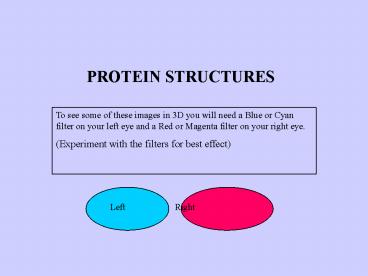
PROTEIN STRUCTURES
To see some of these images in 3D you will need a
Blue or Cyan filter on your left eye and a Red or
Magenta filter on your right eye. (Experiment
with the filters for best effect)
Left Right
2
Proteins are formed from Amino Acids joined in a
chain
AMINO ACID
Amino end
Carboxyl end
All Amino Acids have an alkaline Amino group
(-NH2) and an acid Carboxyl group (-COOH) There
are just 20 naturally occurring Amino Acids, each
with a different -R group.
3
The 20 naturally occurring Amino Acids. The
shaded box is the same for all Amino acids. It is
an single Carbon atom bonded to an Amino group, a
Carboxyl Group and a Hydrogen atom...
Disulphide Bridge
R NH2 - C - COOH
H
4
The bonds between Amino Acids are Peptide
Bonds Peptide bonds are formed by Condensation
and broken by Hydrolysis
5
A PEPTIDE BOND O H - C - N -
One Amino Acid
Another Amino Acid
- Formed by Condensation
- Broken by Hydrolysis (Heat in acid condition)
6
PRIMARY STRUCTURE
Primary structure is described by the sequence
of Amino Acids in the chain
This diagram shows the primary structure of PIG
INSULIN, a protein hormone as discovered by
Frederick Sanger. He was given a Nobel prize in
1958.
7
SECONDARY STRUCTURE
Secondary structure describes the way the chain
folds.
Some chains fold in a repeating spiral e.g.
Alpha Helix Some chains fold in a series of zig
zags e.g. Beta Pleated Sheets Some chains have
other combinations, as well as both those above.
8
The Alpha Helix
Left handed
Right handed
9
The dotted lines are hydrogen bonds which bind
the strands together, making this a strong flat
fibrous protein. In Silk, these sheets stack
together like a pile of corrugated iron. H-bonds
between the sheets make silk a very strong
structural protein
Beta Pleated Sheet
10
Poly-L-Proline These strands contain repeating
units of the Amino Acid, Proline. When three
such strands wrap around each other like rope
they make a strong fibrous protein, similar to
Collagen. Collagen is found in skin, ligaments
and tendons
11
COLLAGEN An important component of animal
connective tissue. Collagen is made from three
strands consisting mainly of Glycine and
Proline... -Gly-Pro-Pro- repeatedly wrapped into
a rope, the whole forming an Helix.
Natural Collagen may also have some other Amino
Acids inserted along parts of the chain.
12
COLLAGEN - an important animal connective tissue
Left - Collagen Fibres from the tendon of a
birds neck. Right - Collagen Fibres from a rats
tail
13
TERTIARY STRUCTURE
Tertiary Structure describes the shapes which
form when the secondary spirals of the protein
chain further fold up on themselves.
QUATERNARY STRUCTURE
Quaternary structure describes any final
adjustments to the molecule before it can become
active. For example, pairs of chains may bind
together or other inorganic substances may be
incorporated into the molecule. (Prosthetic
Groups)
14
MYOGLOBIN - An oxygen carrier in muscle similar
to Haemoglobin
Tertiary Stucture Spot the Tertiary
folding. Quaternary Structure Spot the Haem
group (Side chains have been omitted for
clarity.)
15
The Haem group looks like this...
Haem - a prosthetic group
Fe
Haem is an inorganic molecule called a Porphryn
Ring. In Haemoglobin and Myoglobin it has an Iron
ion at its centre and is a vital for the carriage
of Oxygen. Such an inorganic structure, required
to make a protein active, is called Prosthetic
Group and this is part of the Quaternary
structure of Myoglobin and Haemoglobin. A similar
Porphryn Ring, with Magnesium at its centre is
the essential part of a Chlorophyll molecule.
16
MYOGLOBIN - An oxygen carrier in muscle Here is
another way of visualising tertiary the structure
Tertiary Stucture Spot the Tertiary
folding. Quaternary Structure Spot the Haem
group
17
FIBROUS PROTEINS Fibrous proteins like Collagen,
Silk, Keratin (hair) and Chitin (insect
exoskeleton) tend to be insoluble and strong and
so they have a structural role for support or
protection.
GLOBULAR PROTEINS Proteins which fold into a ball
or globule like Myoglobin are called Globular
Proteins. They tend to be soluble. The most
common group of Globular Proteins are ENZYMES
which control the reactions in living cells.
(They work because their Globular shape includes
an Active Site).
18
LYSOZYME An enzyme found in tears and mucus which
acts as a strong anti-bacterial agent because it
digests bacteria cell walls. It has 129 Amino
Acids folded in both alpha helices and
anti-parallel hairpins. Can you see the active
site? (Side chains omitted for clarity)
19
LYSOZYME Including the Side chains. Can
you see any active site now?
20
CHYMOTRYPSIN A protein digesting enzyme in your
stomach. Only the carbon backbone is shown so
the secondary structure can be seen. Some parts
are twisted into an Alpha Helix. Other parts (in
bold) are the straight Beta strands. Spot the
Active Site
21
ENZYME-SUBSTRATE COMPLEX
This shows Subtilisin - a bacterial enzyme
binding with its substrate. The substrate is a
short polysaccharide made from six sugar
molecules, which is part of the bacterial cell
wall. The substrate (in bold) is wedged into its
active site and held in place by the H-bonds
shown as dotted lines. This is the Enzyme-
substrate complex which forms for a brief moment
during any enzyme controlled reaction.
Recommended
