
Overview of the Cellular Basis of Life PowerPoint PPT Presentation
1 / 37
Title: Overview of the Cellular Basis of Life
1
Chapter 3 Cells and Tissues
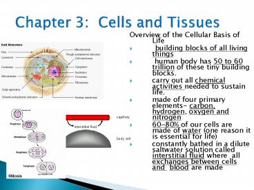
- Overview of the Cellular Basis of Life
- building blocks of all living things
- human body has 50 to 60 trillion of these tiny
building blocks. - carry out all chemical activities needed to
sustain life. - made of four primary elements- carbon, hydrogen,
oxygen and nitrogen - 60-80 of our cells are made of water (one reason
it is essential for life) - constantly bathed in a dilute saltwater solution
called interstitial fluid where all exchanges
between cells and blood are made
2
II. Anatomy of a Cell
- Anatomy of a Cell
- Cells are organized into three main regions
- 1. Nucleus that contains DNA
- 2. Cytoplasm
- 3. Plasma Membrane (cell membrane)
- a. barrier for cells contents
- b. double phospholipids bilayer
- c. Selectively permeable- regulates what enters
and leaves the cell
3
d. Plasma membrane junctions the meeting of two
adjacent cell membranes. There are three types
- Tight junctions- fit together like a zipper
forming an impermeable junction. These exist in
cells lining the digestive tract to keep
digestive enzymes and microorganisms from seeping
into the bloodstream. - Desmosomes- anchoring junctions that prevent cell
separation. These are abundant in tissues that
are subjected to great mechnical stress, such as
skin cells. - Gap junctions- the cells are connected by hollow
cylinders (connexons) which allows chemical
substances to pass between cells. These are
abundant in heart muscle tissue where the
movement of ions from cell to cell to synchronize
the heart rhythm.
4
Cell Diversity
- All cells share the same general structures a
cell membrane, a nucleus and cytoplasm. However,
their function in the body is different. See
examples below
Secretory vs. Absorptive Epithelial Picture
5
Eukaryotic Cell
6
- 4 Tissue types found in the body
7
III. Body Tissues
- Tissues are groups of cells with similar
structure and function. - Four primary tissues types
- 1. Epithelium (covering)
- 2. Connective (support)
- 3. Nervous Tissue (control)
- 4. Muscle (movement)
8
IV. Epithelium
- Found in different areas of the body, such as
body coverings, body linings, and glandular
tissue. - Functions are for protection (skin), absorption
(small intestine), filtration (kidneys), and
secretion (glands). - Characteristics of epithelial tissue include
- 1. Cells fit closely together
- 2. Tissue layer always has one free surface (the
apical surface) that is exposed to the cavity of
an internal organ or the bodys exterior. - 3. The lower surface is bound by a basement
membrane. - 4. Avascular (these tissues have no blood supply
of their own) - 5. Regenerate well if nourished.
- .
9
Classification of epithelium- not in your notes
but may want to write
- Number of cell layers
- simple- one layer
- stratified- more than one layer
- Shape of cells
- squamous- flattened
- cuboidal- cube shape
- columnar- column- like
10
- SIMPLE SQUAMOUS - single layer (simple) of very
thin, flattened cells (squamous). - Function diffusion and filtration. Found in air
sacs of lungs, walls of capillaries.
11
- SIMPLE CUBOIDAL - single layer, cube-shaped
cells. Function Secretion and absorption. Found
Lining of kidney tubules, ducts of glands,
covering surface of ovaries
12
- SIMPLE COLUMNAR - single layer, elongated cells
with their nuclei in about the same position in
each cell (usually near the basement membrane).
Protection, secretion, absorption. Found in the
lining of digestive tract and uterus- contains
scatter goblet cells functioning in the secretion
of mucus- some columnar cells (involved in
absorption) have tiny finger-like processes from
their free surface called microvilli (increases
surface area)
13
- STRATIFIED SQUAMOUS - muli-layered, squamous
cells. Thicker tissue.Functions in protection.
Found lining body cavities like the mouth and
outer layer of skin
14
- PSEUDOSTRATIFIED COLUMNAR - appear "stratified"
but really a single layer with nuclei at various
levels giving the appearance of layered cells.
Usually ciliated (tiny, hair-like projections for
sweeping materials along a surface). Contains
goblet cells.- Function secretion and
cilia-aided movement- Location lining air
passages like the trachea and tubes of the
reproductive system
15
- TRANSITIONAL EPITHELIUM - thick, layered cuboidal
cells. "Stretchable" tissue, also forms barrier
to block diffusion. Found lining of urinary
bladder.
16
Types of Epithelium
Thin for gas exchange between alveoli and
capillaries
Can secrete mucus and have cilia to help clean air
Some cells Shorter and nuclei Appear at
different Heights above membrane
Contain lots of golgi and ER to manufacture and
secrete or absorb
Many layers of cells to replace those lost when
swallowing
Common in glands
Adapted for secretion (goblet cells) of mucus
and ciliated to propel food
Stretches as bladder fills with urine
Cells at basal layer Are cuboidal or columnar
and vary at the free surface
17
Connective Tissue
- A Found everywhere in the body, the most abundant
and widely distributed tissue. - Functions include binding tissues together,
support, and protection - Characteristics of connective tissue
- 1. Variations in blood supply- some tissue types
are well vascularized (have good blood supply),
while some have a poor blood supply (tendons and
ligaments). Cartilage is avascular.
18
Connective Tissue
- 2. Extracellular matrix- the nonliving material
that surrounds the tissue. (This is what makes
connective tissue so different from other
tissues.) - Matrix is composed of a ground substance (water,
protein, and other molecules) and fibers
(collagen, elastic, reticular). - The matrix allows connective tissue to act as a
soft packing tissue around organs (adipose
tissue), to bear weight, or withstand stretching
and abrasion (bones, tendons and ligaments).
- Mast cells (prevents clots)
- ? Macrophages (consumers)
- ? Fibroblasts (produce fibers)
19
Connective Tissue D. Connective tissue types
- Bone (osseous) - composed of bone cells, hard
matrix of calcium salts, large numbers of
collagen fibers. - used to protect and support the body
20
Connective Tissue D. Connective tissue types
- Hyaline Cartilage- most common type of cartilage,
composed of collagen and matrix - Entire fetal skeleton is hyaline cartilage, but
by the time of birth, most cartilage is replaced
by bone.
21
V. Connective Tissue D. Connective tissue
types
- Elastic cartilage- provides elasticity
- Example- supports the external ear
- Fibrocartilage- highly compressible
- Example- forms cushion-like discs between
vertebrae
22
V. Connective Tissue D. Connective tissue types
- Dense connective tissue- main matrix element is
collagen fibers which form strong rope-like
structures, (the collagen producing cells are
called fibroblasts) - Example- tendons (attach muscle to bone),
ligaments (attach bone to bone)
23
V. Connective Tissue D. Connective tissue types
- Areolar connective tissue- most widely
distributed connective tissue that serves as a
kind of universal packing material between other
tissues - contains all fiber types,
- can soak up excess fluid (this is the tissue that
swells causing edema) - universal packing tissue and glue that holds
internal organs together
24
V. Connective Tissue D. Connective tissue types
- Adipose tissue- commonly called fat
25
V. Connective Tissue D. Connective tissue types
- Blood- blood cells surrounded by a fluid matrix
- fibers are visible during clotting
- functions as the transport vehicle for materials
26
IV. Muscle Tissue
- Functions to produce movement
- Three types are
- Skeletal muscle- voluntary, striated
- Smooth muscle involuntary, surrounds organs
- Cardiac muscle- involuntary, only in heart,
striated - Intercalated disks are the junctions that allow
heart cells to rapidly conduct electrical
impulses through the heart.
27
VII. Nervous Tissue
- Composed of neurons and nerve support cells
- Functions to send and receive impulses to other
areas of body - Located in nervous system structures such as the
brain, spinal cord and nerves.
28
How can we tell the difference between
connective, muscle, epithelial and nervous tissue?
29
What are mast cells?
- Mast cell A connective tissue cell whose normal
function is unknown but which is frequently
injured in allergic reactions, releasing
chemicals including histamine that are very
irritating and cause itching, swelling, and fluid
leakage from cells. These allergic chemicals may
also cause muscle spasm and lead to lung and
throat tightening, as in asthma. Also known as a
mastocyte or labrocyte.
30
Wound healing videos
- http//www.youtube.com/watch?vFraKUUetOpc
31
3 Phases of Wound Healing
- Inflammatory-
- Redness, swelling, pain are symptoms
- Increase in capillary permeability
- Clotting occurs (platelets)
- Phagocytes (type of WBC) moves in
- Growth factors attract fibroblasts (cells that
make matrix and collagen)
32
Proliferative Phase
- Granulation tissue forms, this involved
fibroblast cells actively secreting collagen - Angiogensis- new blood vessels form
- Endothelial cells from granulation tissue- which
is the foundation of scar tissue development - Epithelialization- new epithelial cells
- White blood cells leave, swelling goes down
33
Remodeling Phase
- Final scar tissue formed
- Scar becomes avascular
34
VIII. Tissue Repair (Wound Healing) A. Two
types of tissue repair 1. Tissue regeneration
is the replacement of destroyed tissue by the
same kind of cells 2. Fibrosis occurs when
repair by dense fibrous connective tissue
called scar tissue forms. Fibrosis occurs in
cardiac and nervous tissues of the body. B.
The type of tissue repair depends on the type of
tissue damage and the severity of the injury.
35
C. Steps in Tissue repair 1. Capillaries become
very permeable. 2. Clotting proteins, platelets,
macrophages, and other substances seep into the
injured area. 3. A clot is constructed to wall
off the injured area (when the clot dries and
hardens this forms the scab). 4. Formation of
granulation tissue (delicate tissue that is
made of new capillaries that grow into the
damaged area). Replaces the clot! a. this
tissue also contains fibroblasts that synthesize
collagen fibers that bridge the gap 5. Surface
epithelium regenerates this covers an
underlying layer of fibrosis (the scar).
36
(No Transcript)
37
- D. The regeneration of tissue
- 1. Tissues that regenerate easily epithelial,
fibrous connective, and bone - 2. Tissues that regenerate poorly skeletal
muscle - 3. Tissues that are replaced largely with scar
tissue cardiac and nervous tissue within the
brain and spinal cord. Scar tissue lacks the
normal flexibility of tissues which hinders the
functioning. - As we age there is a decrease in mass and
viability of most tissues. The epithelia thin,
the amount of collagen in the body declines which
makes tissue repair less efficient, and nervous
tissues begins to atrophy.
CNS neurons cannot regenerate, but PNS neurons
can!
Recommended
