
Chapter 7 Body Systems PowerPoint PPT Presentation
1 / 55
Title: Chapter 7 Body Systems
1
(No Transcript)
2
(No Transcript)
3
Introduction- Chapter 6
- Skin (integument) is bodys largest organ
- Approximately 1.6 to 1.9 m2 in average-sized
adult - Integumentary system describes the skin and its
appendages the hair, nails, and skin glands
4
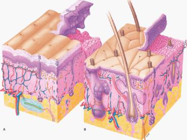
Structure of the Skin
- Skin classified as a cutaneous membrane
- Two primary layersepidermis and dermis joined
by dermal-epidermal junction - Hypodermis lies beneath dermis
5
Structure of the Skin
- Thin and thick skin
- Thin skincovers most of body surface
- 1 to 3 mm thick
- Thick skinsoles and palms
- 4 to 5 mm thick
- Makes fingerprints
- No hair
6
Structure of the Skin
- Epidermis
- Cell types
- Keratinocytes
- Melanocytes
- Langerhans cells
7
Keratinocytes
- constitute over 90 of cells present
- principal structural element of the outer skin
- Arranged in strata, or layers
8
Melanocytes
- pigment-producing cells (5 of the total)
- contribute to skin color
- filter ultraviolet light decrease the amount of
UV light that can penetrate into the deeper
layers of the - skin
9
Langerhans cells
- they play a role in immune response
- Originate in the bone marrow
- Function with specialized leukocytes called
helper T cells, to trigger immune response
10
Structure of the Skin
- Epidermis
- Cell layers- 5 Layers (deep ? superficial)
- Stratum germinativum (growth layer)describes the
stratum spinosum and stratum basale together - Stratum basale (base layer)
- Stratum spinosum (spiny layer)
- Stratum granulosum (granular layer)
- Stratum lucidum (clear layer)
- Stratum corneum (horny layer)
11
Structure of the Skin
- Epidermis
- The cells form in the basale layer and degenerate
and fill with keratin as they move up to the
surface - This is keratinization
12
section reveals epidermis containing basal,
spinous, granular keratinocytes and stratum
corneum. Dermis contains numerous viable
fibroblasts. 400X)
13
1. Stratum basale (base layer- deepest)
- single layer of columnar cells
- only these cells undergo mitosis
- ? migrate through the other layers until they
are shed - The renewal of the human
- epidermis takes
- about 3 to 4 weeks.
14
2. Stratum spinosum (spiny layer)
- cells arranged in 8 to 10 layers with desmosomes
(bridges) that pull cells into spiny shapes - cells rich in RNA ? protein synthesis to produce
keratin
15
3. Stratum granulosum (granular layer)
- cells arranged in two to four layers
- filled with granules ? makes keratin
- contain high levels of lysosomal enzymes ?
theyre starting to degenerate - Nuclei absent
- May not be in regions of thin skin
16
4. Stratum lucidum (clear layer)
- Closely packed and clear keratinocytes
- cells filled with eleidin (ay lee din) ? blocks
water penetration or loss (eventually makes
keratin) - absent in thin skin (in thick skin- hands and
feet) - Nuclei absent
17
5. Stratum corneum (horny layer)
- Dead, squamous cells
- most superficial layer
- dead cells filled with keratin ? water-repellent
protein - Cell membranes are thick and chemically resistant
- Also called barrier area because it functions to
prevent water loss
18
Structure of the Skin
- Thick Skin
- Thin Skin
19
Wrinkled Fingers and Toes
- FYI
- dead cells in stratum corneum absorb water
- (deeper layers do NOT)
- When swollen water-filled cells expand over the
normal sized cells below, the skin wrinkles
20
Structure of the Skin
- Epidermis
- Epidermal growth and repair
- Turnover or regeneration time refers to time
required for epidermal cells to form in the
stratum basale and migrate to the skin surface
about 35 days - Shortened turnover time will increase the
thickness of the stratum corneum and result in
callus formation
21
Structure of the Skin
- Epidermal growth and repair continued
- Normally 10 to 12 of all cells in stratum
basale enter mitosis daily - Each group of 8 to 10 basal cells in mitosis with
their vertical columns of migrating keratinocytes
is called an epidermal proliferating unit, or EPU
22
Structure of the Skin
- Dermal-epidermal junction
- Definite basement membrane
- glues the epidermis to the dermis below
- Partial barrier to the passage of some cells and
large molecules
Specific staining of the indicated protein is
displayed as a red band localized at the
dermal/epidermal junction.
23
Blisters
- FYI
- Caused by injury to cells in epidermis OR
- From separation of dermal-epidermal junction
24
Structure of the Skin
- Dermis (corium)
- Sometimes called true skinmuch thicker than
the epidermis and lies beneath it - Gives strength to the skin
Sunburn cell formation in EpiDerm-FT following
solar UV-irradiation. HE stained paraffin
sections were prepared from EpiDerm-FT 24 hr
after irradiation. Sunburn cells are indicated by
arrows.
25
Structure of the Skin
- Dermis (cont.)
- Contains various structures
- Arrector pili muscles and hair follicles
- Sensory receptors
- Sweat and sebaceous glands
- Blood vessels
- Rich vascular supply plays a critical role in
temperature regulation? homeostasis
26
Structure of the Skin
- Dermis (cont.)
- 2 Layers of dermis
- Papillary layer
- Reticular layer
27
Dermis-Papillary Layer
- composed of dermal papillae that project into the
epidermis - ? fingerprints- used for grasping and gripping
tools - contains loose connective and elastic fibers
- contains the dermal-epidermal junction
- Like egg crate foam mattress pad
28
Dermis- Reticular Layer
- contains interlacing collagenous fibers and
elastic fibers - ? make the skin tough yet stretchable
- when processed from animal skin, produces leather
- Attachment point for muscle fibers (smooth and
skeletal) - Contains arrector pili muscles
29
(No Transcript)
30
Arrector pili muscles
- Bundle of smooth muscles on each hair follicle
- Makes the hair stand on end
- Occurs due to fright or cold
- Causes erection of nipples and elevation of testes
31
- Where are the arrector pili?
- Where is the adipose tissue?
- Is adipose deep or superficial to the stratum
lucidum?
32
Dermis
- Dermal growth and repair
- The dermis does NOT continually shed and
regenerate - During wound healing, begins forming a dense mass
of new connective fibers ? scar - If elastic fibers in dermis stretch too much,
they TEAR and form ? stretch marks - Eventually lose color- NOT due to cocoa butter
33
FYI
- If an incision cuts across cleavage lines
(Langers lines), stress tends to pull the cut
edges apart and may retard healing. - Surgical incisions parallel to cleavage lines are
subjected to less stress and tend to heal more
rapidly.
34
Hypodermis
- Also called subcutaneous layer or superficial
fascia - Deep to the dermis
- forms connection between the skin and other
structures - NOT part of the skin
35
Skin Color
- Melanin
- Basic determinant of skin color
- Melanin formed by melanocytes
- Albinism
- congenital
- absence of
- melanin
36
(No Transcript)
37
Functions of the Skin (Table 6-1 Page 171)
- Protection
- Physical barrier
- Prevents dehydration
- Sensation
- Pain
- Heat and cold
- Pressure and touch
38
Functions of the Skin contd (Table 6-1 Page 171)
- Movement growth
- Imagine moving without elastic skin
- Endocrine (hormones)
- vitamin D production
- Excretion (minor role)
- Water Urea/ammonia/uric acid
39
Functions of the Skin contd (Table 6-1 Page 171)
- Immunity
- Phagocytic cells (phago- to eat) (-cyte-
cell) - Langerhans cells
- Temperature Regulation
- Heat loss or retention
40
Temperature Regulation- Vasodilation
- Heat lossapproximately 80 of heat loss occurs
through the skin - Increased blood flow to epidermis
- Decreased blood flow to organs
- Redness after exercise
41
Temperature Regulation- Vasoconstriction
- Heat retention
- Decreased blood flow to skin
- Increased blood flow to organs
- Turning white
- Vasoconstriction
42
How do we sense touch?
- MANY receptors in our skin (mostly dermis) allow
for different touch stimuli - Meissners corpuscle- detects light touch-
superficial dermis - Pacinian corpuscle- detects pressure- deep dermis
- ?Receptors send signals to the brain (those
signals were called?)
43
Somatosensory Cortex in Brain
- Receptors send action potentials to specialized
part of brain ? somatosensory cortex (soma-
body) (cortex- outermost, superficial) - Each part of the body
- corresponds with a
- particular area in the
- cortex
- Also a corresponding
- Motor cortex
44
Homunculi (Page 394)
- Both sensory motor cortex have homunculi
(little man) - Hands and face- large part of cortical area
- Most important for survival
45
Two point discrimination
- The density of receptors varies
- If the hands and the face have the most cortical
area, where do you think the densest area of
receptors will be?
46
Skin Glands
- 3 Types
- Sweat (2 types)
- Sebaceous
- Ceruminous
47
Skin Glands- Sweat Glands
- Eccrine glands
- Most numerous sweat glands very small
- Distributed over total body surface except a few
places (ear - canal, lips)
- Simple, coiled, tubular glands
- Secrete sweat ? constant
- core temperature
48
Skin Glands- Sweat Glands
- 2. Apocrine glands
- Located deep in subcutaneous layer
- Limited distributionaxilla (armpit), areola of
breast, and around anus - Large (gt 5 mm in diameter)
- Connected to hair follicle
- Begin to function at puberty
49
Skin Glands
- Sebaceous glands
- Secrete sebum (oil)
- Hair and skin
- prevents excessive water loss from the skin
- antifungal activity
- dermis except in palms and soles
- Secretion increases in adolescence
- Oxidated sebum accumulations ? blackheads
Stages of acne. (A) Normal follicle (B) open
comedo (blackhead) (C) closed comedo
(whitehead) (D) (E) pustule v
50
Blackhead
Whitehead
Papules
51
Skin Glands
- Ceruminous glands (se ROO mi nus)
- Modified apocrine sweat glands
- Mixed secretions of sebaceous and ceruminous
glands called cerumen (wax) - protects from dehydration
52
Cycle of Life Skin
- Children
- Skin is smooth, unwrinkled, and characterized by
elasticity and flexibility - Few sweat glands
- Rapid healing
53
Cycle of Life Skin
- Adults
- Development and activation of sebaceous and
sweat glands - Increased sweat production
- Body odor
- Increased sebum production
- Acne
54
Cycle of Life Skin
- Old age
- Decreased sebaceous and sweat gland activity
- Wrinkling
- Decrease of bodys ability to cool itself
55
The Big Picture Skin and the Whole Body
- Skin is a major component of the bodys
structural framework - Skin defines the internal environment of the
body - Primary functions are support and protection
Recommended
