The Upper Limb - in context - PowerPoint PPT Presentation
1 / 33
Title: The Upper Limb - in context
1
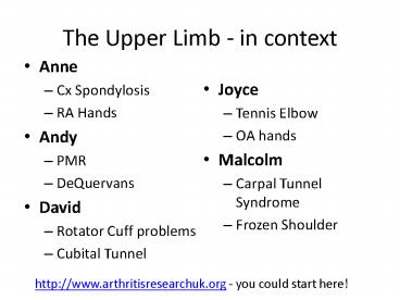
The Upper Limb - in context
- Anne
- Cx Spondylosis
- RA Hands
- Andy
- PMR
- DeQuervans
- David
- Rotator Cuff problems
- Cubital Tunnel
- Joyce
- Tennis Elbow
- OA hands
- Malcolm
- Carpal Tunnel Syndrome
- Frozen Shoulder
http//www.arthritisresearchuk.org - you could
start here!
2
The Upper Limb - in context
- Bring laptops / other IT equipment to prepare a
2-3 slide thumbnail about the condition - Features in Diagnosis
- Possible helpful tests and
- Management options
3
Objectives
- Upper limb disorders are common in general
practice 3rd most common M/Sk presentation and
shoulder pain alone accounts of 5 of all GP
encounters.... - By the end of this session we hope you will be
able - To approach upper limb disorder diagnosis with
more confidence - To examine the upper limb with greater competence
and efficiency - To be able to recommend evidence-based sensible
options for up to date management - To work together as a team to produce
well-focused presentations
4
Afternoon programme
- 220 work in groups prepare 2 x short power
points on the 2 conditions per cluster
respectively - 250 work in groups of 4 on cases until tea.
Hand in diagnosis as soon as group certain. - Diagnosis correct no extra prompts 10 points
- Diagnosis correct 1 extra prompt
8 points - Diagnosis correct 2 extra prompts 5 points
- Diagnosis correct - 3 extra promts
3 points - 320 tea
- 340 Presentations broken up by use of DVD and
final cases - 450 Big Prize for highest-scoring group and
round up - 500 - End
5
Case 1
- Mr Desai is 56 is a non-smoker and a type 2
diabetic. He has been a rare visitor at the
surgery over the years until 10 months ago when
he had an MI. He had successful stenting and is
on the usual medications and has done cardiac
rehab well, now doing regular exercise himself. A
month ago he began to complain of pain in his
right shoulder which you have examined and now
his left is causing him the same pain. He cant
recollect any trauma. The pain is in the deltoid
area, prevents him reaching up, reaching forward
and is keeping him awake.
6
Additional info, case 1
- The onset was insidious
- He has pain and significant, solid limitation on
external rotation of the gleno-humeral joint,
both actively and passively. - X rays of the shoulder are normal
7
Shoulder pain
Shoulder pain is the third most common reason
for musculoskeletal consultation in general
practice, after back and neck pain. Adults
present to primary care with shoulder pain with a
prevalence of 2.36 and incidence of 1.47
Shoulder pain accounts for 5 of all GP
encounters. In a study of adults consulting for
shoulder pain in a UK primary care setting a,
peaking at 50 years and showing a linear increase
with age. Self-reported prevalence of shoulder
pain is between 16 and 26 with a lifetime
prevalence of over 30 in adults.
8
Adhesive capsulitis
- Prevalence of about 3 in the adult population.
10- 35 diabetics - Usually sixth decade of life, and onset before
the age of 40 is very uncommon. The peak age is
56, and the condition occurs slightly more often
in women than men - 39 full recovery, 54 clinical limitation
without functional disability, and 7 functional
limitation - Three phases of clinical presentation
- Painful freezing phase
- Duration 10-36 weeks. Pain and stiffness around
the shoulder with no history of injury. A nagging
constant pain is worse at night, with little
response to non-steroidal anti-inflammatory drugs
- Adhesive phase
- Occurs at 4-12 months. The pain gradually
subsides but stiffness remains. Pain is apparent
only at the extremes of movement. Gross reduction
of glenohumeral movements, with near total
obliteration of external rotation - Resolution phase
- Takes 12-42 months. Follows the adhesive phase
with spontaneous improvement in the range of
movement. Mean duration from onset of frozen
shoulder to the greatest resolution is over 30
months
9
Adhesive capsulitis
- Summary points
- True frozen shoulder is a clinical diagnosis
- The three hallmarks of frozen shoulder are
insidious shoulder stiffness severe pain, even
at night and near complete loss of passive and
active external rotation of the shoulder - Lab tests are normal
- Frozen shoulder is rare under the age of 40 the
peak age is 56 - Frozen shoulder progresses through three clinical
phases - It lasts about 30 months, but recovery can be
accelerated by simple measures - Physiotherapy alone is of little benefit,
although steroid injection is effective and best
combined with physiotherapy - Refractory cases can be referred for manipulation
under anaesthesia and, rarely, arthroscopic
release - Nearly all patients recover, but normal range of
movement may never return
10
Rotator cuff problems
- Mechanical impingement is the most common
recognisable source of recurring rotator cuff
pain and disability in the active population - Tearing of the rotator cuff as a function of
age is a common occurrence, and may be clinically
silent - Often, the diagnosis can be made by history and
clinical examination alone
11
Rotator cuff problems
USS/ MRI scans for those anticipating shoulder
surgery can be helpful in evaluating tears and
muscle atrophy and in establishing the presence
of co-morbidities
- Most patients with symptomatic rotator cuff
disease respond to non-operative treatment - Early surgical management should be considered
for acute rotator cuff tears in physiologically
young and active individuals
12
- Subacromial impingement is defined as shoulder
pain resulting from the catching of the rotator
cuff under the coracoacromial arch of the
shoulder. Repeated impingement can lead to a tear
in the tendon of rotator cuff muscles which can
be either partial (partial-thickness tear) or
complete (full-thickness tear). - Sensitivity and specificity of various diagnostic
modalities in establishing a diagnosis of - rotator cuff disorders.
- Sensitivity () Specificity
() - Clinical examination 90 50
- Ultrasound 85 92
- Magnetic resonance 86 90
- imaging
- Magnetic resonance 92 97
- arthrography
13
Case 2
- Ellen Bridges, aged 74, comes in to see you with
her son, who is concerned about her. She has been
getting a lot of aching all over recently,
especially in her shoulders. She has been feeling
low in energy, and her appetite has not been as
good as usual. She has not lost any weight. She
has started to ask her son to walk her little
dog, as she is finding it too much now. She lives
alone, but is struggling to do her own washing
and shopping over the last few weeks.
14
- PMH OA knees 2002
- Hypothyroidism 2000
- Widowed 2008
- DH Co-codamol 2 qds
- Levothyroxine 100mcg daily
- SH lives alone
- son lives 10 minutes away by car
15
Extra information, case 2
- Full range of movement, but pain on shoulder
abduction - On exam tender over upper arms on squeezing,
but no weakness or atrophy of muscles - ESR98, CRP86
16
PMR
- Polymyalgia (poly many myalgia aching
muscles) rheumatica (PMR) is an inflammatory
rheumatic condition. - It affects around 4 per 1000 people over the age
of 50. The usual onset is after age 60. Symptoms
can start abruptly, or they can come on over a
week or two. - Both men and women are equally affected but women
slightly more than men. It's common in Caucasians
and rare in Asians and Afro-Caribbeans.
17
PMR
- predominantly occurs in patients over 60 years
old. Incidence of PMR is approximately 20/100,000
(more than 50/100,000 in patients over 50 years
old) in the UK. The age-adjusted incidence of
diagnosed PMR has increased by 35 between 1990
and 2001 more common in females to males
(31)more common in Caucasians, especially those
of Scandinavian extraction association with
HLA-DR4 association between malignancy and PMR
18
Case 3
- Graham is 46 and has come to see you. You know
him as a mountain bike enthusiast who has needed
various trips to AE because of various fractures
wrists and on one occasion his right elbow. He
has begun to have problems with painful tingling
in his right hand and arm when playing the guitar
which can actually be quite painful on occasions.
What has really pressured him was that he found
he could not use the hand properly because it was
a bit weak at the end of the last session. Now
the pain disturbs him at night and goes all the
way past the elbow.
19
Additional Information, case 3
- Froments sign is positive
- There is no tenderness or reproduction of pain on
pressure over the ulnar portion of the wrist or
hypothenar eminence - There is tenderness similar to golfers elbow but
moreso between this and the olecranon
20
Cubital Tunnel Syndrome
- Ulnar nerve palsy causes wasting and weakness of
the small muscles of the hand and partial clawing
of the ring and little finger. - The extent of the deformity and disability
depends on the site of the lesion. - numbness and tingling along the little finger and
ulnar half of the ring finger - weakness of grip, and particularly when the
patient rests on or flexes the elbow. - pain and tenderness at the level of the cubital
tunnel. - The severity of pain is very variable and the
distribution of pain may spread proximally and/or
distally. - Symptoms may be intermittent at first and then
become more constant. - Patients with chronic ulnar neuropathy may
complain of loss of grip and pinch strength and
loss of fine dexterity. - Severe prolonged compression may present with
intrinsic muscle wasting and clawing or abduction
of the little finger.
21
- Palpate the cubital tunnel region to exclude mass
lesions. - Tinel sign
- Tapping over the cubital tunnel causes pain,
tingling or shock-like sensation down the arm
into the fingers. - A positive Tinel sign finding is typically
present in cubital tunnel syndrome. However the
Tinel sign may be positive in asymptomatic
people. - The elbow flexion test
- Is the most diagnostic test for cubital tunnel
syndrome. - The patient flexes the elbow past 90 degrees,
supinating the forearm, and extending the wrist. - Result is if discomfort is reproduced or
paraesthesia occurs within 60 seconds. - The addition of shoulder abduction may enhance
the sensitivity of the test. - Froment's sign
- The patient holds a piece of paper between the
thumb and the side of the adjacent index finger
as the paper is pulled away. - A patient with an ulnar nerve palsy will flex the
thumb at the interphalangeal joint to try to keep
hold of the paper.
22
- Guyon canal at the wrist. Causes of ulnar nerve
lesions at the wrist include compression by
tumour or ganglion, blunt trauma, fractures. - Other causes of neurological dysfunction along
the C8-T1 distribution. - Syringomyelia, Pancoast tumour (apical lung
cancer). - Carpal tunnel syndrome.
- Polyneuropathy, e.g. diabetes, renal disease,
multiple myeloma, amyloidosis, chronic
alcoholism, malnutrition, leprosy.
23
- Investigations
- e.g. fasting glucose for diabetes.
- Elbow x-rays evidence of arthritis, traumachest
x-ray for Pancoast tumour, neck x-ray for
evidence cervical spondylosis. - Nerve conduction studies will confirm the site of
the lesion.3 - Ultrasound of cubital tunnel there is a
correlation between the stage of ulnar nerve
palsy and the diameter of the major axis.4 - MRI scan is sensitive and specific for diagnosis
of ulnar nerve lesions at the elbow. - Management
- Physiotherapy, splinting, non-steroidal
anti-inflammatory drugs, surgical transposition
of the nerve, and surgical decompression for
cubital tunnel syndrome. The treatment depends on
the site and severity of the lesion - Avoidance of aggravating factors such as full
elbow flexion and pressure on the elbow may be
sufficient in mild cases. - Decompression of the nerve may be necessary in
more severe cases. - It may be necessary to transfer the nerve to the
front of the medial epicondyle. - Recovery may be slow and incomplete often the
symptoms are temporarily exacerbated.
24
Case 4
- Stella Jones is 75, and comes in to see you with
pain in her hands. Several joints are affected
and she has had to take her rings off. She is
struggling to hold heavy things e.g. a kettle,
and is struggling to use her computer keyboard. - She has been suffering for 3 weeks, and has tried
ibuprofen and paracetamol, which have helped a
bit, but she is worried about it.
25
Extra information, case 4
- On exam - Some swelling in hand joints and tender
on squeezing - Early morning stiffness, about 1 hour
- Rheumatoid factor positive ESR73, CRP52
26
RA
- RA exists all over the world, although the more
severe cases are found more often in Northern
Europe. More than 350,000 people in Britain have
rheumatoid arthritis. It can happen in people of
any age, from children to those in their 90s, but
the most common age for the disease to start is
between 40 and 50. About three times as many
women as men are affected. There is some evidence
that lifestyle factors are associated with
rheumatoid arthritis e.g. smoking, red meat etc
27
Case 5
- Muhammed Saqib is 55 and comes in with a 4 weeks
history of pain in his right elbow, which is
relieved partially by rest, but aggravated by
work. He works as a plasterer for a building
firm. He has had to have the last 2 days off work
as the job is making the pain unbearable. He has
tried paracetamol, but it didnt seem to help
much. He used to row for a rowing club, but gave
up 10 years ago.
28
Extra information, case 5
- Pain worse when wrist extended against resistance
- Tender over right lateral epicondyle
- ESR and CRP normal
29
Tennis elbow
- Tennis elbow is caused by inflammation of the
common extensor origin, at the lateral epicondyle
of the humerus. There may also be concommittant
rupture of aponeuritic fibres. It is a frequent
cause of elbow pain. - Tennis elbow is a common problem in primary care
with an incidence of between four and seven per
1,000 people per year.
30
Case 6
- Whilst examining a 9 month old baby Liam with
bronchiolitis his grandmother (Rose44 years) who
has brought him asks you to take a quick look at
her wrist. You know Rose as she has had some
previous hip pain. - Its become a problem over the last week or two
with throbbing pain at the base of the thumb/
that portion of the wrist. Picking up Liam is now
agony as is gripping anything with that thumb
she is dropping some things - with sometimes
tingling around the base of the thumb too. It is
painful to text on her mobile She has tried pain
killers and even nurofen which only help a bit
it is keeping her awake. She thinks there is
now some wrist swelling as well which you too can
immediately see on that aspect of the wrist.
31
Additional Information case 6
- Finklesteins test is positive
- 1st metacarpal grind test is negative
- The radial styloid is swollen and tender.
32
- The tendons of the abductor pollicis longus and
the extensor pollicis brevis are tightly secured
against the radial styloid by the overlying
extensor retinaculum. Any thickening of the
tendons from acute or repetitive trauma restrains
gliding of the tendons through the sheath.
Efforts at thumb motion, especially when combined
with radial or ulnar deviation of the wrist,
cause pain and perpetuate the inflammation and
swelling. - inflammation causes thickening stenosis of
synovial sheath of first compartment pain w/
tendon movement - most common in women
between 30 and 50 years - pts develop pain
over radial styloid process - ( sometimes forearm thumb)
33
- - swelling palpable thickening of fibrous
sheath - sharp tenderness over styloid
process of radius - Finkelstein's test
- pt makes fist over thumb, and ulnarly
deviating wrist - ulnar deviation
stress is applied to index metacarpal
- positive test is indicated by exquisite pain
in region of radial styloid this test may also be
positive in pts w/ CMC DJD - sharp
pain at this site is also produced by active
extension abduction of the thumb against
resistance - Diff Dx of Radial Wrist Pain - DJD of CMC
joint - grind test will be negative in
DeQuervain's but positive in DJD
- performed by forcefully pushing thumb against
CMC joint, while also rotating it slightly, to
cause a grinding motion - typically,
the pain will be located on volar side of the
wrist - Intersection Syndrome -
tendons of first compartment may cross over the
tendons of the second compartment (ECRL/B), just
proximal to the extensor retinaculum -
caused by irritation at the intersection of the
outrigger muscles, ie. between (APL, EPB) and the
(ECRL/ECRB), about 4 cm proximal to wrist
joint - resultant tenosynovitis
occurs mainly in the second compartment, and
steroid injections into this compartment relieve
most symptoms - Wartenberg's Syndrome
- isolated neuritis of the superficial radial
nerve - may have positive Tinel sign
- may be caused by tight jewelry

