
Raman Spectroscopy - PowerPoint PPT Presentation
Title:
Raman Spectroscopy
Description:
Raman Spectroscopy Spectrum is defined by: position of the peaks Intensity of the peaks Peak positions are a function of the force constants, and are ~constant for a ... – PowerPoint PPT presentation
Number of Views:1287
Avg rating:3.0/5.0
Title: Raman Spectroscopy
1
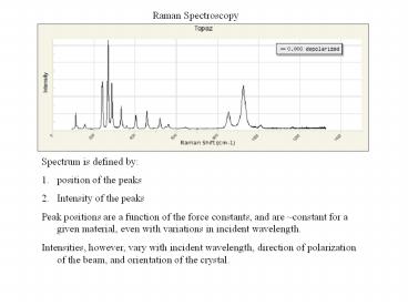
Raman Spectroscopy
- Spectrum is defined by
- position of the peaks
- Intensity of the peaks
- Peak positions are a function of the force
constants, and are constant for a given
material, even with variations in incident
wavelength. - Intensities, however, vary with incident
wavelength, direction of polarization of the
beam, and orientation of the crystal.
2
Laser parallel to c
Laser parallel to a, polarized
parallel to c at angle 0
3
- Intensities are difficult to compute
- Arm waving explanation, you can get Raman
intensity if the vibration of the atoms causes a
change in the polarization of the electron
density at the macro scale. Of course every
vibration of an atom causes a change in the
polarization of the electron density at the
atomic scale. - Important case, if an atom is located on a point
of inversion, then any vibration in one direction
is associated with another vibration in the exact
opposite, resulting in no change in macro scale
polarization, and therefore, no peak intensity
associated with the vibration of that atom. - Consequences, rocksalt and ccp elements have no
Raman spectra because every atom is on a center
of inversion. - Eg. Calcite, CaCO3. Ca is on a center of
inversion, so Raman is only associated with CO3
vibrational modes.
4
Calcite, peak at 280 cm-1, laser along a,
horizontal is // b, vertical is // c, rotate
crystal in 5 increments
Distance from center of plot is proportional to
intensity. Vibrational mode is rocking of planar
CO3 group about a axis.
5
Laser parallel to c
- In this case, there is no variation in intensity
for any of the different vibrational mode as the
polarization of the beam is varied. - That is because we are shooting down an axis of
4-fold symmetry, so the optical ellipsoid is
circular. All properties of a crystal show no
variation as a function of polarization when
examined along the axes of 3-, 4-, or 6-fold
symmetry. - In particular, there is no change in the
polarizability of the electron density in rutile
when the beam is directed along the c-axis.
6
Crystal structure of rutile, TiO2, looking along
the a axis. Note that maximum in 450 cm-1 peak is
when polarization direction is nearly parallel to
the TiO bond
7
Topaz, Al2SiO4F2, orthorhombic Lets examine the
Si-O bond stretching modes. There are 3
non-equivalent SiO bonds, R(SiO1) 1.636
Å R(SiO2) 1.648 Å R(SiO3) 1.640 Å twice
SiO3
SiO2
SiO1
8
b
Red short Green long Blue medium x 2
c
Laser along a-axis, 0 b-axis, 90 c-axis
9
b
Red short Green long Blue medium x 2
a
Laser along c-axis, 0 a-axis, 90 b -axis
10
c
Red short Green long Blue medium x 2
a
Laser along b-axis, 0 c-axis, 90 a-axis
11
In general, tetrahedral groups produce strong
peaks, while octahedral groups do not. Question,
why is this so?