
Bacteriophage T4 - PowerPoint PPT Presentation
1 / 31
Title:
Bacteriophage T4
Description:
Bacteriophages contain Nucleic Acid and Protein. ... Surrounded by a contractile sheath. Base Plates. Tail fibers. The Parts. A package of DNA/RNA ... – PowerPoint PPT presentation
Number of Views:543
Avg rating:3.0/5.0
Title: Bacteriophage T4
1
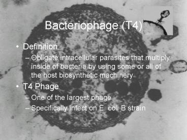
Bacteriophage (T4)
- Definition
- Obligate intracellular parasites that multiply
inside of bacteria by using some or all of the
host biosynthetic machinery - T4 Phage
- One of the largest phage
- Specifically infect on E. coli B strain
2
Composition
- Bacteriophages contain Nucleic Acid and Protein.
- Nucleic Acid can be DNA or RNA, depending upon
the phage - Proteins function in infection and to protect the
nucleic acid from nuclease in the environment.
3
Structure (T4)
- Size
- Approximately 200 nm long and 80-100nm wide
- Head or Capsid
- Icosahedral
- Protective covering for the nucleic acid
- Tail
- A hollow tube through which the nucleic acid
passes during infection. - Surrounded by a contractile sheath.
- Base Plates
- Tail fibers
4
The Parts
- A package of DNA/RNA
- A transport tube
- An attachment device
5
Infection of Host Cells
6
Infection of Host Cells
- Adsorption
- The tail fiber attach to specific receptors on
the bacterial cell. - Irreversible attachment
- Sheath Contraction
- Nucleic Acid Injection
7
Infection of Host Cells
- Replication
- Transcription of Phage DNA to Phage RNA
- Translation of Phage mRNA to Proteins
- Replicate more Phage DNA
8
Infection of Host Cells
- Eclipse period
- During the eclipse phase, no infectious phage
particles can be found either inside or outside
the bacterial cell. - The phage nucleic acid takes over the host
biosynthetic machinery and phage specified
m-RNA's and proteins are made.
9
Infection of Host Cells
- Assembly
- Maturation stage
- Intracellular Accumulation Phase
10
Infection of Host Cells
- Lysis
- After a while the bacteria begin to lyse due to
the accumulation of the phage lysis protein and
intracellular phage are released into the medium.
The number of particles released per infected
bacteria may be as high as 1000.
11
Infection of Host Cells
12
Viral Growth Curve
- Adsorption
- Attachment
- Maturation period
- Assembly of phage particles
- Eclipse period or Latent Period
- No mature phage particles
- Time between initial infection and release of
mature phage
13
Viral Growth Curve
- Rise period
- Host cells begin to burst
- Plateau
- Constant number of phage near the end of
reproduction cycle - Burst size
- Average number of phage particles released by
each infected bacterium
14
Viral Growth Curve
15
Plaques
- Plaques clear areas on a lawn of host cells
that represent the point at which a single
infectious virus particle was deposited - As a result of subsequent lytic cycles, the host
cells in the region are destroyed.
16
Plaques
- Plaque formation is analogous to the plate count
of bacteria in which each plaque (colony)
represents a single infectious virus (viable
bacterium) in the initial inoculum. - A bacteriophage plaque count can be used to
determine the titer, the concentration of phage
particles, in the suspension provided.
17
Experiment Tips
- Assign job duties to each group member
- Top agar has been dispensed for you.
- READ YOUR JOB DUTIES BEFORE YOU START THE LAB!!!
- Keep close track of the time
- Be careful when mixing- vigorous vortexing will
shear the phage off the bacteria - Remove ALL samples from the dilution tube
18
Experiment Tips
- Pour the lawns QUICKLY- the agar solidifies fast
- Go over flow chart- 1 plate per time point
- 15, 25 and 30 minute time points- use top row of
chart - 35, 40, 45, and 50 minute time points- use bottom
row - The next week we will count the number of plaques
per plate and graph them to determine burst size. - Worksheet due at the end of class
19
Experiment
- Set-Up
- Use Figure 7.1 to arrange and label 15 culture
tubes - Label w/ time and row
- Label dilution tube and place in water bath
- Transfer appropriate amount of LB broth to each
tube - Label plates (min., initials, section, date,
experiment)
20
Experiment
Figure 7.1. Pattern of dilution tubes
21
Experiment
- Adsorption
- Place phage in 37C water bath for 1-2 minutes
- Vortex E. coli culture
- Transfer 2.9 ml if E. coli into ADS tube w/ phage
- THIS IS TIME ZERO!!!!!
- Finger vortex ADS tube
- Fill in time table in lab book
22
Experiment
- Dilution to stop adsorption
- After five minutes gently mix ADS tube
- NEVER VORTEX ANY TUBE CONTAINING PHAGE!!!!!!!
- Transfer 0.1 ml from ADS tube to Dilution tube
- Return to 37C water bath until next time point
- This step dilutes the phage and E. coli to the
point that there are very few collisions and
adsorption does not occur
23
Only Finger Vortex or You will Destroy the Phage!
24
Experiment
- Taking 15, 25, and 30 min. Samples
- After five minutes gently mix Dilution tube
- Transfer 0.1 ml from Dilution tube into the 1st
tube in your rack - Gently mix
- Transfer 1.0 ml from 1st tube to the tube behind
it - Gently mix
25
Experiment
- Get a tube of soft agar from 47C water bath
- Add 0.3 ml of E. coli B lawn cells to soft agar
and vortex BRIEFLY - Transfer 0.1 ml from 2nd row tube to soft agar,
finger vortex - Pour IMMEDIATELY on to plate labeled
- 15 min and spread
26
Experiment
- Taking 35, 40, 45 and 50 min. Samples
- After five minutes gently mix Dilution tube
- Transfer 0.1 ml from Dilution tube into the 1st
tube in your rack - Gently mix
- Transfer 0.1 ml from 1st tube to the tube behind
it - Gently mix
27
Experiment
- Get a tube of soft agar from 47C water bath
- Add 0.3 ml of E. coli B lawn cells to soft agar
and vortex BRIEFLY - Transfer 0.1 ml from 2nd row tube to soft agar
- Pour IMMEDIATELY on to plate
28
Experiment
- Allow all plates to cool, tape together and
incubate at 37ºC for one week. - Plaques will be counted the next week.
29
Experiment The Step by Step
0.1 ml
0.1 ml
1.0ml
0.1 ml
35
40
15
25
30
11,000,000
110,000,000
30
Experiment
31
Completing the Worksheet
- Fill out worksheet as you go.
- Due at the end of lab the week we count the
plates.