
The ear PowerPoint PPT Presentation
1 / 65
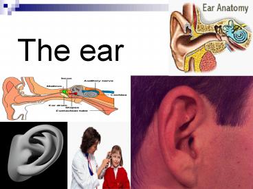
Title: The ear
1
The ear
2
(No Transcript)
3
(No Transcript)
4
Overview
- The ear is a sensory organ with dual
functionshearing and balance. The sense of
hearing is essential for normal development and
maintenance of speech as well as the ability to
communicate with others. Balance, or equilibrium,
is essential for maintaining body movement,
position, and coordination. The delicate
structure and function of the ear make early
detection and accurate diagnosis of disorders
necessary for preservation of normal hearing and
balance. Among the professionals involved in the
diagnosis and treatment of these disorders are
otolaryngologists, pediatricians, internists, and
nurses.
5
Anatomic and Physiologic Overview
- The cranium encloses and protects the brain and
surrounding structures, providing attachment for
various muscles that control head and jaw
movements. Eight bones form the cranium the
occipital bone, the frontal bone, two parietal
bones, two temporal bones, the sphenoid bone, and
the ethmoid bone. Some of these bones contain
sinuses, which are cavities lined with mucous
membranes and connected to the nasal cavity. The
ears are located on either side of the cranium at
approximately eye level
6
(No Transcript)
7
Anatomy of the External Ear
- The external ear includes the auricle (pinna) and
the external auditory canal .The external ear is
separated from the middle ear by a disk-like
structure called the tympanic membrane (eardrum). - Auricle The auricle, attached to the side of the
head by skin, is composed mainly of cartilage,
except for the fat and subcutaneous tissue in the
earlobe. The auricle collects the sound waves and
directs vibrations into the external auditory
canal.
8
- External Auditory Canal The external auditory
canal is approximately 2.5 cm long. The lateral
third is an elastic cartilaginous and dense
fibrous framework to which thin skin is attached.
The medial two thirds is bone lined with thin
skin. The external auditory canal ends at the
tympanic membrane. The skin of the canal contains
hair, sebaceous glands, and ceruminous glands,
which secrete a brown, waxlike substance called
cerumen (ear wax). The ear's self-cleaning
mechanism moves old skin cells and cerumen to the
outer part of the ear. Just anterior to the
external auditory canal is the temporomandibular
joint. The head of the mandible can be felt by
placing a fingertip in the external auditory
canal while the patient opens and closes the
mouth.
9
Anatomy of the Middle Ear
- The middle ear, an air-filled cavity, includes
the tympanic membrane laterally and the otic
capsule medially. The middle ear cleft lies
between the two. The middle ear is connected by
the eustachian tube to the nasopharynx and is
continuous with air-filled cells in the adjacent
mastoid portion of the temporal bone. The
eustachian tube, which is approximately 1 mm wide
and 35 mm long, connects the middle ear to the
nasopharynx. Normally, the eustachian tube is
closed, but it opens by action of the tensor veli
palatini muscle when the person performs a
Valsalva maneuver, yawns, or swallows. It drains
normal and abnormal secretions of the middle ear
and equalizes pressure in the middle ear with
that of the atmosphere.
10
- Tympanic Membrane The tympanic membrane
(eardrum), about 1 cm in diameter and very thin,
is normally pearly gray and translucent. It
consists of three layers of tissue an outer
layer, continuous with the skin of the ear canal
a fibrous middle layer and an inner mucosal
layer, continuous with the The remaining 20
lacks the middle layer and is called the pars
flaccida. The absence of this fibrous middle
layer makes the pars flaccida more vulnerable to
pathologic disorders than the pars tensa.
Distinguishing landmarks of the tympanic membrane
include the annulus, the fibrous border that
attaches the eardrum to the temporal bone the
short process of the malleus the long process of
the malleus the umbo of the malleus, which
attaches to the tympanic membrane in the center
the pars flaccida and the pars tensa
11
- The tympanic membrane protects the middle ear and
conducts sound vibrations from the external canal
to the ossicles. The sound pressure is magnified
22 times as a result of transmission from a
larger area to a smaller one. - Ossicles The middle ear contains the three
smallest bones (the ossicles) of the body the
malleus, the incus, and the stapes. The ossicles,
which are held in place by joints, muscles, and
ligaments, assist in the transmission of sound.
Two small fenestrae (oval and round windows),
located in the medial wall of the middle ear,
separate the middle ear from the inner ear. The
footplate of the stapes sits in the oval window,
secured by a fibrous annulus (ring-shaped
structure). The footplate transmits sound to the
inner ear. The round window, covered by a thin
membrane, provides an exit for sound vibrations
12
(No Transcript)
13
Anatomy of the Inner Ear
- The inner ear is housed deep within the temporal
bone. The organs for hearing (cochlea) and
balance (semicircular canals), as well as cranial
nerves VII (facial nerve) and VIII
(vestibulocochlear nerve), are all part of this
complex anatomy .The cochlea and semicircular
canals are housed in the bony labyrinth. The bony
labyrinth surrounds and protects the membranous
labyrinth, which is bathed in a fluid called
perilymph. - Membranous Labyrinth The membranous labyrinth
is composed of the utricle, the saccule, the
cochlear duct, the semicircular canals, and the
organ of Corti. The membranous labyrinth contains
a fluid called endolymph.
14
- The three semicircular canalsposterior,
superior, and lateral, which lie at 90-degree
angles to one anothercontain sensory receptor
organs, arranged to detect rotational movement.
These receptor end organs are stimulated by
changes in the rate or direction of a person's
movement. The utricle and saccule are involved
with linear movements. - Organ of Corti The organ of Corti is located
in the cochlea, a snail-shaped, bony tube about
3.5 cm long with two and a half spiral turns.
Membranes separate the cochlear duct (scala
media) from the scala vestibuli, and the scala
tympani from the basilar membrane. The organ of
Corti is located on the basilar membrane
stretching from the base to the apex of the
cochlea. As sound vibrations enter the perilymph
at the oval window and travel along the scala
vestibuli, they pass through the scala tympani,
enter the cochlear duct
15
- and cause movement of the basilar membrane. The
organ of Corti, also called the end organ for
hearing, transforms mechanical energy into neural
activity and separates sounds into different
frequencies. This electrochemical impulse travels
through the acoustic nerve to the temporal cortex
of the brain to be interpreted as meaningful
sound. In the internal auditory canal, the
cochlear (acoustic) nerve, arising from the
cochlea, joins the vestibular nerve, arising from
the semicircular canals, utricle, and saccule, to
become the vestibulocochlear nerve (cranial nerve
VIII). This canal also houses the facial nerve
and the blood supply from the ear to the brain.
16
Function of the Ears
- Hearing Hearing is conducted over two pathways
air and bone. Sounds transmitted by air
conduction travel over the air-filled external
and middle ear through vibration of the tympanic
membrane and ossicles. Sounds transmitted by bone
conduction travel directly through bone to the
inner ear, bypassing the tympanic membrane and
ossicles. Normally, air conduction is the more
efficient pathway. However, a defect in the
tympanic membrane or interruption of the
ossicular chain disrupts normal air conduction,
which results in a conductive hearing loss.
17
- Sound Conduction and Transmission Sound enters
the ear through the external auditory canal and
causes the tympanic membrane to vibrate. These
vibrations transmit sound through the lever
action of the ossicles to the oval window as
mechanical energy. This mechanical energy is then
transmitted through the inner ear fluids to the
cochlea, stimulating the hair cells, and is
subsequently converted to electrical energy. The
electrical energy travels through the
vestibulocochlear nerve to the central nervous
system, where it is interpreted in its final form
as sound. Vibrations transmitted by the tympanic
membrane to the ossicles of the middle ear are
transmitted to the cochlea, located in the
labyrinth of the inner ear. The stapes rocks,
causing vibrations (waves) in fluids contained in
the inner ear. These fluid waves cause movement
of the basilar membrane, stimulating the hair
cells of the organ of Corti in the cochlea to
move in a wavelike manner.
18
- The movements of the tympanic membrane set up
electrical currents that stimulate the various
areas of the cochlea. The hair cells set up
neural impulses that are encoded and then
transferred to the auditory cortex in the brain,
where they are decoded into a sound message. The
footplate of the stapes receives impulses
transmitted by the incus and the malleus from the
tympanic membrane. The round window, which opens
on the opposite side of the cochlear duct, is
protected from sound waves by the intact tympanic
membrane, permitting motion of the inner ear
fluids by sound wave stimulation. For example, in
the normally intact tympanic membrane, sound
waves stimulate the oval window first, and a lag
occurs before the terminal effect of the stimulus
reaches the round window. However, this lag phase
is changed when a perforation of the tympanic
membrane allows sound waves to impinge on the
oval and round windows simultaneously
19
- This effect cancels the lag and prevents the
maximal effect of inner ear fluid motility and
its subsequent effect in stimulating the hair
cells in the organ of Corti. The result is a
reduction in hearing ability. - Balance and Equilibrium Body balance is
maintained by the cooperation of the muscles and
joints of the body (proprioceptive system), the
eyes (visual system), and the labyrinth
(vestibular system). These areas send their
information about equilibrium, or balance, to the
brain (cerebellar system) for coordination and
perception in the cerebral cortex. The brain
obtains its blood supply from the heart and
arterial system. A problem in any of these areas,
such as arteriosclerosis or impaired vision, can
cause a disturbance of balance. The vestibular
apparatus of the inner ear provides feedback
regarding the movements and the position of the
head and body in space.
20
Assessment
- Assessment of hearing and balance involves
inspection of the external, middle, and inner
ear. Evaluation of gross hearing acuity also is
included in every physical examination. - Inspection of the External Ear Inspection of
the external ear is a simple procedure, but it is
often overlooked. The external ear is examined by
inspection and direct palpation the auricle and
surrounding tissues should be inspected for
deformities, lesions, and discharge, as well as
size, symmetry, and angle of attachment to the
head. Manipulation of the auricle does not
normally elicit pain. If this maneuver is
painful, acute external otitis is suspected.
Tenderness on palpation in the area of the
mastoid may indicate acute mastoiditis or
inflammation of the posterior auricular node.
21
- Occasionally, sebaceous cysts and tophi
(subcutaneous mineral deposits) are present on
the pinna. A flaky scaliness on or behind the
auricle usually indicates seborrheic dermatitis
and can be present on the scalp and facial
structures as well. - Otoscopic ExaminationThe tympanic membrane is
inspected with an otoscope and indirect palpation
with a pneumatic otoscope. To examine the
external auditory canal and tympanic membrane,
the otoscope should be held in the examiner's
right hand, in a pencil-hold position, with the
examiner's hand braced against the patient's face
.This position prevents the examiner from
inserting the otoscope too far into the external
canal. Using the opposite hand, the auricle is
grasped and gently pulled back to straighten the
canal in the adult. If the canal is not
straightened with this technique, the tympanic
membrane is more difficult to visualize because
the canal obstructs the view.
22
- The speculum is slowly inserted into the ear
canal, with the examiner's eye held close to the
magnifying lens of the otoscope to visualize the
canal and tympanic membrane. The largest speculum
that the canal can accommodate (usually 5 mm in
an adult) is guided gently down into the canal
and slightly forward. Because the distal portion
of the canal is bony and covered by a sensitive
layer of epithelium, only light pressure can be
used without causing pain. The external auditory
canal is examined for discharge, inflammation, or
a foreign body. The healthy tympanic membrane is
pearly gray and is positioned obliquely at the
base of the canal. The following landmarks are
identified, if visible .the pars tensa, the umbo,
the manubrium of the malleus, and its short
process. A slow, circular movement of the
speculum allows further visualization of the
malleolar folds and periphery.
23
- The position and color of the membrane and any
unusual markings or deviations from normal are
documented. The presence of fluid, air bubbles,
blood, or masses in the middle ear also should be
noted. Proper otoscopic examination of the
external auditory canal and tympanic membrane
requires that the canal be free of large amounts
of cerumen. Cerumen is normally present in the
external canal, and small amounts should not
interfere with otoscopic examination. If the
tympanic membrane cannot be visualized because of
cerumen, the cerumen may be removed by gently
irrigating the external canal with warm water
(unless contraindicated). If adherent cerumen is
present, a small amount of mineral oil or
over-the-counter cerumen softener may be
instilled within the ear canal, and the patient
is instructed to return for subsequent removal of
the cerumen and inspection of the ear.
24
- Proper otoscopic examination of the external
auditory canal and tympanic membrane requires
that the canal be free of large amounts of
cerumen. Cerumen is normally present in the
external canal, and small amounts should not
interfere with otoscopic examination. If the
tympanic membrane cannot be visualized because of
cerumen, the cerumen may be removed by gently
irrigating the external canal with warm water
(unless contraindicated). If adherent cerumen is
present, a small amount of mineral oil or
over-the-counter cerumen softener may be
instilled within the ear canal, and the patient
is instructed to return for subsequent removal of
the cerumen and inspection of the ear. The use of
instruments such as a cerumen curette for cerumen
removal is reserved for otolaryngologists and
nurses with specialized training because of the
danger of perforating the tympanic membrane or
excoriating the external auditory canal. Cerumen
buildup is a common cause of hearing loss and
local irritation.
25
Evaluation of Gross Auditory Acuity
- A general estimate of hearing can be made by
assessing the patient's ability to hear a
whispered phrase or a ticking watch, testing one
ear at a time. The Weber and Rinne tests may be
used to distinguish conductive loss from
sensorineural loss when hearing is impaired.
These tests are part of the usual screening
physical examination and are useful if a more
specific assessment is needed, if hearing loss is
detected, or if confirmation of audiometric
results is desired. - Whisper Test To exclude one ear from the
testing, the examiner covers the untested ear
with the palm of the hand. Then the examiner
whispers softly from a distance of 1 or 2 feet
from the unoccluded ear and out of the patient's
sight. The patient with normal acuity can
correctly repeat what was whispered.
26
- Weber Test The Weber test uses bone conduction
to test lateralization of sound. A tuning fork
(ideally, 512 Hz), set in motion by grasping it
firmly by its stem and tapping it on the
examiner's knee or hand, is placed on the
patient's head or forehead. A person with normal
hearing hears the sound equally in both ears or
describes the sound as centered in the middle of
the head. A person with such as from
otosclerosis or otitis media, hears the sound
better in the affected ear. A person with
resulting from damage to the cochlear or
vestibulocochlear nerve, hears the sound in the
better-hearing ear. The Weber test is useful for
detecting unilateral hearing loss
27
(No Transcript)
28
- Rinne Test In the Rinne test (pronounced ),
the examiner shifts the stem of a vibrating
tuning fork between two positions 2 inches from
the opening of the ear canal (for air conduction)
and against the mastoid bone (for bone
conduction). As the position changes, the patient
is asked to indicate which tone is louder or when
the tone is no longer audible. The Rinne test is
useful for distinguishing between conductive and
sensorineural hearing loss. A person with normal
hearing reports that air-conducted sound is
louder than bone-conducted sound.
29
- A person with a conductive hearing loss hears
bone-conducted sound as long as or longer than
air-conducted sound. A person with a
sensorineural hearing loss hears air-conducted
sound longer than bone-conducted sound.
30
- Middle Ear Endoscopy With endoscopes with very
small diameters and acute angles, the ear can be
examined by an endoscopist specializing in
otolaryngology. Middle ear endoscopy is performed
safely and effectively as an office procedure to
evaluate suspected perilymphatic fistula and
new-onset conductive hearing loss, the anatomy of
the round window before transtympanic treatment
of Ménière's disease, and the tympanic cavity
before ear surgery to treat chronic middle ear
and mastoid infections. The tympanic membrane is
anesthetized topically for about 10 minutes
before the procedure. Then, the external auditory
canal is irrigated with sterile normal saline
solution. With the aid of a microscope, a
tympanotomy is created with a laser beam or a
myringotomy knife, so that the endoscope can be
inserted into the middle ear cavity. Video and
photo documentation can be accomplished through
the scope.
31
Hearing Loss
- More than 28 million people in the United States
have some form of hearing impairment . Hearing
loss is greater in men than in women. By the year
2050, about one of every five people in the
United States, or almost 58 million people, will
be 55 years of age or older. Of this population,
almost half can expect to have a hearing
impairment. Approximately 10 million people in
the United States have irreversible hearing loss
. It is estimated that more than 30 million
people are exposed on a daily basis to noise
levels that produce hearing loss. Occupations
such as carpentry, plumbing, and coal mining have
the highest risk of noise-induced hearing loss.
Hearing loss is an important health issue, and
studies indicate that as people age, hearing
screening and treatment are indicated. Conductive
hearing loss usually results from an external ear
disorder, such as impacted cerumen, or a middle
ear disorder, such as otitis media or
otosclerosis. In such instances.
32
- the efficient transmission of sound by air to the
inner ear is interrupted. A sensorineural loss
involves damage to the cochlea or
vestibulocochlear nerve. - Mixed hearing loss and functional hearing loss
also may occur. Patients with mixed hearing loss
have conductive loss and sensorineural loss,
resulting from dysfunction of air and bone
conduction. A functional (or psychogenic) hearing
loss is nonorganic and unrelated to detectable
structural changes in the hearing mechanisms it
is usually a manifestation of an emotional
disturbance.
33
Clinical Manifestations
- Early manifestations of hearing impairment and
loss may include tinnitus, increasing inability
to hear when in a group, and a need to turn up
the volume of the television. Hearing impairment
can also trigger changes in attitude, the ability
to communicate, the awareness of surroundings,
and even the ability to protect oneself,
affecting a person's quality of life. In a
classroom, a student with impaired hearing may be
uninterested and inattentive and have failing
grades. A person at home may feel isolated
because of an inability to hear the clock chime,
the refrigerator hum, the birds sing, or the
traffic pass. A pedestrian who is
hearing-impaired may attempt to cross the street
and fail to hear an approaching car. People with
impaired hearing may miss parts of a
conversation. Many people are unaware of their
gradual hearing impairment.
34
- Often, it is not the person with the hearing loss
but the people with whom he or she is
communicating who recognize the impairment first.
For various reasons, some people with hearing
loss refuse to seek medical attention or wear a
hearing aid. Others feel self-conscious wearing a
hearing aid. Insightful people generally ask
those with whom they are trying to communicate to
let them know whether difficulties in
communication exist. These attitudes and
behaviors should be taken into account when
counseling patients who need hearing assistance.
The decision to wear a hearing aid is a personal
one that is affected by these attitudes and
behaviors.
35
Prevention
- Many environmental factors have an adverse effect
on the auditory system and, with time, result in
permanent sensorineural hearing loss. The most
common is noise. Noise (unwanted and unavoidable
sound) has been identified as one of today's
environmental hazards. The volume of noise that
surrounds us daily has increased into a
potentially dangerous source of physical and
psychological damage. Loud, persistent noise has
been found to cause constriction of peripheral
blood vessels, increased blood pressure and heart
rate (because of increased secretion of
adrenalin), and increased gastrointestinal
activity. Although research is needed to address
the overall effects of noise on the human body, a
quiet environment is more conducive to peace of
mind. A person who is ill feels more at ease when
noise is kept to a minimum.
36
- Numerous factors contribute to hearing loss.
refers to hearing loss that follows a long period
of exposure to loud noise (eg, heavy machinery,
engines, artillery). refers to hearing loss
caused by a single exposure to an extremely
intense noise, such as an explosion. Usually,
noise-induced hearing loss occurs at a high
frequency (about 4000 Hz). However, with
continued noise exposure, the hearing loss can
become more severe and include adjacent
frequencies. The minimum noise level known to
cause noise-induced hearing loss, regardless of
duration, is about 85 to 90 dB. Noise exposure is
inherent in many jobs (eg, mechanics, printers,
pilots, musicians) and in hobbies such as
woodworking and hunting. The Occupational Safety
and Health Administration requires that workers
wear ear protection to prevent noise-induced
hearing loss when exposed to noise above the
legal limits. Ear protection against noise is the
most effective preventive measure available.
There are no medications that protect against
noise-induced hearing loss hearing loss is
permanent because the hair cells in the organ of
37
Nursing Management
- Nurses who understand the different types of
hearing loss are more successful in adopting a
communication style to fit the needs and
preferences of every patient. Trying to speak in
a loud voice to a person who cannot hear
high-frequency sounds only makes understanding
more difficult. However, strategies such as
talking into the less-impaired ear and using
gestures and facial expressions can help major
issue for many people who are deaf or
hearing-impaired is that they have other health
problems that often do not receive attention, in
large part because of communication barriers with
their health care practitioners. To meet the
health care needs of these patients,
practitioners are legally obligated to make
accommodations for a patient's inability to hear.
Providing interpreters for those who can
communicate through sign language is essential in
many situations so that the practitioner can
effectively communicate with the patient.
38
- During health care and screening procedures, the
practitioner (eg, dentist, physician, nurse) must
be aware that patients who are deaf or
hearing-impaired are unable to read lips, see a
signer, or read written materials in the dark
rooms required during some diagnostic tests. The
same situation exists if the practitioner is
wearing a mask or not in sight (eg, x-ray
studies, magnetic resonance imaging MRI,
colonoscopy). Nurses and other health care
practitioners must work with patients who are
deaf or hearing-impaired and their families to
identify practical and effective means of
communication. Nurses can serve as catalysts
throughout the health care system to ensure that
accommodations are made to meet the communication
needs of these patients.
39
Conditions of the External Ear
- Cerumen Impaction Cerumen normally accumulates
in the external canal in various amounts and
colors. Although wax does not usually need to be
removed, impaction occasionally occurs, causing
a sensation of fullness or pain in the ear, with
or without a hearing loss. Accumulation of
cerumen as a cause of hearing loss is especially
significant in the elderly population. Attempts
to clear the external auditory canal with
matches, hairpins, and other implements are
dangerous because trauma to the skin, infection,
and damage to the tympanic membrane can occur.
40
Management
- Cerumen can be removed by irrigation, suction, or
instrumentation. Unless the patient has a
perforated eardrum or an inflamed external ear
(ie, otitis externa), gentle irrigation usually
helps remove impacted cerumen, particularly if it
is not tightly packed in the external auditory
canal. For successful removal, the water stream
must flow behind the obstructing cerumen to move
it first laterally and then out of the canal. To
prevent injury, the lowest effective pressure
should be used. However, if the eardrum behind
the impaction is perforated, water can enter the
middle ear, producing acute vertigo and
infection. If irrigation is unsuccessful, direct
visual, mechanical removal can be performed on a
cooperative patient by a trained health care
provider. Instilling a few drops of warmed
glycerin, mineral oil, or half-strength hydrogen
peroxide into the ear canal for 30 minutes can
soften cerumen before its removal. Ceruminolytic
agents, such as peroxide in glyceryl (Debrox),
are available
41
- however, these compounds may cause an allergic
dermatitis reaction. Using any softening solution
two or three times a day for several days is
generally sufficient. If the cerumen cannot be
dislodged by these methods, instruments, such as
a cerumen curette, aural suction, and a binocular
microscope for magnification, can be used.
42
Foreign Bodies
- Some objects are inserted intentionally into the
ear by adults who may have been trying to clean
the external canal or relieve itching or by
children who introduce peas, beans, pebbles,
toys, and beads. Insects may also enter the ear
canal. In either case, the effects may range from
no symptoms to profound pain and decreased
hearing. - Management
- Removing a foreign body from the external
auditory canal can be quite challenging. The
three standard methods for removing foreign
bodies are the same as those for removing
cerumen irrigation, suction, and
instrumentation. The contraindications for
irrigation are also the same. Foreign vegetable
bodies and insects tend to swell thus,
irrigation is contraindicated. Usually, an insect
can be dislodged by instilling
43
) External Otitis (Otitis Externa
- refers to an inflammation of the external
auditory canal. Causes include water in the ear
canal (swimmer's ear) trauma to the skin of the
ear canal, permitting entrance of organisms into
the tissues and systemic conditions, such as
vitamin deficiency and endocrine disorders.
Bacterial or fungal infections are most
frequently encountered. The most common bacterial
pathogens associated with external otitis are
and species. The most common fungus isolated in
both normal and infected ears is External otitis
is often caused by a dermatosis such as
psoriasis, eczema, or seborrheic dermatitis. Even
allergic reactions to hair spray, hair dye, and
permanent wave lotions can cause dermatitis,
which clears when the offending agent is removed.
44
Clinical Manifestations
- Patients usually report pain, discharge from the
external auditory canal, aural tenderness
(usually not present in middle ear infections),
and occasionally fever, cellulitis, and
lymphadenopathy. Other symptoms may include
pruritus and hearing loss or a feeling of
fullness. On otoscopic examination, the ear canal
is erythematous and edematous. Discharge may be
yellow or green and foul-smelling. In fungal
infections, hairlike black spores may even be
visible. - Medical Management
- The principles of therapy are aimed at relieving
the discomfort, reducing the swelling of the ear
canal, and eradicating the infection. Patients
may require analgesics for the first 48 to 92
hours. If the tissues of the external canal are
edematous, a wick should be inserted to keep the
canal open so that liquid medications (eg,
Burow's solution, antibiotic otic preparations)
can be introduced.
45
- These medications may be administered by dropper
at room temperature. Such medications usually
combine antibiotic and corticosteroid agents to
soothe the inflamed tissues. For cellulitis or
fever, systemic antibiotics may be prescribed.
For fungal disorders, antifungal agents are
prescribed. - Nursing Management
- Nurses should instruct patients not to clean the
external auditory canal with cotton-tipped
applicators and to avoid events that traumatize
the external canal such as scratching the canal
with the fingernail or other objects. Trauma may
lead to infection of the canal. Patients should
also avoid getting the canal wet when swimming or
shampooing the hair. A cotton ball can be covered
in a water-insoluble gel such as petroleum jelly
and placed in the ear as a barrier to water
contamination. Infection can be prevented by
using antiseptic otic preparations after swimming
(eg, Swim Ear, Ear Dry), unless there is a
history of tympanic membrane perforation or a
current ear infection
46
Masses of the External Ear
- are small, hard, bony protrusions found in the
lower posterior bony portion of the ear canal
they usually occur bilaterally. The skin covering
the exostosis is normal. It is believed that
exostoses are caused by an exposure to cold
water, as in scuba diving or surfing. The usual
treatment, if any, is surgical excision.
Malignant tumors also may be found in the
external ear. Most common are basal cell
carcinomas on the pinna and squamous cell
carcinomas in the ear canal. If untreated,
squamous cell carcinoma may spread through the
temporal bone, causing facial nerve paralysis and
hearing loss. Carcinomas must be treated
surgically.
47
Conditions of the Middle Ear
- Tympanic Membrane Perforation
- Perforation of the tympanic membrane is usually
caused by infection or trauma. Sources of trauma
include skull fracture, explosive injury, or a
severe blow to the ear. Less frequently,
perforation is caused by foreign objects (eg,
cotton-tipped applicators, bobby pins, keys) that
have been pushed too far into the external
auditory canal. In addition to tympanic membrane
perforation, injury to the ossicles and even the
inner ear may result from this type of action.
Attempts by patients to clear the external
auditory canal should be discouraged. During
infection, the tympanic membrane can rupture if
the pressure in the middle ear exceeds the
atmospheric pressure in the external auditory
canal.
48
Medical Management
- Although most tympanic membrane perforations heal
spontaneously within weeks after rupture, some
may take several months to heal. Some
perforations persist because scar tissue grows
over the edges of the perforation, preventing
extension of the epithelial cells across the
margins and final healing. In the case of a head
injury or temporal bone fracture, a patient is
observed for evidence of cerebrospinal fluid or
a clear, watery drainage from the ear or nose,
respectively. While healing, the ear must be
protected from water. - Surgical Management
- Perforations that do not heal on their own may
require surgery. The decision to perform a
(surgical repair of the tympanic membrane) is
usually based on the need to prevent potential
infection from water entering the ear or the
desire to improve the patient's hearing.
49
- Performed on an outpatient basis, tympanoplasty
may involve a variety of surgical techniques. In
all techniques, tissue (commonly from the
temporalis fascia) is placed across the
perforation to allow healing. Surgery is usually
successful in closing the perforation permanently
and improving hearing. - Acute Otitis Media
- Ear infections can occur at any age however,
they are most commonly seen in children.
Approximately three out of four children
experience an ear infection by the time they are
3 years of age. (AOM) is an acute infection of
the middle ear, usually lasting less than 6
weeks. The pathogens that cause acute otitis
media are usually and
50
- which enter the middle ear after eustachian tube
dysfunction caused by obstruction related to
upper respiratory infections, inflammation of
surrounding structures (eg, sinusitis, adenoid
hypertrophy), or allergic reactions (eg, allergic
rhinitis). Bacteria can enter the eustachian tube
from contaminated secretions in the nasopharynx
and the middle ear from a tympanic membrane
perforation. A purulent exudate is usually
present in the middle ear, resulting in a
conductive hearing loss. - Clinical Manifestations
- The symptoms of otitis media vary with the
severity of the infection. The condition, usually
unilateral in adults, may be accompanied by
otalgia. The pain is relieved after spontaneous
perforation or therapeutic incision of the
tympanic membrane.
51
- Other symptoms may include drainage from the ear,
fever, and hearing loss. On otoscopic
examination, the external auditory canal appears
normal. The tympanic membrane is erythematous and
often bulging. Patients report no pain with
movement of the auricle. - Medical Management
- The outcome of AOM depends on the efficacy of
therapy (the prescribed dose of an oral
antibiotic and the duration of therapy), the
virulence of the bacteria, and the physical
status of the patient. With early and appropriate
broad-spectrum antibiotic therapy, otitis media
may resolve with no serious sequelae. If drainage
occurs, an antibiotic otic preparation is usually
prescribed. The condition may become subacute
(lasting 3 weeks to 3 months), with persistent
purulent discharge from the ear. Rarely does
permanent hearing loss occur.
52
- Secondary complications involving the mastoid and
other serious intracranial complications, such as
meningitis or brain abscess, although rare, can
occur. - Surgical Management
- An incision in the tympanic membrane is known as
or The tympanic membrane is numbed with a local
anesthetic such as phenol or by iontophoresis
(ie, electrical current flows through a
lidocaine-and-epinephrine solution to numb the
ear canal and tympanic membrane). The procedure
is painless and takes less than 15 minutes. Under
microscopic guidance, an incision is made through
the tympanic membrane to relieve pressure and to
drain serous or purulent fluid from the middle
ear.
53
Otosclerosis
- involves the stapes and is thought to result
from the formation of new, abnormal spongy bone,
especially around the oval window, with resulting
fixation of the stapes. The efficient
transmission of sound is prevented because the
stapes cannot vibrate and carry the sound as
conducted from the malleus and incus to the inner
ear. Otosclerosis is more common in women and
frequently hereditary, and pregnancy may worsen
it. - Clinical Manifestations
- Otosclerosis may involve one or both ears and
manifests as a progressive conductive or mixed
hearing loss. The patient may or may not complain
of tinnitus. Otoscopic examination usually
reveals a normal tympanic membrane. Bone
conduction is better than air conduction on Rinne
testing. The audiogram confirms conductive
hearing loss or mixed loss, especially in the low
frequencies.
54
- Medical Management
- There is no known nonsurgical treatment for
otosclerosis. However, some physicians believe
the use of sodium fluoride can mature the
abnormal spongy bone growth and prevent the
breakdown of the bone tissue. Amplification with
a hearing aid also may help. - Surgical Management
- One of two surgical procedures may be performed,
the stapedectomy or the stapedotomy. A
stapedectomy involves removing the stapes
superstructure and part of the footplate and
inserting a tissue graft and a suitable
prosthesis .The surgeon drills a small hole into
the footplate to hold a prosthesis. The
prosthesis bridges the gap between the incus and
the inner ear, providing better sound conduction.
55
- Stapes surgery is very successful
approximately 95 of patients experience
resolution of conductive hearing loss. Balance
disturbance or true vertigo, which rarely occurs
in other middle ear surgical procedures, may
occur during the postoperative period for several
days. Long-term balance disorders are rare
56
Middle Ear Masses
- Other than cholesteatoma, masses in the middle
ear are rare. Glomus jugulare tumor is a tumor
that arises from the jugular vein. A
histologically identical tumor that arises from
Jacobson's nerve (in the temporal bone of the
skull) and remains limited to the middle ear is
known as a glomus tympanicum. On otoscopy, a red
blemish on or behind the tympanic membrane is
indicative of a glomus tumor. Glomus jugulare
tumors are rarely malignant however, due to
their location, treatment may be necessary to
relieve symptoms. The treatment for glomus tumors
is surgical excision, except in poor surgical
candidates, in whom radiation therapy is used. A
facial nerve neuroma is a tumor on cranial nerve
VII, the facial nerve. These types of tumors are
usually not visible on otoscopic examination but
are suspected when a patient presents with a
facial nerve paresis. X-ray evaluation is used to
identify the site of the tumor along the facial
nerve. The treatment is surgical removal.
57
Conditions of the Inner Ear
- Motion Sickness
- Motion sickness is a disturbance of equilibrium
caused by constant motion. For example, it can
occur aboard a ship, while riding on a
merry-go-round or swing, or in the back seat of a
car. - Clinical Manifestations
- The syndrome manifests itself in sweating,
pallor, nausea, and vomiting caused by vestibular
overstimulation. These manifestations may persist
for several hours after the stimulation stops.
58
- Management
- Over-the-counter antihistamines such as
dimenhydrinate (Dramamine) or meclizine
hydrochloride (Antivert) may provide some relief
of nausea and vomiting by blocking the conduction
of the vestibular pathway of the inner ear.
Anticholinergic medications, such as scopolamine
patches, may also be effective because they
antagonize the histamine response. These must be
replaced every few days. Side effects such as dry
mouth and drowsiness may occur. Potentially
hazardous activities such as driving a car or
operating heavy machinery should be avoided if
drowsiness occurs.
59
Tinnitus
- Tinnitus is a symptom of an underlying disorder
of the ear that is associated with hearing loss.
This condition affects approximately 10 of the
U.S. population between 40 and 70 years of age.
The severity of tinnitus may range from mild to
severe. Patients describe tinnitus as a roaring,
buzzing, or hissing sound in one or both ears.
Numerous factors may contribute to the
development of tinnitus, including several
ototoxic substances .Underlying disorders that
contribute to tinnitus may include thyroid
disease, hyperlipidemia, vitamin B12 deficiency,
psychological disorders (eg, depression,
anxiety), fibromyalgia, otologic disorders
(Ménière's disease, acoustic neuroma), and
neurologic disorders (head injury, multiple
sclerosis).
60
- A physical examination should be performed to
determine the cause of tinnitus. Diagnostic
testing determines if hearing loss is present. An
audiograph speech discrimination test or a
tympanogram may be used to help determine the
cause. Some forms of tinnitus are irreversible
therefore, patients may need teaching and
counseling about ways of adjusting to their
treatment and dealing with tinnitus in the
future. - Ototoxicity
- A variety of medications may have adverse effects
on the cochlea, vestibular apparatus, or cranial
nerve VIII. All but a few, such as aspirin and
quinine, cause irreversible hearing loss. At high
doses, aspirin toxicity can produce bilateral
tinnitus. IV medications, especially the
aminoglycosides, are the most common cause of
ototoxicity, and they destroy the hair cells in
the organ of Corti
61
- To prevent loss of hearing or balance, patients
receiving potentially ototoxic medications should
be counseled about the side effects of these
medications. These medications should be used
with caution in patients who are at high risk for
complications, such as children, the elderly,
pregnant patients, patients with kidney or liver
problems, and patients with current hearing
disorders. Blood levels of the medications should
be monitored, and patients receiving long-term IV
antibiotics should be monitored with an audiogram
twice each week during therapy.
62
Hearing Aids
- A hearing aid is a device through which speech
and environmental sounds are received by a
microphone, converted to electrical signals,
amplified, and reconverted to acoustic signals.
Many aids available for sensorineural hearing
loss depress the low frequencies, or tones, and
enhance hearing for the high frequencies. - Implanted Hearing Devices
- Three types of implanted hearing devices are
commercially available or in the investigational
stage the cochlear implant, the bone conduction
device, and the semi-implantable hearing device.
63
the cochlear implant device
64
the bone conduction device
65
the semi-implantable hearing device.
Recommended
