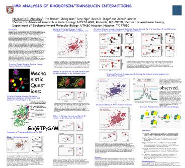
NMR ANALYSIS OF RHODOPSINTRANSDUCIN INTERACTIONS - PowerPoint PPT Presentation
1 / 1
Title: NMR ANALYSIS OF RHODOPSINTRANSDUCIN INTERACTIONS
1
NMR ANALYSIS OF RHODOPSIN/TRANSDUCIN
INTERACTIONS Najmoutin G. Abdulaev, Eva
Ramon, Xiang Mao,Tony Ngo, Kevin D. Ridge and
John P. Marino Center for Advanced Research in
Biotechnology, NIST/UMBI, Rockville, MD 20850,
Center for Membrane Biology, Department of
Biochemistry and Molecular Biology,
UTHSC-Houston, Houston, TX 77030
Correlated Changes between the Guanine Nucleotide
Binding Site and the C-terminal Cys-347 and Gly5
mutant proteins and the amino-terminal Ala-3,
Lys-10 Mutant Proteins
Monitoring Structural Changes through
Perturbations of 15N,1HN Chemical Shifts in Ga
Introduction Interaction of the G protein
transducin with light-activated rhodopsin (R) in
detergent solution, as well as a soluble mimic of
R, has been probed using high-resolution NMR
methods, with a particular emphasis on developing
detailed models for the structural changes in the
G protein a-subunit (Ga ) that accompany signal
transfer from R. We have shown that 1) an
15N-labeled Ga chimera (ChiT) displays a
relatively well-dispersed 2D spectrum with
uniform line widths and undergoes aluminum
fluoride (AlF4-) induced chemical shift
perturbations for resonances associated with
switch II (Trp-207) and the C-terminus (Phe-350)
2) similar perturbations in these regions are
also evident upon heterotrimer formation, likely
providing kinetic advantages in R/G protein
coupling 3) R- and G protein bg-subunit
(Gbg)-released and exchanged ChiT displays
further C-terminal perturbation and increased
conformational flexibility of switch II, which
may be important for Ga/effector interactions and
GTP hydrolysis 4) the GDP-released, R-bound
state of ChiT shows severe line-broadening
suggestive of a dynamic intermediate that results
from changes in the R-interacting N- and
C-termini 5) N-terminal truncation results in
a perturbation of the chemical shift for Trp-207
in the ground state and greater apparent
heterogeneity upon AlF4- adduct formation,
suggesting that the N-terminus either affects the
switch II region directly or through an
allosteric mechanism. Phe-350 did not shift in
this mutant, so truncation of the N-terminus
appears to have an inhibitory effect on the
C-terminal structural change and 6) ChiT with a
high-affinity C-terminal sequence shows a small
perturbation in the chemical shift for Trp-207 in
the ground state with expected shifts for all
Trp signals in the presence of AlF4-. Phe-350
shows signal in both the ground and activated
positions in the 'ground state' thereby allowing
receptor binding in the absence of Gbg.
Overlay of 15N-HSQC spectra of GDP/Mg2 (blue)
and GDP-AlF4-/Mg2 (red) bound ChiT (in NMR
buffer at 303K) acquired using a Bruker 600 MHz
NMR Cryoprobe system. Differences in the
conformations are manifested in a number of
changes in chemical shifts of the NH cross-peaks.
Boxes 1 and 2 highlight changes in the chemical
shifts for the assigned cross peaks of the three
tryptophan indoles and carboxyl-terminal
phenylalanine, respectively.
Expansion of the tryptophan indole (left panel)
and Phe-350 (right panels) resonance regions of
the HSQC spectra of the GDP/Mg2- (blue) and
GDPAlF4-/Mg2-bound (red) forms of the ChiT
mutants. Assignments for the 1HN, 15N cross peaks
of the tryptophan indoles and Phe-350 are
indicated. Note that for the C347W and K10W
mutants cross peaks are observed at both ground
and activated state positions in GDP/Mg2-
bound form. In contrast, all mutants display no
shift in the Phe-350 amide correlation upon
addition of AlF4-.
G protein Coupled Receptor Signaling through
Guanine Nucleotide Exchange
Mechanistic Questions What are the contact
points between an agonist activated GPCR (R) and
the G protein? How are changes at the
cytoplasmic surface of R transmitted to surface
regions on the G protein? How do structural
changes in the surface regions of the G protein
correlate with guanine nucleotide exchange?
Changes in Trp-207 and Trp-254 correlate with
Structural Changes in the Activated State
Structural/Functional Consequences of Mutating
the Carboxyl terminal sequence to a
High-Affinity sequence
(A) Overlay of 15N-HSQC spectra of GDP/Mg2 (red)
ChiT and GDP/Mg2 (blue) bound ChiT-HAP1 (in NMR
buffer at 303K) acquired using a Bruker 600 MHz
NMR Cryoprobe system. Only a few perturbations
are observed in chemical shifts of the NH
cross-peaks. Boxes 1 and 2 highlight changes in
the chemical shifts for the assigned cross peaks
of the three tryptophan indoles and
carboxyl-terminal phenylalanine, respectively.
Notably, the F350 residue is observed to
partially shift to the activated position in
the ChiT-HAP1 construct. (B) Eleven amino acid
native and high-affinity ChiT carboxyl terminal
sequences. (C) Binding of ChiT-HAP1 to activated
disk membranes monitored as a function of
broadening of the 1D amide proton spectrum of
ChiT-HAP1 in response to light.
Structural Coupling between the Guanine
Nucleotide Binding Site and the Amino- and
Carboxyl-terminal regions of G?
Conformational differences in switch II between
the GDP/Mg2 bound (left, PDB code 1TAG) and
activated GDP-AlF4-/Mg2 bound (right, PDB code
1TAD) forms of G? as observed in the crystal
structures at 1.8 Å and 1.7 Å , respectively
(Sondek et al., 1994). The backbones are shown
in yellow ribbon, with the switch II regions in
red and shown in CPK to highlight the
conformational changes this region undergoes as a
result of guanine nucleotide exchange.
Tryptophan residues 207 and 254 are in light blue
and shown in CPK.
C-terminus (C347,HAP1,5Gly)
Structural effects of Amino-terminal Truncation
N-terminus (A3, K10, D25N)
Schematic Representation of Ga Structural Changes
Effect of Carboxyl-terminal and Switch II mutants
on Ga Structure and Dynamics
Ga(GTPgS/Mg2)
References N. G. Abdulaev, T. Ngo, E. Ramon, D.
M. Brabazon, J. P. Marino and K. D. Ridge (2006)
The receptor bound empty pocket state of the
heterotrimeric G-protein ?-subunit is
conformationally dynamic. Biochemistry
4512986-97. Ridge KD, J. P. Marino, T. Ngo, E.
Ramon, D. M. Brabazon, and N. G. Abdulaev (2006)
NMR Analysis of Rhodopsin-Transducin
Interactions. Vis. Res. 464482-92. Ridge KD,
Abdulaev NG, Zhang C, Ngo T, Brabazon DM, Marino
JP. (2006) Conformational changes associated with
receptor stimulated guanine nucleotide exchange
in a heterotrimeric G-protein ?-subunit NMR
analysis of GTPgS-bound states. J. Biol. Chem.
281 7635-48. Abdulaev NG, Ngo T, Zhang C, Dinh
A, Brabazon DM, Ridge KD, Marino JP. (2005)
Heterotrimeric G-protein a-subunit adopts a
"preactivated" conformation when associated with
??-subunits. J. Biol. Chem. 28038071-80. N.
G. Abdulaev, C. Zhang, A. Dinh, T. Ngo, P. N.
Bryan, D. M. Brabazon, J. P. Marino, and K. D.
Ridge (2005) Bacterial expression and one-step
purification of an isotope-labeled heterotrimeric
G-protein a-subunit. J. Biomol. NMR, 32, 31-40.
Assignment of Tryptophan Indole and Phe-350 Amide
15N,1HN Correlations
Overlay of 15N-HSQC spectra of GDP/Mg2 (blue)
and GDP-AlF4-/Mg2 (red) bound ChiT-D25N (in NMR
buffer at 303K) acquired using a Bruker 600 MHz
NMR Cryoprobe system. Differences in the
conformations are manifested in a number of
changes in chemical shifts of the NH cross-peaks.
Boxes 1 and 2 highlight changes in the chemical
shifts for the assigned cross peaks of the three
tryptophan indoles and carboxyl-terminal
phenylalanine, respectively. These spectra
suggest that the conformation of Switch II is
more heterogeneous in this mutant and the
truncation of the N-terminus impacts the
conformation change normally observed in the
C-terminus upon activation.
An overlay of the 15N-HSQC of the GDP/Mg2 bound
forms of ChiT and ChiT mutants F350A and W207F.
Note that all three spectra are relatively well
dispersed and for each spectrum 340 of the 345
non-proline NH resonances can be identified
(backbone side chain), indicating that all
three proteins are properly folded. Assignment
for the W207 (indole NH) cross peak is obtained
by the absence of a single indole cross peak in
the W207F mutant spectra (red contours).
Likewise, assignment for the carboxyl terminal
F350(NH) is obtained by the absence of a single
cross peak and appearance of a new cross peak in
the F350A spectrum (green contours).
Summary
Using 15N,1H chemical shift perturbation mapping,
certain mutations in the amino- and carboxyl-
termini, as well as chemical modification of
residue Cys-347 in the carboxyl-terminus, have
been shown to have long range consequences on the
conformation of Ga and Switch II in particular.
These observations provide direct evidence that
these receptor interacting regions of Ga are
structurally coupled to the guanine nucleotide
binding pocket and suggests that both regions may
work in tandem to facilitate GPCR catalyzed
GDP/GTP exchange.
Expansion of the tryptophan indole (left panel)
and Phe-350 (right panel) resonance regions of
the HSQC spectra of the GDP/Mg2- (blue) and
GDPAlF4-/ Mg2-bound (red) forms of the ChiT
mutants. Assignments for the 1HN, 15N cross peaks
of the tryptophan indoles and Phe-350 are
indicated and the changes in chemical shifts
between ground and transition/activated states
are indicated by arrows.
Acknowledgements This work was supported by NIH
Grant EY-016493 (K.D.R. and J.P.M.), the Welch
Foundation (K.D.R.), and the Spanish Ministry of
Science (E.R.). NMR instrumentation was supported
by NIH/NCRR, NIST and the W.M. Keck Foundation.

