Treatment of PeriImplant Fractures of the Femur - PowerPoint PPT Presentation
1 / 85
Title:
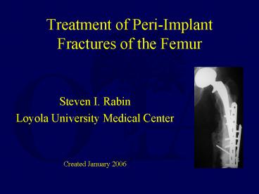
Treatment of PeriImplant Fractures of the Femur
Description:
Garbuz DS, Masri BA, Duncan CP. Instr Course Lect. ... and knee fractures: the scope of the problem.Younger AS, Dunwoody I, Duncan CP. Am J Orthop. ... – PowerPoint PPT presentation
Number of Views:810
Avg rating:3.0/5.0
Title: Treatment of PeriImplant Fractures of the Femur
1
Treatment of Peri-Implant Fractures of the Femur
- Steven I. Rabin
- Loyola University Medical Center
- Created January 2006
2
Fractures around Implants
- Pose Unique Fixation Challenges
3
Number of Implants in the Femur are Increasing
- Population is Aging
- Joint Replacement - Indicated More Often
- Fracture Fixation - Indicated More Often
4
Increasing Number of Implants in the Femur
- Over 123,000 Total Hip Replacements
- Over 150,000 Total Knee Replacement
- each year in the United States
- Numbers Expected To Increase with
Aging Population
5
Increasing Number of Implants in the Femur
- Over 300,000 Hip Fractures
- each year in the United States
- almost all are treated surgically with
internal fixation or prosthetic replacement
6
- As the Number of Implants Placed Increases
- the Number of Associated Fractures will Increase
7
Fractures around Implants Unique Fixation
Challenges
- Original Placement of the Implant may predispose
to later fracture - Long Term Presence of the Implant may change the
structure of bone and increase susceptibility of
fracture - Implant Itself may interfere with healing or the
placement of fixation devices
8
Peri-Implant Fractures May be Caused by Technical
Problems During Implant Placement
9
Technical Problems during Implant Placement
include
- Notching Anterior Femoral Cortex during Knee
Replacement - Cracking Calcar during Hip Replacement
- Penetrating Shaft during Hip Replacement
- Cracks between Screw Holes during Internal
Fixation
10
Notching Anterior Femoral Cortex During Knee
Replacement
- May have 40 fracture rate at 8 years
- Figgie et. al. J. Arthroplasty 1990
11
- Incidence of Supracondylar Femur Fracture after
Total Knee Replacement - .6 to 2.5
12
Fracture Associated with Implant Placement
- Fracture of the Femoral Neck may occur with
Antegrade Intramedullary Rodding - Stress Riser at Insertion Site
13
Calcar May Fracture During Hip Arthroplasty
- If the prosthesis or trials are not properly sized
14
Femoral Stem may Perforate the Femoral Shaft
- During
- Hip Replacement especially if the femur is bowed
- 3.5 fracture rate during Primary Total Hip
Replacement - Shaw Greer, 1994
15
The Bone Can Crack Between Screw Holes During
Internal Fixation
- Especially in osteoporotic bone
16
Stress Risers During Internal Fixation
- Any Drill Hole up to 20 of the bones diameter
will weaken bone by 40 - 90 of fractures around fixation implants occur
through a drill hole - Koval et. al. 1994
17
Stress Risers During Internal Fixation
- Fractures Tend to Occur at the End of Implants
where weaker bone meets the rigid device
18
Fractures can occur Postoperatively
- Incidence of 0.6 2.5
19
Fractures Associated with Implant Removal
- During Prosthetic Revisions
- 17.6 fracture rate compared to 3.5 during
primary hip replacements - (5 times the rate for primary hip replacement)
- through osteoporotic bone or osteolytic defects
20
Fractures Associated with Implant Removal
- Zickel IM Nails are associated with
Subtrochanteric Fractures after Removal - Plates Stress Shield
- Cortical bone - increased rate of fractures after
removal (especially forearm)
21
Problems with Treating Peri-Implant Fractures
- Implants may block new fixation devices
- Stems, rods, and bone cement may fill the
medullary canal preventing IM fixation of
fractures - Stems and rods may also block screw fixation
through the medullary canal to hold plates on
bone - Implants may impair healing due to endosteal
ischemia - Defects in bone from Osteolysis, Osteoporosis,
and Implant Motion may compromise fixation
22
Peri-Implant Fracture Fixation Methods
- Follow Standard Principles of Fixation
- Must Achieve Stable Anatomic Fixation while
Preserving Soft Tissue Attachments - Indirect Reduction Techniques
- Careful Preoperative Planning
- Intra-Operative Flexibility/Creativity
- Choose the Device That Fits the Patient
23
Periprosthetic Femur Fractures
- Treatment Options are
- Long-stem revision arthroplasty
- Cortical strut allografting
- Plate fixation with screws
- Plate fixation with cables
- Intramedullary Devices
24
Treatment Options
- Most
- Important Factor
- in Treating
- Peri-Implant Fractures is the
- Status of the Implant
25
- When the Implant is Loose, Mal-aligned or
Deformed - Consider Revision/Replacement
26
- When the Implant is Stable, and Well Aligned with
Good Quality Bone - Consider Fixation
27
Implant Revision/Replacement
- Avoids potential difficulties of fixation
- does not have to avoid the implant
- does not require stable fixation in poor bone
- Avoids potential complications of malunion or
nonunion - Indicated if Implant is Loose, Mal-Aligned,
Deformed or there is Poor Bone Quality
28
Case Example 1 Revision of Loose Prosthesis
Complicated by Fracture
- 82 y/o F
- Pre-existing LOOSE Hip Replacement
- Fell sustaining Peri-Prosthetic Femoral Shaft
Fracture - X-ray Findings Osteolysis, Subsidence
29
Case Example 1 Revision of Loose Prosthesis
Complicated by Fracture
- 82 y/o F
- Treatment Prosthesis Removal, Strut Medial
Allograft, and Long Stem Femoral Revision - Follow-up - allograft incorporated and
prosthesis stable with healed fracture at 6 months
30
Case Example 2 Hip Replacement after Fracture
at Tip of DHS Implant
- Elderly M
- DHS for Intertrochanteric Hip Fracture Fixation
31
Case Example 2 Hip Replacement after Fracture
at Tip of DHS Implant
- Elderly M
- Intertrochanteric Fracture Healed
- Fell 1 year later sustaining Femoral Neck
Fracture at tip of lag screw - X-rays showed poor bone stock
32
Case Example 2 Hip Replacement after Fracture
at Tip of DHS Implant
- Elderly M
- Treatment Hardware Removal, Hemiarthroplasty
- Follow-up Functioning well at 6 months
33
Fixation Around An Implant
- Avoids Difficulties of Implant Removal
- may be technically difficult
- may be time-consuming
- may cause further fracturing of bone
- Indicated if Implant is Stable, Well Aligned, and
Bone Quality is Good
34
Peri-Implant Fracture Fixation
- A Wide Selection of Devices Must be Available
- Special Plates with Cerclage Wires
- Curved Plates to Match the anterior Bow of the
Femur are Now Available. - Flexible Intramedullary Rods
- Rigid Intramedullary Rods
35
Plating Techniques for Peri-Implant Fractures
- Advantages of Plates
- Allow Direct Fracture Reduction and Exact
Anatomic Alignment - Less Chance of Later Prosthetic Loosening due to
Mechanical Mal-alignment - Allow Interfragmentary Compression and A Rigid
Construct for Early Motion
36
Plating Techniques for Peri-Implant Fractures
- Disadvantages of Plates
- Biologic and Mechanical Disadvantages Compared to
IM devices even with Indirect Techniques - Require Special Plates which accept Cerclage
Wires, and/or allow Unicortical Screws and/or
match the shape of the bone
37
Case Example 3 Fracture at the Proximal End of a
Supracondylar Nail Treated with a Plate
- Elderly F
- Pre-existing healed supracondylar femur fracture
- New fracture at end of rod after MVA
- Treatment ORIF with Plate/wires
- Follow-up Healed after 3 months and still
asymptomatic at 2 years
38
PeriProsthetic Fracture
- For Hip Peri-Prosthetic Fixation
- -Standard is with Plate or Allograft
or
39
Allograft Technique
- Picture/x-ray courtesy of Dr. John Cardea
40
Plate Technique
- Advantages of
- Plate over Allograft
- Less Invasive
- Leaves Medial Soft Tissues Intact
- Avoids Potential Allograft Risks
- Including Donor Infection
- Stronger
- Allograft bone can be Brittle
41
PeriProsthetic Fracture
- Plate or allograft attachment is by Cerclage
Wires or unicortical screws
or
42
Plate Techniques May Use Cables to attach the
plate to the bone
- Cables
- Require Extensive Exposure
- And are Technically Demanding
- So the fewer Used, the Better To decrease
operative trauma and operating time
- Pictures courtesy of Dr. John Cardea
43
Plate Techniques Can Also Use Screws to Attach
the Plate to Bone
- Screws
- Can be Placed Easier than Cables
- And Can be Placed Percutaneously with less soft
tissue trauma than Cables - So using Screws instead of Cables should decrease
operative trauma and operating time
44
Use of plates with cablesThere are many reports
- Examples
- -Ogden and Rendall, Orthop Trans, 1978
- -Zenni, et al, Clin Orthop, 1988
- -Berman and Zamarin, Orthopaedics, 1993
- -Haddad, et al, Injury, 1997
- But none of these address the question how
many cables are necessary?
45
Cables
- Cables resist bending loads
- -Mihalko, et al, J Biomechanics, 1992
- BUT Cables resist torsional loads poorly compared
to screws - -Schmotzer, et al, J Arthroplasty, 1996
- The Use of Screws should improve Rotational
Stability
46
PeriProsthetic Fracture
- Cerclage Wires are Less Mechanically Sound than
Unicortical Screws - Lohrbach Rabin MidAmerica Orthopedic Assoc.
Annual Meeting 2002
47
Conclusions
- A unicortical screw significantly increases
torsional and A-P stability and should be added
to cable-plate constructs - At least six cables are needed in the absence of
a unicortical screw to improve A-P and rotational
stability
Lohrbach Rabin MidAmerica Orthopedic Assoc.
Annual Meeting 2002
48
Case Example 4 Fracture at Distal End of Hip
Replacement Stem Treated with a Standard Plate
- Elderly F
- Pre-existing Asymptomatic Hip Arthroplasty
- Fell out of a car sustaining fracture at tip of
stem - X-rays showed a solid prosthesis
49
Case Example 4 Fracture at Distal End of Hip
Replacement Stem Treated with a Standard Plate
- Elderly F
- Treatment DCP plate w. screws/cerclage wires
- Follow-up Healed/Asymptomatic at 3 years
50
Case Example 5 Peri-Prosthetic Fracture Treated
with Locking Compression Plate
- 73y/o M
- Healthy
- 3 previous platings
51
Case Example 5 Peri-Prosthetic Repair with
Locking Plates
- Treatment Double Locked Compression Plate,
electrical stimulator, Hardware removal - Locking Screw Plates are Ideal because they
provide stable fixed angled unicortical fixation
52
Case Example 5 Peri-Prosthetic Repair with
Locking Plates
- Clinically painless by 6 weeks
- Radiographically appeared healed at 2 months
- Follow-up 13 months
- Complication S. epi post-op infection required
ID e-stim removal at 3 months
53
Case Example 6 Peri-Prosthetic Repair with LISS
Plate
- 49 y/o F
- Healthy Fracture at end of Hip Stem
- 3 previous platings,
- 1 previous retrograde rod
54
Case Example 6 Peri-Prosthetic Repair with LISS
- Treatment LISS locking plate, electrical
stimulator, bone graft - (LISS less invasive stabilization system)
55
Case Example 6 Peri-Prosthetic Repair with LISS
- Follow-up 19 mo.
- No Pain by 2 mo.
- Bridging 5 mo.
56
Case Example 7 Fracture Distal to Hip Stem
Treated with Curved Locking Plate
- 72 y/o Male with Hip Replacement for Arthritis
- X-ray from Routine Annual Follow-up (6 months
prior to fracture)
57
Case 7 Treatment with Curved Plate
- Fracture
58
Case 7 Curved Plate
- Intra-op
- Curved Plate Matches Bow of Femur
59
Case 7 Curved Plate Example
- Healed at 6 months
60
Flexible Intramedullary Rods(Zickel, Enders etc.)
- Flexible Rods Advantages
- can be placed via minimal incisions
- act as internal splints until fracture healing
61
Flexible Intramedullary Rods
- Flexible Rods Disadvantages
- require external protection (cast or brace)
- rarely allow early motion or weight-bearing
- must be enough space in the medullary canal for
implant and rod
62
Case Example 8 Distal Femur Fracture w.
Proximal Hip Replacement Treated with Flexible IM
Rod
- Elderly F s/p MI
- Pre-existing Asymptomatic Hip Hemiarthroplasty
- Fall sustaining distal femur shaft fracture
- X-rays showed wide medullary canal and
osteoporosis
63
Case Example 8 Distal Femur Fracture w.
Proximal Hip Replacement Treated with Flexible IM
rod
- Elderly F s/p MI
- Treatment Zickel Supracondylar Device
- Follow-up Healed Asymptomatic at 3yrs
64
Rigid Intramedullary Rods(Antegrade,
Supracondylar, Retrograde)
- Rigid Rod Advantages
- Do Not Require External Support
- Provide Rigid Fixation
- Biologic Mechanical Advantages of
Intramedullary Position
65
Rigid Intramedullary Rods
- Rigid Rod Disadvantages
- Cannot be used with a pre-existing stemmed implant
66
Case Example 9 Fracture at the End of a Blade
Plate Treated with a Retrograde Nail
- Young M
- 2 yrs after healed subtrochanteric hip fracture
with retained blade plate - In a High Speed Motor Vehicle Accident, sustained
a fracture at the distal end of the plate
67
Case Example 9 Fracture at the End of a Blade
Plate Treated with a Retrograde Nail
- Young M
- 2 yrs after healed subtrochanteric hip fracture
with retained blade plate - Treatment Retrograde Rodding
- Follow-up at 2 years healed and asymptomatic
68
Case Example 10 Fracture Above a Total Knee
Replacement Treated w. an Antegrade Nail
- Elderly F
- Bilateral Knee Replacements
- Sustained Bilateral Distal Femur Fractures
Proximal to Knee Replacements after MVA
69
Case Example 10 Fracture Above a Total Knee
Replacement Treated w. an Antegrade Nail
- Elderly F
- Bilateral Knee Replacements
- Treatment Bilateral Antegrade Rodding
- Follow-up at 3 years Fractures healed and both
knees asymptomatic
70
Summary
- If the prosthesis or implant is Loose, or Bone
Quality is Poor - then the implant should be
revised while fixing the fracture - If the prosthesis or implant is Stable and Bone
Quality is Adequate for Fixation - then the
implant should be retained while the fracture is
fixed following standard principles
71
Remember
- If Fixation is chosen Follow Principles of Good
Fracture Care
72
Case Example 11 Revision of Fixation Requiring
Osteotomy
- 78 y/o Female
- X-rays from 7 years ago after treatment of
infected intertrochanteric nonunion - Asymptomatic in interim
73
Example 11 Revision of Fixation
- Femoral Neck Fracture
- (Vertical Shear Pattern)
74
Example 11 Revision of Fixation
- Fixation of fracture with Valgus
Intertrochanteric Osteotomy restores leg length
and converts shear forces across the femoral neck
fracture into compressive forces
75
Example 11 Revision of Fixation
- Healing at 3 months
- (Plans to shorten blade)
76
Warning!
- The Bone Quality Must be Adequate to Hold
Fixation in addition to Stability of the Implant
if Fixation is chosen instead of
revision/replacement.
77
Example 12 Stable Prosthesis But Poor Bone
Quality
- 90 year old Female with asymptomatic
Hemi-arthroplasty at annual follow-up
78
Example 12 Stable Prosthesis But Poor Bone
Quality
- Fracture
- 2 months later
79
Example 12 Stable Prosthesis But Poor Bone
Quality
- Stable Prosthesis so Fixation with curved locked
plate with Uni-cortical screws Chosen for
Treatment
80
Example 12 Stable Prosthesis But Poor Bone
Quality
- Plate Failure At 3 months
81
Example 12 Stable Prosthesis But Poor Bone
Quality
- Salvage with Proximal Femoral Replacement
82
Conclusions
- Surgeon must carefully Evaluate Stability of the
Implant - Loose Fixation Implants will allow motion at the
fracture site that interferes with healing and
gets in the way of more stable fixation devices - Loose Prosthetic Implants will be painful and
also interfere with adequate fixation
83
Conclusions
- If the prosthesis or implant is Loose, or Bone
Quality is Poor - - the implant should be revised while fixing the
fracture
84
Conclusions
- If the prosthesis or implant is Stable and Bone
Quality is Adequate for Fixation - the implant should be retained while the
fracture is fixed following standard principles
85
Selected References
- Orthop Clin North Am. 1999 Apr30(2)249-57The
treatment of periprosthetic fractures of the
femur using cortical onlay allograft struts.Brady
OH, Garbuz DS, Masri BA, Duncan CP. - Instr Course Lect. 199847237-42.Periprosthetic
fractures of the femur principles of prevention
and management. Garbuz DS, Masri BA, Duncan CP. - Instr Course Lect. 199847251-6. Periprosthetic
hip and knee fractures the scope of the
problem.Younger AS, Dunwoody I, Duncan CP. - Am J Orthop. 1998 Jan27(1)35-41 One-stage
revision of periprosthetic fractures around loose
cemented total hip arthroplasty.Incavo SJ, Beard
DM, Pupparo F, Ries M, Wiedel J. - Instr Course Lect. 200150379-89.Periprosthetic
fractures following total knee arthroplasty.
Dennis DA - Orthop Clin North Am. 2002 Jan33(1)143-52,
ix.Periprosthetic fractures of the femur. Schmidt
AH, Kyle RF - J Arthroplasty. 2002 Jun17(4 Suppl
1)11-3.Management of periprosthetic fractures
the hip.Berry DJ. - Clinical Orthopaedics Related Research.
(420)80-95, March 2004.Periprosthetic Fractures
Evaluation and Treatment. Masri, Bassam Meek, R
M. Dominic Duncan, Clive P
If you would like to volunteer as an author for
the Resident Slide Project or recommend updates
to any of the following slides, please send an
e-mail to ota_at_aaos.org
Return to Lower Extremity Index
E-mail OTA about Questions/Comments


