
What do all of these things have in common PowerPoint PPT Presentation
1 / 10
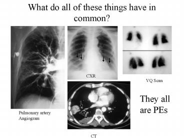
Title: What do all of these things have in common
1
What do all of these things have in common?
CXR
VQ Scan
They all are PEs
Pulmonary artery Angiogram
CT
2
Why do we care about this?
- The dreaded saddle embolus
- PE has a mortality rate of 30 without treatment1
- An accurate diagnosis w/ anticoagulant therapy
decreases this rate to 2-8
Pulmonary embolism mortality in the United
States, 1979-1998 an analysis using
multiple-cause mortality data. AUHorlander KT
Mannino DM Leeper KV SOArch Intern Med 2003 Jul
28163(14)1711-7.
3
Symptoms and signs of acute pulmonary embolism
4
PIOPED
- BEAT (OR FLOG) A DEAD HORSE
- British politician John Bright thought the
Reform Bill of 1867, which called for more
democratic representation, would never be passed
by Parliament. - Trying to rouse Parliament from its apathy on the
issue, he said in a speech, would be like trying
to 'flog a dead horse' to make it pull a load. - This is the first recorded use of the expression,
which is still common for 'trying to revive
interest in an exhausted issue
Encyclopedia of Word and Phrase Origins" by
Robert Hendrickson (Fact on File, New York, 1997)
5
PIOPED in brief
Angiography
VQ scan
vs
- The challenger
- Non-invasive
- Good for patients with dye allergy
- Good for patients with renal disease
- Low risk
- Gold standard
- Invasive
- Patients must not be allergic to dye or contrast
- Cant be used in patients with renal disease
- Some MM
6
PIOPED in brief
- Your pre-test clinical suspicion matters
- Diagnostic accuracy was greatest when the V/Q
scan was combined with clinical probability - In patients with a low pre-test suspicion, having
a low or near normal scan result was helpful,
missing only 4 and 2 respectively - In patients with a high pre-test suspicion, a
HIGH scan probability detected 96 of PEs, but a
LOW probability scan detected roughly 40 of PEs - So if you have a high suspicion, a low
probability scan is not that helpful to you
Pioped investigators. Value of the
Ventilation/Perfusion Scan in Acute Pulmonary
Embolism. JAMA May 23/30 1990 vol 263 no 20
7
Quantifying your clinical suspicion
Modified Wells criteria clinical assessment for
pulmonary embolism
Data from van Belle, A, et al. JAMA 2006
295172.
8
Clinical Question
- What are the options if you still are still
worried about PE in your patient, but the VQ scan
was low prob?
9
2 options Take your pick
- At study by Kearon, et al found that it was safe
to withhold anticoagulation after obtaining
normal results on initial US, followed by repeat
US 1 week later - Study design randomized, multi-center
- 810 patients w/ normal initial compression US
studies for proximal veins - Interventions D-dimer testing followed by no
further testing if the result was negative and
venography if the result was positive
(experimental) or ultrasonography - US repeated after 1 week in all patients
(control). - Measurements Symptomatic DVT diagnosed initially
and DVT during 6 months of follow-up - Conclusion In outpatients with suspected deep
venous thrombosis who initially had normal
results on ultrasonography of the proximal veins,
a strategy based on D-dimer testing followed by
no further testing if the result was negative and
venography if the result was positive had
acceptable safety and did not differ from the
safety of a strategy based on withholding
anticoagulant therapy and routinely repeating
ultrasonography after 1 week.
Kereon, et al. A Randomized Trial of Diagnostic
Strategies after Normal Proximal Vein
Ultrasonography for Suspected Deep Venous
Thrombosis D-Dimer Testing Compared with
Repeated Ultrasonography. Ann Intern Med.
2005142490-496
10
More literature on this
- Study Design retrospective chart review on 662
PIOPED patients - Results Single noninvasive leg test in patients
with nondiagnostic VQ scans picks up DVT in 11
of patients who would otherwise require
angiography - Serial noninvasive leg tests in patients w/
normal initial test either picks up the DVT or
excludes it in 47 of patients - Using serial dopplers decreases the need for
invasive angiography to 29 from 63 for 1
Doppler study - Additionally, using NO Doppler study has a
baseline angiography requirement in 71 of
patients - Clearly 1 is not good enough and 2 is a lot
better - Conclusion A noninvasive strategy that includes
VQ scans, single noninvasive leg tests, and
serial noninvasive leg tests would permit a
diagnosis of thromboembolic disease or a safe
exclusion of thromboembolic disease in 71 of
patients with suspected acute pulmonary embolism.
Stein, Paul, et al. Strategy That Includes Serial
Noninvasive Leg Tests for Diagnosis of
Thromboembolic Disease in Patients With Suspected
Acute Pulmonary Embolism Based on Data From
PIOPED. Arch Int Med. Volume 155(19), 23 October
1995, pp 2101-2104.
Recommended
