
CYTOLOGY - PowerPoint PPT Presentation
1 / 89
Title:
CYTOLOGY
Description:
Transfer the details to the cytopathology register. ... Record should be transferred to a cytopathology register. Internal quality control Procedures: ... – PowerPoint PPT presentation
Number of Views:14987
Avg rating:5.0/5.0
Title: CYTOLOGY
1
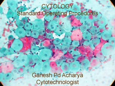
CYTOLOGY Standard Operating Procedures Gan
esh Pd Acharya Cytotechnologist
2
- INTRODUCTION
- Cytology
- It is the branch of Medical sciences, which deals
with the study of cells which includes - Shape and size (Morphological study).
- Chemical constituents (cyto-chemical study)
- Maturation process (Metabolic activity study)
- Specialized ultra structures of cell surface and
their functions. (Electron Microscopic study) - Cytogenetics (chromosomal study)
- Cancer marker (Immuno-cyto-chemical study).
3
- Cyto-preparation techniques include
- Methods of specimen collection,
- Fixation and fixatives,
- Preservation of fluid specimens prior to
processing, - Preparation of materials for microscopic
examination, - Staining and mounting of the smears,
- Transportation of prepared slides to the defined
center, - Registration and recording of the reports,
- Distribution of reports to the respective
department /clinic / patient,
4
- Methods of collection
- In general, material for cytological examination
is obtained either in the form of smears prepared
by examining physician, gynecologist, surgeon or
their assistants at the time of clinical
examination e.g. cervical smears. - OR
- In the form of fluid specimens which are
forwarded to the laboratory for further
processing e.g. Body fluids such as - Pleural fluid
- Ascitic fluid
- Peritoneal fluid
- Pericardial fluid
- Joint fluid
- Cystic fluid (Breast/tumor)
- Sputum
- Urine
- Cerebrospinal fluid (CSF)
- Gastro-intestinal aspirates etc.
5
- SMEARS
- Prior to the preparation of smears, it is
important to secure the necessary materials and
lay them out on a suitable, conveniently located
surface within the reach of the operator - Instrument(s) used to obtain smear,
- Clean, new microscopic slides,
- Suitable marker (diamond pencil) for the
identification of slides, - Paper clips (plastic coated or copper) used to
separate the slides from each other if liquid
fixatives are used, - Fixatives,
- Laboratory form with clear identification of the
patient and appropriate history minimum data
required for each patient is listed on the form,
6
- Preparation of smears
- For most diagnostic purposes, well-prepared and
well-fixed smears are required. - Air-drying of smears should be avoided, if
prepared for wet fixation. - Monolayer preparation is suitable for almost all
processing techniques. - Considerable skill and practice are required to
prepare excellent smears by single swift motion
without loss of material or air-drying. - Excessive crushing of the material must be
avoided. - A competent help must be secured in advance.
7
- Fixation
- Immediate fixation of smears is essential for
the correct interpretation. - Most of the fixatives are alcohol-based e.g. 95
ethanol, 95 rectified spirit, 80 iso-propanol
or propanol, absolute methanol, Ether95 Ethanol
mixture (11), Carnoy's fixative. - Air dried smears are required or desirable in
special situations (special stains e.g. MGG stain)
8
- Fixatives
- Two types of fixatives are commonly used.
- Fluid fixatives
- Spray fixatives
- Fluid fixatives
- Are prepared in bottles of suitable sizes,
provided with caps or coplin jars with covers. - Fixatives solution must be new for each batch of
fixation. - Filtration of fixatives should be done in case of
repeated use.
9
(No Transcript)
10
- 2.Spray fixatives
- Contain 2-10 carbo wax in 95 alcohol
- Protect the smears from drying by forming an
invisible film on the surface of the slides - May be used in lieu of fluid fixatives i.e.
immediately after the process of smear
preparation has been completed. - Correct use of spray fixative calls for several
precautions such as - Spray must be smooth and steady
- Distance between spray nozzle and smear must be
10-12 inches (25-30 cm) - Smears coated with spray fixatives must be
air-dried before mailing
11
(No Transcript)
12
- Fluid specimens
- May be obtained from a variety of body sites
such as - Respiratory tract (sputum)
- Gastro-intestinal tract (endoscopic aspirates)
- Urinary tract (Urine / barbotage samples)
- Effusion fluids (body cavity fluids-pleural/
peritoneal/pericardial) should be collected in
anticoagulants container (1 ammonium oxalate in
the ratio of 91 i.e. 9 parts of fluid and 1 part
anticoagulant)
13
Cytology STANDARD OPERATING PROCEDURE NPHL, Teku,
Kathmandu
14
PROCEDURE Principle- The
smears of body fluids, which contain exfoliated
cells and/or cells produced by transudation,
which are then concentrated by centrifugation, so
as to make the screening process efficient.
15
- Method
- Take two centrifuge tubes.
- Label the centrifuge tubes.
- Mix the fluid properly by inverting the container
ten times. - Put the centrifuge tubes in the centrifuge
machine at the opposite sides so as to balance
while centrifugation. - Add 10 ml each of well-mixed fluid to the labeled
centrifuge tubes. - Set the centrifuge machine at 1500 rpm for 15-20
minutes.
16
- Remove the centrifuge tubes once the machine
stops and discard the supernatant in the
disinfectant container with the help of a
Pasteur pipette. - Label four clean slides with the help of
diamond pencil. - Transfer the sediment on the clean slides 2 cm
away from the end with the help of a Pasteur
pipette. - Make the smears immediately by holding the slide
with one hand and spreading with the help of the
flat surface of another slide.
17
- If the sample is haemorrhagic (reddish in
colour), smear should be prepared from the
buffy- coat layer of the sediment. In such
condition FISH TAIL smear preparation is
suggested. - Three slides are immediately fixed (while still
wet) in the alcoholic fixative for 20-30
minutes. - One slide is left to dry in open air at room
temperature
18
(No Transcript)
19
- Internal Quality Control Procedures
- The proportion of fluid and anticoagulant should
be maintained (91). - In case of delay in processing, the fluid should
be stored in a refrigerator. (DO NOT FREEZE) - Supernatant and remaining fluid should always be
discarded in a disinfectant solution. - Protective clothing such as gloves, apron and
mask should be worn while processing. - Smears should be prepared so as to give monolayer
of cells for easy differentiation. - Fixation should be done immediately after
preparation of the smears so as to prevent
changes in cell morphology.
20
(No Transcript)
21
- PROCEDURE
- Principle
- Fixation prepares the samples from different
sites of the body for the purpose of preserving
and maintaining the existing form and structure
of all constituent elements. - Method
- Four slides (urine), three slides (body
fluids/sputum), 1-2 slides (cervical smear) and
one slide (CSF) should be immediately put in the
coplin jar/container with 95 Ethanol (alcohol)
or its equivalent fixative. - Leave the slides in the fixative for a minimum of
30 minutes. - Take out the slides with the help of forceps and
leave to dry in a slide rack. - One slide for all specimens except urine and
cervical smear is not fixed in the alcoholic
solution but dried at room temperature.
22
- Internal Quality Control Procedures
- Grease-free, clean new slides are to be used for
all types of specimens. - Proper fixative and time of fixation are to be
strictly maintained. - Use of fresh fixatives is recommended. If the
fixative is to be re-used it should be filtered
(Whatman filter paper) for every batch of slide
fixation.
23
(No Transcript)
24
- PROCEDURE
- Principle
- Properly filled request form, properly labelled
samples and proper registration of the patient
identification with their clinical parameters are
the pre-requisites for Quality results. - Method
- Match the request form and labelled sputum
samples. - Check the request form for patient's full name,
age, short clinical history and other additional
parameters. - Enter the detail in register.
- Label the sample and the request form with the
running number with permanent marker. - Make them ready for further processing.
25
- DOCUMENTATION
- Date of sputum sample reception.
- Name age of patient
- Name of the lab personnel who receives the smear.
- Transfer the detailed to the cytopathology
register.
26
- Internal Quality Control Procedures
- The sputum sample should be received in a wide
mouth bottle with proper labeling. - Accept only completely filled request form.
- Details of patient should be documented in the
register.
27
(No Transcript)
28
- MATERIALS REQUIRED
- Sputum sample
- New glass slides
- Wooden spatula
- Wooden racks
- Coplin jar
- 80 propanol
- Diamond Pencil
29
- PROCEDURE
- Principle
- The neoplastic cells are exfoliated and are
expectorated in sputum. The exfoliated cells are
spread over the slide in a manner which will make
cytological study possible. Similarly, other
cells may exfoliate and may indicate the other
underlying pulmonary pathology. - Method
- Clean the working bench
- Put on protective clothing, gloves.
- Place specimen on the working bench. If specimen
has been refrigerated, bring to room temperature.
- Label 4 slides with patient identification
laboratory acquisition number. - Examine specimen carefully. This may require that
the specimen to be transferred to a petridish
placed on a dark background to visualize
suspicious areas.
30
- Look for fresh or old blood stained areas,
discolored portion of sample tissue fragments. - Transfer suspected areas with a disposable stick
or applicator to a glass slide. Spread material
across surface of glass slide. - Take a second glass slide.
- Spread them gently between the 2 slides until an
even distribution of material is obtained. - Fix slides in 95 ethanol for 15-30 minutes
31
(No Transcript)
32
- DOCUMENTATION
- Date of sputum sample collected.
- Date of sputum sample received in the lab.
- Date of sputum sample processed.
- Gross appearance
- Fixatives used or not
- Total no of smears.
- Type of fixative used.
- Clinical details clinical diagnosis.
- Name of the technician handling the sample.
- Name of the pathologist who is going to report.
- Transfer the material to cytopathology register
33
- Internal Quality Control Procedures
- Not to receive any broken or dirty slides.
- Accept only completely filled request form
(Request forms must be filled by the concerned
clinician) - Details of documents should be registered.
34
(No Transcript)
35
- MATERIALS REQUIRED
- Properly labeled and fixed smeared cervical
slides. - Request formfilled by the clinician with
identification of both patient and smears with
short clinical history and minimum data
requirements. - Permanent marker for labeling.
- Cervical cytology register.
36
- PROCEDURE
- Principle
- Properly filled request form, properly labeled
and fixed cervical smears and proper registration
of the patient's identification with their
clinical parameters are the pre-requisites for
quality results. - Method
- Match the labeled slide number with the request
form. - Check the request form for patient's full name,
age, LMP (Last menstrual period), address, short
clinical history and other additional parameters. - Enter the details in the Cervical cytology
register. - Label the slides and the request form with the
running laboratory number with a permanent
marker. - Store the received smears in a proper place.
- Make them ready for mailing.
37
- DOCUMENTATION
- Date of smear reception.
- Numbers of slides received.
- Date of LMP.
- Name and age of patients.
- Name of the clinician who took the smear.
- Name of the Lab. personnel who received the
smear. - Transfer the details to the cytopathology
register. - Check the details on a form, which will be mailed
with the slides for reporting.
38
- Internal Quality control procedures
- Do not receive any broken or dirty slides.
- Clinician must properly fill the request form. Do
not accept any improperly filled such form having
no significant reason. - Details of all documents should be register
properly.
39
(No Transcript)
40
- MATERIALS REQUIRED
- 95 Ethanol / 95 rectified spirit /80
iso-propanol or propanol / absolute methanol /
Ether95 Ethanol mixture (11) /Spray coating
fixatives (2-10 Carbowax in 95 Ethanol)/
Carnoy's fixative for haemorrhagic fluids. - Grease-free, clean new slides
- Slide racks
- Forceps
- Coplin jars /suitable containerplastic coated
paper clips - 0.5 Sodium hypochlorite solution / 1 phenolic
solution container - Diamond pencil (Slide marker)
- Filter paper (Whatman)
- Funnel
41
PROCEDURE Principle Fixation prepares the
samples from different sites of the body for the
purpose of preserving and maintaining the
existing form and structure of all constituent
elements.
42
- Method
- Four slides (urine), three slides (body
fluids/sputum), 1-2 slides (cervical smear) and
one slide (CSF) should be immediately put in the
coplin jar/container with 95 Ethanol (alcohol)
or its equivalent fixative. - Leave the slides in the fixative for a minimum of
30 minutes. - Take out the slides with the help of forceps and
leave to dry in a slide rack. - One slide for all specimens except urine and
cervical smear is not fixed in the alcoholic
solution but dried at room temperature
43
- DOCUMENTATION
- Date and time of sample collection
- Date and time of sample receipt.
- Amount of fluid received.
- Gross appearance of the fluid.
- Total number of smears prepared.
- Total number of dry smears.
- Total number of clot smears (if any).
- Transfer the details to the cytopathology
register. - Transfer the details on a form, which will be
forwarded with the slides for reporting.
44
- Internal Quality Control Procedures
- Grease-free, clean new slides are to be used for
all types of specimens. - Proper fixative and time of fixation are to be
strictly maintained. - Use of fresh fixatives is recommended. If the
fixative is to be re-used it should be filtered
(Whatman filter paper) for every batch of slide
fixation.
45
(No Transcript)
46
- MATERIALS REQUIRED
- Cervical smears
- 95 Ethanol /80 iso-propanol / absolute methanol
for fixation/hydration - Coplin jars
- Staining rack
- Alcohols 50, 70, 80, 95 and absolute alcohol.
- Staining solutions
- Harris Haematoxylin
- OG-modified
- EA-modified
- Diluted 1Lithium Carbonate (30 drops of 1
Lithium carbonate in 1000 ml of distilled water)
47
- 0.5 HCl
- Tap water/Distilled water
- Xylene
- DPX Mountant
- Coverslips
- Labeling Stickers
48
PROCEDURE Principle- The Papanicolaou staining
procedure was devised for optimal visualization
of cancer cells exfoliated from the epithelial
surface of the body. It is a polychrome staining
reaction, consisting of a water-based nuclear
stain and two alcohol-based cytoplasmic
counterstains, designed to display the many
variations of cellular morphology and to show the
degree of cellular maturity and metabolic
activity.
49
- Method
- Papanicolaou Staining (regressive method)
- Arrange the slides in a staining rack.
- Hydrate the smears by immersing the slides in 95
alcohol for 10-15 minutes followed by 80, 70
and 50 alcohol (2-3 minutes each). - Immerse the slides in distilled water/tap water
for 5 minutes. - Drain the excess water and put in Harris
Haematoxylin solution for 3-5 minutes. - Rinse in tap water for one minute.
- Differentiate in 0.5 HCl (1-2 quick dips)
- Rinse in tap water for 2-3 minutes.
- Immerse the slides in diluted Lithium carbonate
for 1 minute. - Rinse with tap water/distilled water for 1
minute. - Dehydrate by immersing in 70 ethanol for 30
seconds. - Next immerse in 80 ethanol for 30 seconds.
50
- Immerse in two changes of 95 ethanol for 30
seconds each. - Stain in OG-modified solution for one minute.
- Rinse in two changes of 95 ethanol for 30
seconds each. - Stain in EA-modified for 5-11 minutes (depending
on the quality of stain and frequency of
staining). - Immerse in three changes of absolute ethanol for
30 seconds each. - Immerse in three changes of Xylene for 30 seconds
each. - Mount the smears with DPX.
- Label the smears with stickers.
- Submit with the requisition form for microscopy.
51
(No Transcript)
52
- DOCUMENTATION
- Date of smear preparation
- Number of smears received.
- Date of LMP
- Age of the patient.
- Name of the clinician who took the smear.
- Short clinical history.
- Name of the technician/Laboratory personnel who
processed the smear. - Record should be transferred to a cytopathology
register.
53
- Internal quality control Procedures
- Harris Haematoxylin should be filtered everyday
before staining. - Staining time can vary with each batch of new
stains (depending on the quality of the stain and
frequency of staining). - Staining solutions should be prepared in small
quantities to cover a period of three
months (Stains being alcoholic
preparations) - 0.5 HCl and diluted Lithium carbonate should be
prepared freshly each day.
54
(No Transcript)
55
- MATERIALS REQUIRED
- CSF sample
- Cytocentrifuge
- Centrifuge tubes.
- New glass slides.
- Marker pencil
- Normal saline solution.
56
(No Transcript)
57
(No Transcript)
58
(No Transcript)
59
(No Transcript)
60
- PROCEDURE
- Principle
- CSF sample contain either very low cell count or
the total volume of sample is very low the cyto
centrifugation technique is used to obtain
comparatively higher cellular manolayer smear. - Method
- Take a pre-coated glass slide.
- Fix the pre-coated slide into cytocentrifuge
- Take CSF sample add normal saline solution to
make final volume of 5 ml. - In opposite chamber counter balance is done by
adding normal saline. - Put the CSF solution in to the centrifuge chamber
centrifuge 1000 rpm for 4 minutes. - Pick up glass slides from centrifuges head.
- For wet preparation of smear fix the glass slide
immediately into 95 ethanol. - For dry smear leave the slide on rack.
- Send the slides for Papanicolaou stain and MGG
stain
61
- DOCUMENTATION
- Number of slides received.
- No of dry slides/wet slides
- Date of CSF sample collection
- Name of Doctor who collected the sample.
- Name of person who performed the test processing.
- Test result should be entering in the Cyto
pathology Register book.
62
- Internal Quality Control Procedure
- Grease free and clean slides must be used.
- C.S.F should be centrifuged at desired speed for
desired time. - The glass slides should be coated before smear
preparation.
63
(No Transcript)
64
- MATERIALS REQUIRED
- Body fluids in an anticoagulated container (1
Ammonium Oxalate solution--Ratio of fluid to
anticoagulant is 91) - New, grease-free, clean slides
- Graduated centrifuge tubes (Capacity-10 ml)
- Centrifuge (Speed limit not lt1500 rpm)
- Cytocentrifuge (if available)
- Coplin jars
- 95 Ethanol/80 isopropanol/absolute methanol
- Ether/95 Ethanol mixture (11)
- Pasteur pipettes (Glass/plastic)
- 0.5 Sodium hypochlorite solution/1 phenolic
solution container (Disinfectant)
65
- PROCEDURE
- Principle
- The smears of body fluids,
which contain exfoliated cells and/or cells
produced by transudation, which are then
concentrated by centrifugation, so as to make the
screening process efficient. - Method
- Take two centrifuge tubes.
- Label the centrifuge tubes.
- Mix the fluid properly by inverting the container
ten times. - Add 10 ml each of well-mixed fluid to the labeled
centrifuge tubes. - Put the centrifuge tubes in the centrifuge
machine at the opposite sides so as to balance
while centrifugation. - Set the centrifuge machine at 1500 rpm for 15-20
minutes. - Remove the centrifuge tubes once the machine
stops and discard the supernatant in the
disinfectant container with the help of a Pasteur
pipette.
66
- Label four clean slides with the help of diamond
pencil. - Transfer the sediment on the clean slides 2 cm
away from the end with the help of a Pasteur
pipette. - Make the smears immediately by holding the slide
with one hand and spreading with the help of the
flat surface of another slide. - If the sample is haemorrhagic (reddish in
colour), smear should be prepared from the buffy-
coat layer of the sediment. In such condition
FISH TAIL smear preparation is suggested. - Three slides are immediately fixed (while still
wet) in the alcoholic fixative for 20-30 minutes. - One slide is left to dry in open air at room
temperature.
67
(No Transcript)
68
- DOCUMENTATION
- Date and time of sample collection.
- Date and time of sample receipt.
- Amount of fluid received.
- Gross appearance of the fluid.
- Total number of smears prepared.
- Total number of dry smears.
- Total number of clot smears (if any).
- Transfer the details to the cytopathology
register. - Transfer the details on a form, which will be
forwarded with the slides for reporting.
69
- Internal Quality Control Procedures
- The proportion of fluid and anticoagulant should
be maintained (91). - In case of delay in processing, the fluid should
be stored in a refrigerator. (DO NOT FREEZE) - Supernatant and remaining fluid should always be
discarded in a disinfectant solution. - Protective clothing such as gloves, apron and
mask should be worn while processing. - Smears should be prepared so as to give monolayer
of cells for easy differentiation. - Fixation should be done immediately after
preparation of the smears so as to prevent
changes in cell morphology.
70
(No Transcript)
71
- MATERIALS REQUIRED
- Slide racks for air-drying the pre fixed smears
- Slide mailer kit
- Request form-identification of both patient and
sample. - Postal stamps
- Stickers with postal address of the center.
72
(No Transcript)
73
(No Transcript)
74
- PROCEDURE
- Principle
- The smears collected for the diagnosis need to be
sent to a defined center. For this one should
have a thorough knowledge of mailing procedure to
prevent the deterioration of the collected
material, loss of smear and breakage of slides. - Method
- After pre-fixation, slides are taken out of the
fixative and air-dried in a slide rack. - Slides are then arranged in an orderly form.
- Slides are arranged in the slide mailer and the
mailer is closed properly. - The slide mailer along with the properly filled
requisition form is then put in the envelope used
for transportation. - The envelope is sealed and the address of the
diagnostic center is pasted on it along with the
postal stamp. - The envelope is then taken to the post office
promptly.
75
- DOCUMENTATION
- Type of sample.
- Patient/Slide identification number.
- Date and time of sample collection.
- Date and time of sample receipt.
- Amount of fluid received.
- Gross appearance of the fluid.
- Total number of smears prepared.
- Total number of dry smears (Numbering done with a
capital 'D' in front). - Total number of clot smears (if any).
- Total number of smears sent.
- Transfer the details to the cytopathology
register. - Transfer the details on a form, which will be
mailed with the slides for reporting.
76
- Internal Quality Control Procedures
- Check the lock of the slide mailer.
- Check the seal of the envelope.
- Check the mailing address.
77
- PREPARATION OF REAGENTS
- Materials required
- Measuring cylinder100 ml and 1000 ml capacity.
- Conical or flat bottom flasks100ml and 500 ml
capacity. - Beakers250 ml and 500 ml capacity.
- 10 ml graduated pipette or micropipette with
disposable tips. - Reagent bottles (Dark coloured)500 ml and 1000
ml capacity. - Funnels3 inches and 6 inches in diameter.
- Permanent markers for labeling.
- Analytical balance and weights.
- Heater/Stove.
- Tripod stand.
- Thermometer.
- Waterbath.
78
- Preparation of Harris Haematoxylin
- Haematoxylin (Colour index No. 75290) 5 g
- Absolute methanol 50 ml
- Distilled water 1000 ml
- Mercuric Oxide (HgO) 2.5 g
- Aluminium ammonium sulphate or Potassium
- ammonium sulphate (Alum) 100 g
- Glacial acetic acid 40 ml
- Method of preparation
- Dissolve Alum in 1000 ml distilled water and
bring it to boil. - Dissolve haematoxylin in alcohol by warming up to
60C. - Add dissolved haematoxylin to Alum and bring
again to boil. - Remove the flask from heat.
- Immediately add Mercuric oxide.
- Stir this solution till dark purple colour
appears (Not more than 10 seconds). - Plunge flask into water bath (ice cold water) to
cool. - Store in a dark bottle.
- Filter before use.
79
- . Preparation of EA modified
- a. Light green (colour index no 42095) 0.3 g
- b. Eosin Y (colour index no 45380) 4.0 g
- c. Phosphotungstic acid 2.0 g
- d. Distilled water 480 ml
- e. 95 ethanol (alcohol) 500 ml
- f. Glacial acetic acid 20 ml
- Method of preparation
- Mix above listed (abc) dyes and chemicals in
480 ml distilled water in a flask. - Mix well by stirring and warming to 60-80C.
- Cool the mixture to room temperature.
- Add 500 ml of 95 Ethanol and mix well by
stirring. - Add 20 ml glacial acetic acid to the above
mixture. - Store in dark coloured reagent bottle and filter
before use.
80
- Preparation of OG- modified
- a. Orange G 5.0 g
- b. Distilled water 500 ml
- c. Phosphotungstic acid 2.0 g
- d. Absolute ethanol 500 ml
- e. Glacial acetic acid 10 ml
- Method of preparation
- Dissolve abc dye and chemical in distilled
water. - Warm the mixture 60-80C.
- Mix by frequent stirring and cool to room
temperature. - Add methanol and glacial acetic acid.
- Mix by stirring.
- Store in dark bottle.
- Filter before use.
81
- Preparation of 0.5 Hydrochloric acid (HCl)
- Conc. HCl 5 ml
- Distilled water 995ml
- Method of preparation
- Measure 995 ml distilled water and transfer to
1000 ml conical flask. - Add 5 ml of conc. HCl with the help of the
pipette slowly with continuous mixing to the
distilled water. - Transfer the prepared solution in a reagent
bottle with name and date of preparation.
82
- Preparation of 1 Lithium Carbonate
- (Stock solution)
- Lithium Carbonate (LiCO3) 10 g
- Distilled water 1000 ml
- Method of preparation
- Measure 1000 ml distilled water in a conical
flask. - Add 10 g of LiCO3 to the distilled water and mix
till properly dissolved. - Transfer the mixture to a reagent bottle with
name and date of preparation. - Working solution is prepared by adding 30 drops
of stock solution to 1000 ml of distilled water.
83
- PREPARATION OF FIXATIVES
- MATERIALS REQUIRED
- Measuring cylinder1000 ml and 100 ml capacity.
- Conical or flat bottom flasks100 ml and 500 ml
capacity. - Beakers500 ml and 250 ml capacity.
- Reagent bottles500 ml and 1000 ml capacity.
- Funnels6 inches and 3 inches diameter.
- Permanent markers for labeling.
84
- REAGENT PREPARATION
- 95 Ethanol
- Take 950 ml of Absolute Ethanol in 1000 ml
capacity measuring cylinder. - Take 50 ml of distilled or deionised water in
100 ml capacity measuring cylinder. - Take 1000 ml Conical/flat-bottom flask and mix
the Ethanol and distilled water properly in it. - Label a reagent bottle as 95 Ethanol with the
date of preparation and transfer the above
solution in it.
85
- 95 rectified spirit
- Take 950 ml of Rectified spirit in a 1000 ml
capacity measuring cylinder. - Take 50 ml of distilled or deionised water in 100
ml capacity measuring cylinder. - Take 1000 ml Conical/flat-bottom flask and mix
the rectified spirit and distilled water properly
in it. - Label a reagent bottle as 95 Rectified spirit
with the date of preparation and transfer the
above solution in it.
86
- 80 iso-propanol/propanol
- Take 800 ml of iso-propanol/propanol in 1000 ml
capacity measuring cylinder. - Take 200 ml of distilled or deionised water in
1000 ml capacity measuring cylinder. - Take 1000 ml Conical/flat-bottom flask and mix
the alcohol and distilled water properly in it. - Label a reagent bottle as 80 iso-propanol/propano
l with the date of preparation and transfer the
above solution in it.
87
- Ether95 Ethanol Mixture
- Take 100 ml of Ether in 100 ml capacity
measuring cylinder. - Take 100 ml of 95 Ethanol in 100 ml capacity
measuring cylinder. - Take 500 ml capacity conical/flat-bottom flask
and mix the ether and ethanol in it properly. - Label a reagent bottle as Ether95 Ethanol
Mixture with the date of preparation and
transfer the above solution in it.
88
- Carnoy's Fixative
- Take 60 ml absolute Ethanol in 100 ml capacity
measuring cylinder. - Take 30 ml Chloroform in another measuring
cylinder. - Take 10 ml Glacial acetic acid in another
measuring cylinder. - Mix the above solutions in a 500 ml capacity
conical/flat-bottom flask. - Label a reagent bottle as Cornoy's fixative with
the date of preparation and transfer the above
solution in it.
89
Namaste
Recommended
CrystalGraphics Presentations


