
Autoradiography - PowerPoint PPT Presentation
1 / 21
Title:
Autoradiography
Description:
Labeling Techniques Autoradiography Efficient for studying dynamic processes in cells and tissues. Molecules biosynthesized with radioactive elements behave in the ... – PowerPoint PPT presentation
Number of Views:729
Avg rating:3.0/5.0
Title: Autoradiography
1
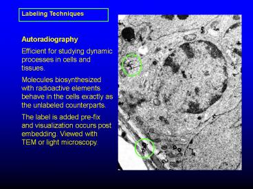
Labeling Techniques
Autoradiography Efficient for studying dynamic
processes in cells and tissues. Molecules
biosynthesized with radioactive elements behave
in the cells exactly as the unlabeled
counterparts. The label is added pre-fix and
visualization occurs post embedding. Viewed with
TEM or light microscopy.
2
History of Radiography
- Niepce de St. Victor observes blackening of
silver iodide and silver chloride emulsions by
uranium nitrate. - 1869 Bequerel observes the same darkening with
opaque paper placed between the uranium nitrate
and the emulsions.
3
1904 London puts a frog immersed in Radium water
on a photographic plate.
1956 Technique scaled down for use in EM by
Liquier-Milward 1980s 14C- and 3H-labeled
leucine are used to measure rates of amino acid
incorporation into proteins. The use of
autoradiography is also proposed for the
measurement of ligand binding and drug
interaction.
4
(No Transcript)
5
Radioactivity - radioisotopes (elements with
higher than normal atomic weight) become more
stable elemental isotopes by emitting charged
particles.
6
If only electron given off - Beta
emission Neutrons and protons - Alpha emissions
7
14C, 3H, 35S, or 125I is detected in tissue by
covering sections with photographic emulsion.
The sections are kept in a light-tight box where
radioactive disintegrations occur to create
latent images. At sites of radioactive material,
the radioactive emission acts on the silver
halide in the emulsion. Subsequent development
and fixation turn radiated silver halide into
black grains
8
Macroautoradiographic Film 1. A flexible base
200 µm thick polyester or triacetate 2. The
photosensitive emulsion 10 to 30 µm thick
composed of silver halide grains dispersed within
gelatin. 3. A protective supercoat 1-10 µm
thick non-photosensitive gelatin)
9
The grains are 1 µm or greater in diameter large
grains facilitate greater sensitivity, small
grains enable finer resolution. The grains
consist of silver, bromine, and iodine ions
arranged in a crystal lattice. Sulfur-containing
compounds are often added in order to form
specks of silver sulfide, which increase
photosensitivity.
10
(No Transcript)
11
TEM 1. Sections are placed on slides then
overlaid with emulsion Incubation for required
time in light tight box 2. Removal of emulsion
and sections from slide 3. Grids placed over
sections 4. Emulsion scored around grids and
picked up
2
1
4
3
12
Commercial emulsion dipping apparatus
13
(No Transcript)
14
(No Transcript)
15
A. Exposure of silver bromide crystal to beta
particle. B. Exposed crystal developed into
filament of silver. Non-exposed silver
dissolved C. Emulsion or gelatin removed.
16
Most radionuclides used in autoradiography are
ß-emitters. Most commonly used ß-emitters are 3H
and 14C. Criteria for choosing nuclide include
path length, specific activity and half-life
17
- 3H--90 of radiation stays within 1 µm of its
point of entry into the emulsion - 14C--90 of radiation stays within 20 µm its
point of entry into the emulsion
Resolution of label
18
Lysosomes labeled with 125I-Albumin and acid
phosphatase rxn product (arrows)
19
Duck reticulocyte incubated with 3H-leucine
20
Endocytosis
125I-albumin taken up in proximal tubules of rat
kidney
21
Defects
Trailing caused by uranium contamination
Carbon film imperfections and lead interference