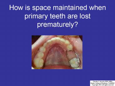
How is space maintained when primary teeth are lost prematurely? - PowerPoint PPT Presentation
1 / 34
Title:
How is space maintained when primary teeth are lost prematurely?
Description:
Permanent Maxillary Incisors Maxillary Central Incisor Calcification & Eruption Schedule Calcification begins 3.5 months Crown completed – PowerPoint PPT presentation
Number of Views:122
Avg rating:3.0/5.0
Title: How is space maintained when primary teeth are lost prematurely?
1
How is space maintained when primary teeth are
lost prematurely?
2
PermanentMaxillary Incisors
Beverly York, D.D.S.
3
Maxillary Central Lateral Incisors
Class Traits
- The incisal two-thirds of the crowns appear
flattened or compressed faciolingually, providing
a long horizontal biting edge or margin - Distinct and rounded protuberances termed
mamelons representing developmental lobes
surmount the incisal margins of all newly erupted
incisors - The marginal ridges of all incisors are located
on the mesial and distal borders of the lingual
surfaces
4
Maxillary Central IncisorCalcification
Eruption Schedule
- Calcification begins 3.5 months
- Crown completed 4.5 years
- Eruption (emergence) .. 7.5 years
- Root completed.. 10 years
5
Maxillary Central IncisorFacial Aspect
- Cervico-Incisal length (10.5 mm) is greater than
the Mesiodistal width (8.5 mm) which is greater
than the Faciolingual depth (7.0 mm) - Crown outline is trapezoidal with the shorter
parallel side at cervix - Crown width at contact area greater than at cervix
Tooth 9 Facial
D
M
6
Maxillary Central IncisorFacial Aspect
- A young tooth may show evidence of mamelons on
the incisal edge - Mesioincisal line angle is square (90 degrees)
Distoincisal angle is rounded - Incisal edge is relatively straight
- Mesial crown outline straighter than the distal
outline - Cervical outline of crown is slightly concave
incisally and the arc of the curvature is said to
be part of a SEMICIRCLE
Tooth 9 Facial
M
D
7
Maxillary Central IncisorFacial Aspect
- Facial surface may show some evidence of shallow
vertical developmental depressions delineating
lobe structure - Facial surface is relatively flat (only slightly
convex) in its incisal two thirds - Mesial contact area is located in the incisal 1/3
of the crown - Distal contact area is located at the junction of
incisal middle 1/3 of the crown - Root cone shaped with blunt apex slightly distal
of center with no developmental depressions
Tooth 9Facial
D
M
8
Maxillary Central IncisorFacial Aspect
- Facial surface may show horizontal ridges at the
cervical one-third representing differing rates
of enamel formation during normal growth patterns - The raised portions of the ridges (positive
anatomy) are termed perikymata and the
horizontal grooves (negative anatomy) are termed
imbrication lines
9
Maxillary Central IncisorFacial Aspect
- Outline of root is cone shaped with a relatively
blunt apex usually located slightly distal to
center line of tooth, with no developmental
depressions - This tooth is easy to extract in that the
clinician can rotate the tooth within the
alveolus, without fear of fracturing the root or
any osseous tissue
10
Maxillary Central IncisorLingual Aspect
- Cervical outline of crown is more concave (toward
the incisal) than what is seen on facial (it is
not a semicircle) - Lingual fossa in incisal ½ of crown surface is
described as a broad, shallow dish-shaped
depression - Lingual fossa is bound by the incisal ridge, the
mesial and distal marginal ridges and the
cingulum. - Lingual fossa is trapezoidal in shape.
- Cingulum Well-developed in cervical ½ of crown.
- The greatest curvature lingually of the cingulum
and the crest of the cervical line gingivally
will be slightly distal to the mesiodistal long
axis bisector of the crown.
Tooth 9Lingual
D
M
11
Maxillary Central IncisorMesial Aspect
Tooth 9 Mesial
- Crown outline is triangular with apex at incisal
- Incisal edge is centered faciolingually over the
root - The incisal edge is lined up with the height of
curvature of the cervical line and the apex of
the root
L
F
12
Maxillary Central IncisorMesial Aspect
Tooth 9Mesial
- The cervical line viewed from the mesial aspect
curves incisally for 1/3 of total crown length. - This is the greatest height of curvature of the
cervical line incisally or occlusally found on
any permanent tooth - Greatest curvature of the crown outline facially
and lingually will be in the cervical 1/3 of the
crown at the crests of the facial and lingual
cervical ridges - These facial and lingual crests are opposite one
another and at the same level
L
F
13
Maxillary Central IncisorMesial Aspect
- The facial outline of the crown from the crest of
the facial cervical ridge to the incisal ridge is
relatively flat (shows very little convexity) - The lingual outline of the crown from the
cingulum to the incisal ridge is slightly concave - The lingual outline of the crown is said to have
an S shaped contour, denoting the convexities
over the cingulum and linguo-incisal ridge and a
concavity over the lingual fossa - The root surface is relatively smoothly convex
and has no developmental root depressions - The root is cone shaped with the blunt apex
centered faciolingually
Tooth 9Mesial
L
F
14
Maxillary Central IncisorDistal Aspect
Tooth 9Distal
- Greatest faciolingual width is in cervical 1/3 of
the crown - Incisal ridge and apex of the root are in line
with each other along the long axis of the tooth - Note that the convexity of the outline of the
cingulum begins approximately halfway between the
incisal ridge and the cervical line on the
lingual - The curvature of the cervical line incisally is
less on the distal than it is on the mesial
surface by 1.0 mm - The distal root surface has no developmental root
depression and is relatively smoothly convex
F
L
15
Maxillary Central IncisorIncisal Aspect
- Mesiodistal width (8.5 mm) is greater than the
faciolingual diameter (7.0 mm) - Interproximal contact areas (greatest curvature
mesially and distally) are centered
faciolingually - Incisal edge (ridge) is relatively straight and
perpendicular (at a 90 angle) to the mesiodistal
bisecting plane - The incisal edge is positioned parallel to the
faciolingual bisecting plane - Note that the mesiofacial line angle is more
developed than the distofacial line angle
Tooth 9Incisal
M
D
16
Maxillary Central IncisorIncisal Aspect
- Crest of the cingulum (greatest curvature on the
lingual) is slightly distal to the mesiodistal
bisecting plane of the crown - Crown outline converges lingually (gets smaller
from facial to lingual) - Surface outline between the mesiofacial and
distofacial outlines is relatively straight - There is slightly more bulk on the mesial half of
the crown than on the distal half
Tooth 9 Incisal
M
D
17
Maxillary Lateral IncisorCalcification
Eruption Schedule
- Calcification begins 1 year
- Crown completed 4.5 years
- Eruption (emergence) .. 8.5 years
- Root completed.. 11 years
18
Maxillary Lateral IncisorFacial Aspect
- Crown length is 1.0 -1.5 mm shorter than the
maxillary central incisor. Crown width is 2.0 mm
less than the maxillary central incisor. This
gives impression that the maxillary lateral
incisor is relatively long and narrow - Crown outline is trapezoidal with the shorter
parallel side at the cervix - Both mesial and distal incisal line angles are
rounded and each is more rounded than the
corresponding incisal line angle of the maxillary
central incisor. The distoincisal line angle is
more rounded than the mesioincisal angle - The mesial proximal contact is at the level of
the junction of the incisal and middle thirds of
the crown - The distal contact is at a level near the middle
of the middle 1/3 of the crown
Tooth 10Facial
M
D
19
Maxillary Lateral IncisorFacial Aspect
- The curvature of the cervical line incisally is
elliptical and not as broad as that of the
maxillary central incisor - The highest point of the curvature of the
cervical line is likely to be slightly distal to
mesiodistal bisector of the crown - Facial crown surface is convex in all directions
- Developmental depressions are evident on the
crown - The root converges evenly toward the apex for the
cervical two thirds of its length - There is usually a characteristic curve of the
root toward the distal in the apical third
Tooth 10Facial
D
M
20
Maxillary Lateral IncisorLingual Aspect
Tooth 10Lingual
- The lingual fossa is relatively deep, triangular
in shape and also cup-shaped - The lingual fossa occupies the incisal two thirds
of the lingual surface of the crown - The anatomical features serving as boundaries for
the fossa include the incisal ridge, the mesial
and distal marginal ridges and the cingulum - The cingulum of the maxillary lateral incisor is
limited to the cervical 1/3 of the crown and the
mesial and distal marginal ridges form a V as
they flow into the cingulum
M
D
21
Maxillary Lateral IncisorLingual Aspect
- There is a very deep depression or even a pit,
deep in the fossa behind the cingulum at the
point of the V - A developmental groove extending out of the
lingual fossa between a marginal ridge and the
cingulum, may be seen on the distal side of the
cingulum - This groove may be deep and extend across the
cementoenamel junction onto the root - It is termed the linguogingival groove and is
unique to the maxillary lateral incisor - Often times, this linguogingival groove is
decayed or fissured and will need to be restored
Tooth 10Lingual
M
D
22
Maxillary Lateral IncisorMesial Aspect
Tooth 10Mesial
- Greatest curvature of both the facial and lingual
crown outlines is in the cervical 1/3 and each is
identified as a cervical ridge - Incisal ridge is in line with mid-point of the
faciolingual diameter - Facial crown outline is convex from facial
cervical ridge crest to incisal ridge - Lingual crown outline is slightly concave from
lingual cervical ridge to the incisal ridge - Curvature of the cervical line toward the incisal
is greater on the mesial surface than on the
distal surface - The curvature of the cervical line extends for
1/3 of the crown length but not as great of a
distance as on the maxillary central incisor
since the lateral incisor has a shorter crown
length - Apex of the root is facial to the faciolingual
long axis bisector - If there is any evidence of a root depression, it
will be slight
F
L
23
Maxillary Lateral IncisorDistal Aspect
Tooth 10Distal
- The incisal ridge and root apex will not be in
line with each another, but the incisal ridge is
centered faciolingually - The facial outline of root is straighter and less
length than the lingual outline when measured
from cervical line to apex of root - The cervical line curves for a shorter distance
incisally on the distal surface than on the
mesial surface
L
F
24
Maxillary Lateral IncisorIncisal Aspect
- Crown wider mesiodistally (6.5 mm) than it is
faciolingually (6.0 mm) - Incisal ridge is centered F-L
- Incisal ridge crosses approximately midway
between the facial and lingual outline however,
it usually shows some curvature with the
convexity toward the facial - The facial outline is more continuously convex
than that of the maxillary central incisor and
the mesiofacial and distofacial line angles are
more rounded (less prominent) - Lingual crown outline converges sharply toward
the lingual. - The crest of the lingual outline of the cingulum
will be slightly to the distal of the mesiodistal
bisector (all cinguli on anterior teeth incline
or point slightly toward the distal)
Tooth 10 Incisal
M
D
25
Maxillary Lateral Incisor
Except for 3rd molars, maxillary lateral incisors
are associated with the most developmental
anomalies.
- Often congenitally missing (aplasia)
26
Maxillary Lateral Incisor
Except for 3rd molars, maxillary lateral incisors
are associated with the most developmental
anomalies.
- Microdontia (abnormally small teeth)
27
Maxillary Lateral Incisor
Except for 3rd molars, maxillary lateral incisors
are associated with the most developmental
anomalies.
- Peg lateral (only middle facial lobe develops)
28
Maxillary Lateral Incisor
Except for 3rd molars, maxillary lateral incisors
are associated with the most developmental
anomalies.
- Dens in dente (tooth within a tooth)
29
Maxillary Lateral Incisor
Except for 3rd molars, maxillary lateral incisors
are associated with the most developmental
anomalies.
- Supernumerary teeth (teeth in addition to normal
construct)
30
Hutchinsons Incisor
- An anomaly associated with the development of
the incisors associated with congenital
syphilis. - Crown of incisors resemble the shape of
screwdrivers. - Also associated with Mulberry Molars as the
permanent 1st molars begin calcification at
birth (to 3 years) and are in progress when the
incisors development is affected (3 months to 5
years)
31
Hutchinsons Incisor
32
Maxillary Central Lateral Incisors
Type Traits
33
Maxillary Central Lateral Incisors
Summary of Comparisons Contrasts
gtCentral is larger than the Lateral gtCentral is
more angular than the Lateral gtCentral has a
straighter root, while the Lateral has a root
that is often curved or pointed to the
distal gtIncisal third of the facial surface of
the Central is fairly flat, while the incisal
third of the lateral exhibits more
convexity gtCentral exhibits minimal variation,
while the Lateral is one of the most variable
permanent teeth gtCrests of the cingulum on both
incisors are offset to the distal gtCentral has a
trapezoidal shaped lingual fossa, while the
Lateral has a triangular shape fossa gtFrom an
incisal view, both incisors display contact areas
that are centered faciolingually gtBoth incisors,
from a proximal view, display an incisal edge
that is centered over the crest of curvature of
the CEJ and their respective root apices gtFrom a
lingual view, the Lateral frequently displays a
fissured or carious lingual pit, while the
Central does not
34
The End
Permanent Maxillary Incisors


