
Skeletal and Muscular System - PowerPoint PPT Presentation
1 / 71
Title:
Skeletal and Muscular System
Description:
Skeletons are either a fluid-filled body cavity, exoskeletons, or internal ... as well as maintain the shape of the animals, such as the sea anemone and worms ... – PowerPoint PPT presentation
Number of Views:3058
Avg rating:3.0/5.0
Title: Skeletal and Muscular System
1
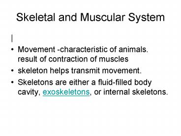
Skeletal and Muscular System
- Movement -characteristic of animals. result of
contraction of muscles - skeleton helps transmit movement.
- Skeletons are either a fluid-filled body cavity,
exoskeletons, or internal skeletons.
2
Just because watch a cut through of the human
body
- http//www.nlm.nih.gov/research/visible/mpeg/umd_v
ideo.mpg - Page 102 study guide. Outline the great
diversity of locomotion in fish earthworm, flying
bird and walking arthropod.
3
Hydrostatic systems
- Hydrostatic skeletons fluid-filled closed
chambers. - Internal pressures generated by muscle
contractions cause movement as well as maintain
the shape of the animals, such as the sea anemone
and worms
4
Exoskeleton
- Exoskeletons are characteristic of the Phylum
Arthropoda. hard segments that cover the muscles
and visceral organs. Muscles for movement attach
to the inner surface of the exoskeleton.
5
Endoskeleton
- Vertebrates -internal mineralized (in most cases)
endoskeleton composed of bone and/or cartilage.
Muscles are on the outside of the endoskeleton.
Cartilage and bone are types of connective
tissue.
6
Functions of Muscles and Bones
- The skeleton and muscles function together as the
musculoskeletal system. - Plays an important homeostatic role allowing the
animal to move to more favorable external
conditions. - Certain cells in the bones produce immune cells
as well as important cellular components of the
blood.
7
- Bone also helps regulate blood calcium levels,
serving as a calcium sink. - Rapid muscular contraction is important in
generating internal heat, another homeostatic
function. - Howstuffworks "Muscle Types Animation"
8
Skeletal Muscle Systems/101 sg
- Vertebrates move by the actions of muscles on
bones. Tendons attach skeletal muscles across
joints, allowing muscle contraction to move the
bones across the joint.
9
- Muscles generally work in pairs to produce
movement when one muscle flexes (or contracts)
the other relaxes, a process known as antagonism.
10
Ligaments
- Ligaments are tough, elastic, connective tissue
joining bone to bone. - Ligaments limit the range of motion at a joint
while providing joint stability.
11
Tendons
- Tendons are thick, dense connective tissues
attaching muscle to bone. They are a continuation
of the fascia. - Tendons are relatively inelastic and transmit the
energy of muscle action to bone.
12
Put it all together in the elbow, page 102 sg
13
Structure of skeletal muscle
- Look at page 101 sg. You will be asked to draw
the structure of skeletal muscles fibers.
Include actin filaments and thick myosin
filaments, sarcoplasmic reticulum and
mitochondria - APC 100
14
Muscles page 101 sg
- Muscles both electrical and chemical activity.
- There is an electrical gradient across the muscle
cell membrane the outside is more positive than
the inside. - Stimulus causes an instantaneous reversal of this
polarity, causing the muscle to contract (the
mechanical characteristic) producing a twitch or
movement.
15
Organization of muscles
16
Skeletal Muscle Structure
- Muscle fibers are multinucleated, with the nuclei
located just under the plasma membrane. Most of
the cell is occupied by striated, thread-like
myofibrils. Within each myofibril there are dense
Z lines.
17
- A sarcomere (or muscle functional unit) extends
from Z line to Z line. Each sarcomere has thick
and thin filaments..
18
- The thick filaments are made of myosin and occupy
the center of each sarcomere. Thin filaments are
made of actin and anchor to the Z line.
19
- Muscles contract by shortening each sarcomere.
- The sliding filament model of muscle contraction
has thin filaments on each side of the sarcomere
sliding past each other until they meet in the
middle. - Myosin filaments have club-shaped heads that
project toward the actin filaments.
20
(No Transcript)
21
(No Transcript)
22
Myosin head
- Myosin heads attach to binding sites on the actin
filaments. - swivel toward the center of the sarcomere
- detach and then reattach to the nearest active
site of the actin filament. - http//www.sciencemag.org/feature/data/1049155s1.m
ov
23
- Each cycle of attachment, swiveling, and
detachment shortens the sarcomere 1. Hundreds of
such cycles occur each second during muscle
contraction.
24
The MYOSIN HEAD has several important
characteristics
- it has ATP-binding sites (ATP represents
potential energy.) - ACTIN-binding sites into which fit molecules of
ACTIN. - it has a "hinge"at the point where it leaves the
core of the thick myofilament. This allows the
head to swivel back and forth, and the
"swivelling" is, as will be described shortly,
what actually causes muscle contraction.
25
Thin myofilaments are composed of 3 types of
protein
- ACTIN
- TROPONIN
- TROPOMYOSIN
- Animation Myofilament Contraction
26
ACTIN
- when actin combines with MYOSIN HEAD the ATP
associated with the head breaks down into ADP.
This reaction released energy that causes the
MYOSIN HEAD to SWIVEL.
27
TROPOMYOSIN
- In a relaxed muscle, the MYOSIN HEADS of the
thick myofilament lie against TROPOMYOSIN
molecules of the thin myofilament. As long as the
MYOSIN HEADS remain in contact with TROPOMYOSIN
nothing happens (i.e., a muscle remains relaxed).
28
(No Transcript)
29
TROPONIN
- Troponin molecules have binding sites for calcium
ions. When a calcium ion fills this site it
causes a change in the shape and position of
TROPONIN. - TROPONIN shifts, it pulls the TROPOMYOSIN to
which it is attached. - When TROPOMYOSIN is moved, the MYOSIN HEAD that
was touching the tropomyosin now comes in contact
with an underlying ACTIN molecule. - Animation Action Potentials and Muscle
Contraction
30
Muscle contraction
- Because skeletal muscle is voluntary muscle,
contraction requires a nervous impulse. - 1 impulse is transferred from a neuron to the
SARCOLEMMA of a muscle cell. - 2 The impulse travels along the SARCOLEMMA and
down the T-TUBULES. From the T-TUBULES, the
impulse passes to the SARCOPLASMIC RETICULUM.
31
(No Transcript)
32
- 3 - As the impulse travels along the Sarcoplasmic
Reticulum (SR), the calcium gates in the membrane
of the SR open. As a result, CALCIUM diffuses out
of the SR and among the myofilaments. - 4 - Calcium fills the binding sites in the
TROPONIN molecules. As noted previously, this
alters the shape and position of the TROPONIN
which in turn causes movement of the attached
TROPOMYOSIN molecule.
33
- 5 - Movement of TROPOMYOSIN permits the MYOSIN
HEAD to contact ACTIN. - 6 - Contact with ACTIN causes the MYOSIN HEAD to
swivel
34
- 7 - During the swivel, the MYOSIN HEAD is firmly
attached to ACTIN. So, when the HEAD swivels it
pulls the ACTIN (and, therefore, the entire thin
myofilament) forward. (Obviously, one MYOSIN HEAD
cannot pull the entire thin myofilament. Many
MYOSIN HEADS are swivelling simultaneously, or
nearly so, and their collective efforts are
enough to pull the entire thin my
35
- Animation Quizzes crossbridge
36
- 8 - At the end of the swivel, ATP fits into the
binding site on the cross-bridge this breaks
the bond between the cross-bridge (myosin) and
actin. The MYOSIN HEAD then swivels back. As it
swivels back, the ATP breaks down to ADP P and
the cross-bridge again binds to an actin
molecule.
37
- 9 - As a result, the HEAD is once again bound
firmly to ACTIN. However, because the HEAD was
not attached to actin when it swivelled back, the
HEAD will bind to a different ACTIN molecule
(i.e., one further back on the thin myofilament).
Once the HEAD is attached to ACTIN, the
cross-bridge again swivels, SO STEP 7 IS
REPEATED.
38
- Skeletal muscle relaxes when the nervous impulse
stops. No impulse means that the membrane of the
SARCOPLASMIC RETICULUM is no longer permeable to
calcium (i.e., no impulse means that the CALCIUM
GATES close
39
- So, under most circumstances, calcium is the
"switch" that turns muscle "on and off"
(contracting and relaxing).
40
Nerves, muscles and movements
- Nerve cells are called neurons.
- Nervous system divided into CNS ( brain and
spinal cord) and PNS ( peripheral nerves).
41
(No Transcript)
42
(No Transcript)
43
(No Transcript)
44
- dendrites provide a large surface area for
connecting with other neurones, and carry nerve
impulses towards the cell body. - A single long axon carries the nerve impulse
away from the cell body. - Most neurones have many companion cells called
Schwann cells, which wrap their cell membrane
around the axon in a spiral to form a thick
insulating lipid layer called the myelin sheath.
45
- Nerve Impulses
- Neurones send messages electrochemically this
means that chemicals cause an electrical impulse.
- Chemicals in the body are electrically charged
when they have an electrical charge, they are
called ions.
46
- Resting Membrane Potential
- When a neurone is not sending a signal, it is at
rest. - The inside of the neurone is negative relative to
the outside. - K can cross through the membrane easily
- Cl- and Na have a more difficult time crossing
- Negatively charged protein molecules inside the
neurone cannot cross the membrane.
47
- Resting Membrane Potential
- The membranes contain sodium-potassium pumps
(NaKATPase). - Uses ATP to simultaneously pump 3 sodium ions out
of the cell and 2 potassium ions in. - Animations
48
- There are also sodium and potassium ion channels
in the membrane. - These channels are normally closed, but even when
closed, they leak, allowing sodium ions to leak
in and potassium ions leak out down their
concentration gradients.
49
- This creates an action potential.
- Chapter 39 Introduction
- Tutorial 44.2 The Action Potential
50
This creates an action potential.
- An action potential is initiated by a stimulus
above a certain intensity or threshold. - Not all stimuli initiate an action potential.
- The stimulus could be a pin prick, light, heat,
sound or an electrical disturbance in another
part of the neuron.
51
Depolarization
- A stimulus causes a gate in the Na Channel to
open. - Since there is a high concentration of Na
outside, Na diffuses into the neuron. - The electrical potential changes to 40 mV.
52
Repolarization
- Depolarization causes the K Channel gate to
immediately open. - K diffuses out of the neuron.
- This reestablishes the initial electrical
potential of -60 mV.
53
Refractory Period
- During this time ( 1 msec), the Na and K
Channels cannot be opened by a stimulus. - The Na/K Pump actively pumps Na out of the
neuron and K into the neuron. This reestablishes
the initial ion distribution of the resting
neuron. Action Potential
54
- Nerve impulse can be passed from the axon of one
neuron to the dendrite of another at a synapse. A
nerve is a discrete bundle of several thousand
neuron axons.
55
The message
- http//www.mind.ilstu.edu/flash/synapse_1.swf
- Animation Function of the Neuromuscular Junction
(Quiz 1)
56
These images illustrate the general process of
synaptic transmission
- Step 1. The neurotransmitter is manufactured by
the neuron and stored in vesicles at the axon
terminal.
57
- Step 2. When the action potential reaches the
axon terminal, it causes the vesicles to release
the neurotransmitter molecules into the synaptic
cleft.
58
- Step 3. The neurotransmitter diffuses across the
cleft and binds to receptors on the post-synaptic
cell. - Step 4. The activated receptors cause changes in
the activity of the post-synaptic neuron.
59
- Step 5. The neurotransmitter molecules are
released from the receptors and diffuse back into
the synaptic cleft
60
- Diffusion back
61
- Step 6. The Neurotransmitter is re-absorbed by
the post synaptic neuron. This process is known
as Reuptake
62
- http//users.rcn.com/jkimball.ma.ultranet/BiologyP
ages/A/autonomic.gif - synaptic transmission
63
Signal Transduction Across the Synapse ( once
again)
- When the wave of Action Potentials reach the end
of the axon the electrical signal is converted
into a chemical signal. - This chemical or neurotransmitter crosses the
space (Synapse) between adjacent neurons and
initiates an Action Potential on another neuron.
64
- The action potential activates a calcium channel
and Ca diffuses into the neuron. - This Ca causes vesicles to fuse with the cell
membrane. Through exocytosis, neurotransmitters
(chemicals) are released into the synapse
65
- These neurotransmitters diffuse across the
synapse and bind to receptors on another neuron.
This causes special Na channels to open and an
action potential is initiated in the next neuron - Once the message has been passed on to the next
neuron, the neurotransmitter is reabsorbed into
the axon, diffuses away or it is destroyed by an
enzyme. - Synapse
66
(No Transcript)
67
vision
- McGraw-Hill Online Learning Center TestltBLURTgt
- We woll do the dissections from this on line
site. http//www.exploratorium.edu/learning_studi
o/cow_eye/coweye.pdf
68
Anatomy of the eye
- Optic Nerve The fatty pad around the eye is
pulled back, revealing parts of the skeletal
muscles (in dark brown) that control the movement
of the eye. The forceps are holding the optic
nerve.
69
Sectioned Eye
- The left section shows the vitreous humor still
attached to the lens ciliary body. The right
section shows retina in the back of the eye.
Alani did this dissection.
70
- The left upper piece is the semi-transparent
cornea - lower left darkly pigmented ciliary body iris
- The pupil is the hole in the center of the iris
71
Lens - capsule peeled back
- Note the thin, fibrous looking epithelial cells
that make up the lens. Hand is holding the lens.
Recommended
CrystalGraphics Presentations


