
PNS Terminology PowerPoint PPT Presentation
1 / 40
Title: PNS Terminology
1
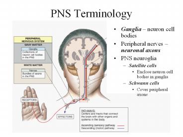
PNS Terminology
- Ganglia neuron cell bodies
- Peripheral nerves neuronal axons
- PNS neuroglia
- Satellite cells
- Enclose neuron cell bodies in ganglia
- Schwann cells
- Cover peripheral axons
2
The Cranial Nerves (PNS)
3
I - Olfactory II - Optic III - Oculomotor IV-Troch
lear V - Trigeminal VI - Abducens VII -
Facial VIII Acoustic/Vestibulocochlear IX -
Glossopharyngeal X - Vagus XI Accessory/Spinal
Accessory XII - Hypoglossal
-cranial nerves 12 pairs -considered part of
the peripheral nervous system (PNS) -olfactory
optic acoustic contain only sensory axons
sensory nerves -some carry motor information
motor nerves e.g. oculomotor, trochlear,
abducens -remaining are mixed nerves both motor
and sensory axons some say my mother bought my
brother some bitter beer my, my
4
Optic Chiasma
5
The Olfactory Nerve (I)
- Carries sensory information
- Sense of smell
- Synapse within olfactory bulbs
6
- The optic nerve (II)
- Carries visual information
7
- The abducens nerve (VI)
- Innervates lateral rectus muscle of eye
- The oculomotor nerve (III)
- Primary source of innervation for extra-ocular
muscles - Move the eyeball
- The trochlear nerve (IV)
- Smallest cranial nerve
- Innervates superior oblique eye muscle
8
The Trigeminal Nerve (V)
- Largest cranial nerve
- Mixed nerve
- sensory touch, pain thermal
- Ophthalmic branch
- sensory upper eyelid, eyeball
- lacrimal glands, side of nose, forehead
- and scalp
- Maxillary branch
- sensory nose, palate, part
- of pharynx, upper teeth, upper
- lip and lower eyelid
- Mandibular branch
- sensory tongue, cheek,
- lower teeth, skin over mandible
- and side of head anterior to ear
- -motor muscles of chewing
9
The Facial Nerve (VII)
- Mixed nerve
- Controls muscles of scalp and face
- Pressure sensations from face
- Taste sensations from tongue
10
The Vestibulocochlear Nerve (VIII)
- Vestibular nerve
- Monitors sense of balance, position and movement
- Cochlear nerve
- Monitors hearing
11
The Glossopharyngeal Nerve (IX)
- Mixed nerve
- Innervates the tongue
- Controls swallowing
12
The Vagus Nerve (NX)
- Mixed nerve
- Vital to autonomic control of visceral function
13
- The accessory nerve (XI)
- Internal branch
- Innervates swallowing muscles
- External branch
- Controls muscles associated with pectoral girdle
- The hypoglossal nerve (XII)
- Voluntary motor control over tongue movements
14
31 Pairs of Spinal Nerves
- Ensheathed by three connective tissue layers
- Outermost epineurium
- Dense network of collagen fibers
- Middle perineurium
- Partitions nerve into fascicles
- Inner endoneurium
- Delicate connective tissue fibers surrounding
each axon - Under the endoneurium is the myelin sheath
outer layer is called the neurilemma - Neurilemma covers the myelin sheath and Schwann
cells - Myelin sheath covers the axon
15
Spinal Nerves
- connected to the spinal cord via roots (bundles
of axons) - Posterior root sensory axons into the posterior
gray horn - Anterior root motor axons from the anterior
gray horn - before the posterior root is the dorsal root
ganglion - cell bodies of incoming sensory
neurons (axons continue on to form the root) - emerge from intervertebral foramina as mixed
nerves
16
Spinal Nerve
- after passing through intervertebral foramina the
spinal nerve branches into three rami - Dorsal ramus
- -sensory/motor innervation to skin and muscles of
back
- Ventral ramus
- - Sensory/motor innervation to ventral and
lateral body surface/skin, body wall structures,
muscles of the upper and lower limbs
17
- rami communicantes
- Third branch from the spinal nerve
- -carries nerves of the ANS
18
Dorsal Root of SN
Ventral Root of SN
SPINAL NERVE
Dorsal Ramus
Ventral Ramus
Rami Communicantes
Signals to and from the ANS VISCERA cardiac
and Smooth muscle
Sensory IN Motor OUT TRUNK LIMBs
Sensory IN Motor OUT SKIN BACK MUSCLES
19
(No Transcript)
20
Nerve Plexuses
- Four major plexuses
- Cervical plexus
- Brachial plexus
- Lumbar plexus
- Sacral plexus
- Joining of ventral rami of spinal nerves to form
nerve networks or plexuses - Found in neck, arm, low back sacral regions
- No plexus in thoracic region
- intercostal nn. innervate intercostal spaces
- T7 to T12 supply abdominal wall as well
21
Cervical Plexus
- Cervical plexus
- C1-C4 ventral rami
- Some fibers from C5
- Innervates muscles of the neck and diaphragm
- Phrenic nerve
22
Brachial Plexus
- Ventral rami of C5-T1
- Innervates pectoral girdle and upper limbs
- Nerves arise from cords or trunks
- Superior, middle and inferior trunks
- Lateral, medial and posterior cords
- Superior and Middle trunk contribute to the
- Lateral cord (SML)
- -Superior, middle and inferior trunk all
- contribute to the Posterior cord (SMIP)
- -inferior trunk continues on as the
- Medial cord (IM)
23
The Brachial Plexus
24
The Cervical and Brachial Plexus
25
Lumbar and Sacral Plexuses
- Lumbar plexus - ventral rami of T12L4
- Sacral plexus ventral rami of L4S4
- Innervate pelvic girdle and lower limbs
26
The Lumbar and Sacral Plexuses,
27
The Lumbar and Sacral Plexuses,
28
Reflex
- Rapid automatic involuntary motor response to
stimuli - Some are inborn (pulling away from heat), others
are learned or acquired - Bypasses the brain integration and processing
occurs in the spinal cord at the level in input
of information Spinal reflex (e.g. pain
response) - If integration occurs in the brain stem Cranial
reflex (e.g. eye tracking) - Somatic reflexes contraction of skeletal
muscles - Autonomic (visceral) reflexes - involuntary
- Preserve homeostasis
- Rapidly adjusts organs or organ systems
29
- Our knowledge of reflexes is largely owed to Sir
Charles Sherrington who has become known as the
Father of the Nervous System. - His book, The Integrative Action of the Nervous
System, circa 1901, became the impetus for study
of primal reflexes.
30
Classification of Reflexes
- By development
- Innate, acquired
- Where information is processed
- Spinal, cranial
- Motor response
- Somatic, visceral
- Complexity of neural circuit
- Monosynaptic
31
- Reflex arc
- Neural wiring of reflex
- Requires 5 functional components 1. sensory
receptor, 2. sensory neuron, 3. integrating
center (SC or BS), 4. motor neuron, 5. effector
32
Spinal Reflexes
- Stretch reflex is monosynaptic - causes
contraction in response to stretch - Regulates skeletal muscle length and tone
- all monosynaptic reflexes are ipsilateral
reflexes - input and output on same side - only one synapse in the CNS - between ad single
sensory and motor neuron - Sensory receptors are found in muscle spindles
- e.g. Patellar reflex muscle spindles in the
quadriceps muscles, hit with a mallet stretches
the quadriceps and its tendon - results in
contraction
33
Spinal Reflexes
- Tendon reflexes - polysynaptic
- controls muscle tension by causing muscle
relaxation before muscle contraction rips tendons - Generally polysynaptic - more than one CNS
synapse involved between more than two different
neurons - sensory synapses with 2 interneurons - one
inhibitory IN synapses with motor neurons and
causes inhibition and relaxation of one set of
muscles, the other stimulatory IN synapses with
motor neurons and causes contraction of the
antagonistic muscle
34
- -Postural reflexes - maintain upright position
- e.g flexor (withdrawl) reflex - polysynaptic
- sensory input -gt interneuron -gt motor neuron
which contracts muscles and pulls limb away - PLUS synapses with motor neurons in adjacent SC
segments -gt contracts muscle - known as an intersegmental reflex arc
- IN ADDITION - the sensory input can cross to the
other side of the SC (via the gray commisure)
where it synapses with and interneuron and motor
neuron to contract the antagonistic muscle group
and maintains balance
withdrawl
crossed extensor
35
-reflexes and clinical significance 1. plantar
flexion - stroke the outer lateral margin of the
sole -curling of toes normal
response -damage to descending motor pathways
alters this reflex 2. Babinski reflex -
stroke the middle of the sole -great toe
extends and the other toes may or may not fan
out - due to incomplete myelination of of
axons in the corticospinal tract -in
children under 18 months reflex is
normal -older than this - results in the
plantar flexion reflex - Newborn babies have
a number of other reflexes which are not seen in
adults, including 1. suckling 2.
hand-to-mouth reflex 3. Grasp reflex 4. Moro
reflex, also known as the startle reflex may
be observed in incomplete form in premature birth
after the 28th week of gestation -normally lost
by the 6th month of life postpartum - a
response to unexpected loud noise or when the
infant feels like it is falling - it is
believed to be the only unlearned fear in human
newborn - origin of this reflex can be found in
that fact that primate infants of our ancestors
clung to their mother's fur soon after birth -if
human babies are falling backward - innate reflex
will be to stretch out the arms to grab and cling
to their mother -the primary significance of
this reflex is in evaluating integration of the
central nervous system (CNS), since the reflex
involves 4 distinct components 1. Startle 2.
abduction of arms spreading out of arms 3.
unspreading the arms 4. Crying (usually)
36
Other reflexes you might want to know about
- sneeze reflex
- a sneeze is a very complicated thing, involving
many areas of the brain - a sneeze is a reflex triggered by sensory
stimulation of the membranes in the nose,
resulting in a coordinated and forceful expulsion
of air through the mouth and nose. - why do some people sneeze when they look at the
sun? - dont know
- involves the "pupillary light reflex". If you
shine a light in your eyes, your pupils get
smaller, or constrict. - in the pupillary light reflex, shining a light in
the eye causes nerve signals to go from the eye
to the brain and then back the eye again, telling
the pupil to constrict. - in the usual sneeze reflex, tickling the nose
causes nerve signals to go from the nose to the
brain and then back out to the nose, mouth, chest
muscles - these nerve signals take complicated routes
through the brain - but usually the pupillary light reflex and sneeze
reflex take different routes. - in 25 of the population - shining a bright
enough light in the eye ALSO sends nerves signals
from the eye to the brain and then back out to
the nose, mouth and chest! - the wires are crossed a little bit in some
people - so shining a light in the eye
"accidentally" activates two different outgoing
pathways. - gag reflex - reflex contraction of the back of
the throat that prevents something from entering
the throat except as part of normal swallowing - helps prevent choking
- also known as a pharyngeal reflex.
- touching the soft palate evokes a strong gag
reflex in most people, - most people can train themselves to resist the
gag reflex, - the afferent limb of the reflex is supplied by
the glossopharyngeal nerve (cranial nerve IX) and
the efferent limb is supplied by the vagus nerve
(cranial nerve X).
37
Divisions of the nervous system
- -the PNS can be divided into
- two divisions
- Somatic motor commands to
- skeletal muscles via cranial
- spinal nerves (sensory information
- in also from these muscles)
- 2. Autonomic involuntary
- motor commands to viscera
- (sensory information in also)
- -divided into
- parasympathetic division
- sympathetic division
38
Somatic nervous system (SNS) of the PNS 1.
sensory division- neurons that convey sensory
information from somatic receptors in the head,
body wall, senses - to the CNS 2. control of
motor output - neurons that conduct voluntary
impulses to skeletal muscles -contributions
from the basal ganglia, cerebellum, brain stem
and SC 3. one neuron pathway somatic motor
neurons synapse directly with the effector (i.e.
one long neuron that emerges from the CNS and
travels to a muscle) 4. neurotransmitter
acetylcholine 5. effectors skeletal
muscles 6. responses - contraction
39
- Autonomic nervous system (ANS) of the PNS
- sensory - neurons that convey info from autonomic
sensory receptors in the visceral organs - to the
CNS - 2. control of motor output - neurons that
conduct impulses from the CNS to - smooth and cardiac muscle glands
- 3. two neuron pathway preganglionic neurons
extend from CNS and synapse with postganglionic
neurons in an autonomic ganglion, postganglionic
neurons that synapse with the effector - -also preganglionic neurons synapse with adrenal
medulla - 4. neurotransmitter preganglionic ACh
- -postganglionic ACh or norepinephrine
- -AD epinephrine and NE
- 5. effectors smooth cardiac muscle, glands,
- 6. responses contraction or relaxation of SM
- -increased or decrease heart contraction
- -increased or decreased gland secretions
40
- motor output branch has two divisions 1.
sympathetic 2. parasympathetic -most organs are
innervated by both divisions which
have opposing functions e.g. sympathetic
increases heart rate parasympathetic
decreases rate
-
Recommended
