
Chronic Periodontitis - PowerPoint PPT Presentation
Title:
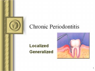
Chronic Periodontitis
Description:
Collagen fibers apical to JE destroyed infiltration of inflammatory cells & edema. Apical migration of junctional epithelium along root. Coronal portion of JE detaches ... – PowerPoint PPT presentation
Number of Views:3909
Avg rating:3.0/5.0
Title: Chronic Periodontitis
1
Chronic Periodontitis
- This presentation will probably involve audience
discussion, which will create action items. Use
PowerPoint to keep track of these action items
during your presentation - In Slide Show, click on the right mouse button
- Select Meeting Minder
- Select the Action Items tab
- Type in action items as they come up
- Click OK to dismiss this box
- This will automatically create an Action Item
slide at the end of your presentation with your
points entered.
- Localized
- Generalized
2
Learning Outcomes
- Describe the development of a periodontal pocket.
- Relate clinical characteristics to the
histopathologic changes for chronic
periodontitis. - Compare the gingival pocket with the periodontal
pocket. - Determine the severity of PD activity using
clinical data.
3
Common Characteristics
- Onset - any age most common in adults
- Plaque initiates condition
- Subgingival calculus common finding
- Slow-mod progression periods of rapid
progression possible - Modified by local factors/systemic
factors/stress/smoking
4
Extent Severity
- Extent
- Localized ?30 of sites affected
- Generalized gt 30 of sites affected
- Severity entire dentition or individual
teeth/site - Slight 1-2 mm CAL
- Moderate 3-4 mm CAL
- Severe ? 5 mm CAL
5
Clinical Characteristics
- Deep red to bluish-red tissues
- Thickened marginal gingiva
- Blunted/cratered papilla
- Bleeding and/or suppuration
- Plaque/calculus deposits
6
Clinical Characteristics
- Variable pocket depths
- Horizontal/vertical bone loss
- Tooth mobility
7
Pathogenesis Pocket Formation
- Bacterial challenge initiates initial lesion of
gingivitis - With disease progression change in
microorganisms ? development of periodontitis
8
Pocket Formation
- Cellular fluid inflammatory exudate ?
degenerates CT - Gingival fibers destroyed
- Collagen fibers apical to JE destroyed ?
infiltration of inflammatory cells edema - Apical migration of junctional epithelium along
root - Coronal portion of JE detaches
9
Pocket Formation
- Continued extension of JE requires healthy
epithelial cells! - Necrotic JE slows down pocket formation
- Pocket base degeneration less severe than lateral
10
Pocket Formation
- Continue inflammation
- Coronal extension of gingival margin
- JE migrates apically separates from root
- Lateral pocket wall proliferates extends into
CT - Leukocytes edema
- Infiltrate lining epithelium
- Varying degrees of degeneration necrosis
11
Development of Periodontal Pocket
12
Continuous Cycle!
- Plaque ? gingival inflammation ? pocket formation
? more plaque
13
Histopathology
- Connective Tissue
- Edematous
- Dense infiltrate
- Plasma cells (80)
- Lymphocytes, PMNs
- Blood vessels proliferate, dilate are engorged
- Varying degrees of degeneration in addition to
newly formed capillaries, fibroblasts, collagen
fibers in some areas
14
Histopathology
- Periodontal pocket
- Lateral wall shows most severe degeneration
- Epithelial proliferation degeneration
- Rete pegs protrude deep within CT
- Dense infiltrate of leukocytes fluid found in
rete pegs epithelium - Degeneration necrosis of epithelium leads to
ulceration of lateral wall, exposure of CT,
suppuration
15
Clinical Histopathologic Features
- Clinical
- Pocket wall bluish-red
- Smooth, shiny surface
- Pitting on pressure
- Histopathology
- Vasodilation vasostagnation
- Epithelial proliferation, edema
- Edema degeneration of epithelium
16
Clinical Histopathologic Features
- Clinical
- Pocket wall may be pink firm
- Bleeding with probing
- Pain with instrumentation
- Histopathology
- Fibrotic changes dominate
- ? blood flow, degenerated, thin epithelium
- Ulceration of pocket epithelium
17
Clinical Histopathologic Features
- Clinical
- Exudate
- Flaccid tissues
- Histopathology
- Accumulation of inflammatory products
- Destruction of gingival fibers
18
Root Surface Wall
- Periodontal disease affects root surface
- Perpetuates disease
- Decay, sensitivity
- Complicates treatment
- Embedded collagen fibers degenerate ? cementum
exposed to environment - Bacteria penetrate unprotected root
19
Root Surface Wall
- Necrotic areas of cementum form clinically soft
- Act as reservoir for bacteria
- Root planing may remove necrotic areas ? firmer
surface
20
Classification of Pockets
- Gingival
- Coronal migration of gingival margin
- Periodontal
- Apical migration of epithelial attachment
- Suprabony
- Base of pocket coronal to height of alveolar
crest - Infrabony
- Base of pocket apical to height of alveolar crest
- Characterized by angular bony defects
21
Periodontal Pocket
- Suprabony pocket
22
Inflammatory Pathway
- Stages I-III inflammation degrades gingival
fibers - Spreads via blood vessels
- Interproximal
- Loose CT ? transseptal fibers ? marrow spaces of
cancellous bone ? periodontal ligament ?
suprabony pockets horizontal bone loss
?transseptal fibers transverse horizontally
23
Inflammatory Pathway
- Interproximal
- Loose CT ? periodontal ligament ? bone ?
infrabony pockets vertical bone loss ?
transseptal fibers transverse in oblique
direction
24
Inflammatory Pathway
- Facial Lingual
- Loose CT ? along periosteum ? marrow spaces of
cancellous bone ? supporting bone destroyed first
? alvoelar bone proper ? periodontal ligament ?
suprabony pocket horizontal bone loss
25
Inflammatory Pathway
- Facial Lingual
- Loose CT ? periodontal ligament ? destruction of
periodontal ligament fibers ? infrabony pockets
vertical or angular bone loss
26
Stages of Periodontal Disease
27
Periodontal Pathogens
- Gram negative organisms dominate
- P.g., P.i., A.a. may infiltrate
- Intercellular spaces of the epithelium
- Between deeper epithelial cells
- Basement lamina
28
Periodontal Pathogens
- Pathogens include
- Nonmotile rods
- Facultative
- A.a., E.c.
- Anaerobic
- P. g., P. i., B.f., F.n.
- Motile rods
- Facultative
- C.r.
- Spirochetes
- Anaerobic, motile
- Treponema denticola
29
Periodontal Disease Activity
- Bursts of activity followed by periods of
quiescence characterized by - Reduced inflammatory response
- Little to no bone loss CT loss
- Accumulation of Gram negative organisms leads to
- Bone attachment loss
- Bleeding, exudate
- May last days, weeks, months
30
Periodontal Disease Activity
- Period of activity followed by period of
remission - Accumulation of Gram positive bacteria
- Condition somewhat stabilized
- Periodontal destruction is site specific
- PD affects few teeth at one time, or some
surfaces of given teeth
31
Overall Prognosis
- Dependent on
- Client compliance
- Systemic involvement
- Severity of condition
- of remaining teeth
32
Prognosis of Individual Teeth
- Dependent on
- Attachment levels, bone height
- Status of adjacent teeth
- Type of pockets suprabony, infrabony
- Furcation involvement
- Root resorption
33
Subclassification of Chronic Periodontitis
Severity Pocket Depths CAL Bone Loss Tooth Mobility Furcation
Early 4-5 mm 1-2 mm Slight horizontal
Moderate 5-7 mm 3-4 mm Sl mod horizontal ? ?
Advanced gt 7 mm ? 5 mm Mod-severe horizontal vertical ? ?