
Images of temporal bone anatomy - PowerPoint PPT Presentation
1 / 21
Title:
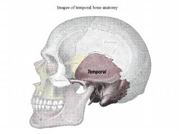
Images of temporal bone anatomy
Description:
Medical education entails learning assisted by images, ... radiological procedure, to define typical or atypical patterns of spread of disease or to ... – PowerPoint PPT presentation
Number of Views:2514
Avg rating:3.0/5.0
Title: Images of temporal bone anatomy
1
Images of temporal bone anatomy
2
A comparison of computer generated visualizations
of temporal bone (Os temporale) with traditional
medical illustrations for medical education
Overview
Medical education entails learning assisted
by images, illustrations and other forms of
traditional drawings. These illustrations are
used to teach a complex surgical or radiological
procedure, to define typical or atypical patterns
of spread of disease or to illustrate normal or
aberrant anatomy. Even though the hand-drawn
illustrations convey meticulous information,
computer generated images and visualizations
start to play the leading role in medical
education. In this project, specific
anatomical features of the temporal bone were
generated by illustrative visualization
techniques. The pros and cons of these
computer-generated images as compared to
traditional medical illustrations in terms of
time, quality, efficiency, clarity and use were
analyzed. These illustrations were used to
develop a comparatively less expensive teaching
module /reference guide for medical education,
training as well as for providing information to
patients. Unsegmented CT scan datasets of
temporal bone were used to generate the images.
3
CT scan dataset of temporal bone of a patient
diagnosed with intra-labyrinthine cochlear
Schwannoma
4
Cropped region of the dataset
5
Mastoid air cells (blue)
Mastoid tip
External ear
Ear canal
Portion of skull
Credit Lakshmi P. Ganapathy, Nikolai Svakhine,
Dr. David S. Ebert Purdue
University Rendering and Perceptualization Lab
6
Credit Lakshmi P. Ganapathy, Nikolai Svakhine,
Dr. David S. Ebert Purdue
University Rendering and Perceptualization Lab
7
Credit Lakshmi P. Ganapathy, Nikolai Svakhine,
Dr. David S. Ebert Purdue
University Rendering and Perceptualization Lab
8
Semicircular canals
Basal turn of cochlea
Apical turn of cochlea
Credit Lakshmi P. Ganapathy, Nikolai Svakhine,
Dr. David S. Ebert Purdue
University Rendering and Perceptualization Lab
9
tissue
Semicircular canals
Middle turn of cochlea
Credit Lakshmi P. Ganapathy, Nikolai Svakhine,
Dr. David S. Ebert Purdue
University Rendering and Perceptualization Lab
10
Semicircular canals
Carotid canal
cochlea
Credit Lakshmi P. Ganapathy, Nikolai Svakhine,
Dr. David S. Ebert Purdue
University Rendering and Perceptualization Lab
11
Credit Lakshmi P. Ganapathy, Nikolai Svakhine,
Dr. David S. Ebert Purdue
University Rendering and Perceptualization Lab
12
Credit Lakshmi P. Ganapathy, Nikolai Svakhine,
Dr. David S. Ebert Purdue
University Rendering and Perceptualization Lab
13
Middle ear entrance to the Eustachian tube
Credit Lakshmi P. Ganapathy, Nikolai Svakhine,
Dr. David S. Ebert Purdue
University Rendering and Perceptualization Lab
14
Credit Lakshmi P. Ganapathy, Nikolai Svakhine,
Dr. David S. Ebert Purdue
University Rendering and Perceptualization Lab
15
Credit Lakshmi P. Ganapathy, Nikolai Svakhine,
Dr. David S. Ebert Purdue
University Rendering and Perceptualization Lab
16
Credit Lakshmi P. Ganapathy, Nikolai Svakhine,
Dr. David S. Ebert Purdue
University Rendering and Perceptualization Lab
17
looking into the vestibule through the anterior
limb of the superiorSCC
part of cochlea near the helicotrema
Credit Lakshmi P. Ganapathy, Nikolai Svakhine,
Dr. David S. Ebert Purdue
University Rendering and Perceptualization Lab
18
Credit Lakshmi P. Ganapathy, Nikolai Svakhine,
Dr. David S. Ebert Purdue
University Rendering and Perceptualization Lab
19
3 auditory ossicles
Credit Lakshmi P. Ganapathy, Nikolai Svakhine,
Dr. David S. Ebert Purdue
University Rendering and Perceptualization Lab
20
Credit Lakshmi P. Ganapathy, Nikolai Svakhine,
Dr. David S. Ebert Purdue
University Rendering and Perceptualization Lab
21
Condyle of mandible
Credit Lakshmi P. Ganapathy, Nikolai Svakhine,
Dr. David S. Ebert Purdue
University Rendering and Perceptualization Lab
Recommended
CrystalGraphics Presentations


