
IMPINGEMENT SYNDROMEROTATOR CUFF LESIONS - PowerPoint PPT Presentation
1 / 51
Title:
IMPINGEMENT SYNDROMEROTATOR CUFF LESIONS
Description:
Minimize vertical displacement of humeral head ... Reverse total shoulder arthroplasty. Elderly ( 70) not young. Acromioplasty (Open or Arthroscopic) ... – PowerPoint PPT presentation
Number of Views:761
Avg rating:3.0/5.0
Title: IMPINGEMENT SYNDROMEROTATOR CUFF LESIONS
1
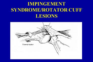
IMPINGEMENT SYNDROME/ROTATOR CUFF LESIONS
2
Gross Anatomy
- Bones
- humerus (greater tuberosity)
- scapula (acromion, coracoid)
- clavicle (distal end) to ligaments
- Bursa subdeltoid (subacromial) bursa
- Ligaments coracoacromial
- Tendons
3
Rotator Cuff Footprint
4
Gross Anatomy
- Tendons
- Supraspinatus (1 tear)
- Infraspinatus (2 tear)
- teres minor
- Subscapularis
- Strongest and Largest
- long head of biceps
- Envelopes 75 of GH articulation
- Multiple layers of collagen (type I)
5
Muscle Functions
- Supraspinatus
- Initiates abduction
- Subscapularis
- Primary IR
- Infraspinatus/Teres minor
- Primary ER
6
Rotator Cuff Function
- Minimize vertical displacement of humeral head
- Provide adequate horizontal compression to
counter shear forces to provide dynamic
stabilization of humeral head - Initiate abduction
7
Primary Impingement (not enough space)
- Definition The abutment of suprahumeral soft
tissues (tendons and bursa) against overlying
structures including the anterior acromion,
acromioclavicular joint, coracoid process, and
coracoacromial ligament with glenohumeral
elevation (flexion/abduction) in 80 - 120o range.
8
Types of Impingement
- Primary mechanical rub
- Secondary internal looseness
- Internal loose anterior ligaments, RC rubbing
on the posterior superior labrum - Associated with posterior glenoid cysts!!
- Tight posterior ligament get internal
impingement - Other
- Postural
- Scapular Dysfunction
9
Predisposing Factors to Primary Impingement
- Acromion
- Morphology - Type I, II, III (Aoki, Bigliani)
- X-ray diagnosis
10
Spur formation
- undersurface
- CA ligament attachment
- calcified
11
Other Primary Impingements
- Coracoacromial ligament
- Hypertrophic bursa
- Acromioclavicular joint spurring (DJD)
- Rotator cuff tendon thickening due to
inflammation, scarring, calcium deposits, partial
tearing (top or bottom)
12
ANDHumerus
- greater tuberosity prominence (after fracture)
- 5mm
13
Secondary ImpingementLigament Problems
- Anterior laxity
- Posterior tightness
14
Secondary Impingement
- Loss of adequate dynamic humeral head
depression/stabilization - rotator cuff/biceps tendon failure
- Posture
- protracted scapular
- forward head
- Position - loss of adequate glenohumeral external
rotation and scapular retraction
15
Instability
- glenohumeral or scapular
- repeated overhead use of shoulder
16
Rotator cuff normal arthroscopic anatomy
- Vascularity
- Critical zone decreased vascularity 1 cm.
proximal to insertion of supraspinatus tendon
(Codman, Rothman, and Parke) - Position dependent - less circulation to critical
zone with shoulder adduction than with abduction
(MacNab and Rathbun)
17
Tendon aging (Brewer)
- Decreased tendon cellularity
- Disorganization of tendon collagen network
- Decreased vascularity
- Increased type III collagen GAG
18
Rotator Cuff Natural History
- Asymptomatic
- 23 in 50-59 y.o.
- 51 in gt80 y.o.
- Supraspinatus
- Greater than 60 of all tears
- Articular surface tears 2-3x greater than bursal
surface
19
Classification of pathology
- Compressive failure - impingement, extrinsic,
bursal side - Tensile failure
- Traction (throwers)
- Incomplete articular
- Tendinosis (tendinitis)
20
Acute - overuse
21
Calcific tendonitis (1o Impingement)
- Degenerative process prior to calcification
- More often in females, dominant shoulder,
supraspinatus
Calcific Tendinitis
Normal
Kidney Stone Ultrasound Protocol
22
Diagnosis (1o Impingement)
- History - repetitive overhead use
- pain with or following activity
- night pain (cannot go to sleep and cannot roll
over)- DONT forget tumors in smokers - Decreased velocity in throwers
23
Physical Exam
- Painful arc of motion (80o - 120o elevation),
decreased range of motion - Greater tuberosity tenderness
- Decreased strength in abduction, external
rotation, internal rotation - Secondary to pain
- Secondary to tendon failure
24
Posterior/inferior capsular tightness
- loss of internal rotation
- Check mobility
- AC and SC joints
25
Positive impingement tests
- Hawkins - flexion at 90o, internal rotation,
horizontal ADD 20o - Active - hand to opposite shoulder, elevate elbow
with shoulder flexion - Passive (Neer) - fix scapula, force shoulder into
full flexion - Biceps tendon Yergasons
- Glenohumeral stability - provocative tests
- elective muscle/tendon recruitment and stretching
26
Diagnostic TestingOutlet view
- X-rays - look for superior migration of humeral
head, greater tuberosity cysts, sclerosis of
underside of acromion, acromial morphology with
outlet view - Arthrogram
- Ultrasound
27
MRI scanning for RC injury
Healthy Supraspinatus With MRI scan notice Dark
color
Tear of supraspinatus Notice white spot
where Dark should be
28
Non-operative Management
- Rest, educate patient on biomechanics
- Treat pain/inflammation
- Injections
- NSAID
- rehab
29
Surgical Considerations
- Age
- Young, active - early repair
- 40 and UP - repair
- Demand related
30
Size of tear
- 1 Outcome determinant
- Partial - acromioplasty
- Small to large - repair most, dominant arm
- Massive(gt 4cm 50 failure)
- Acromioplasty
- Marginal convergence
- Stabilize the remaining tissue
- Possible allograft
- Possible Restore (pig mucosa)
31
Surgical Goals
- Remove impinging tissue
- Remove impaired cuff
- Address Rotator Cuff pathology
- Recreate footprint of supraspinatus
25 mm long
12 mm wide
32
Surgical Procedures
33
Arthroscopy - role
- Diagnostic
- Rotator cuff tears - partial - undersurface,
complete - size - Glenohumeral clean out
- Bursal hypertrophy
- PASTA
- Acromioclavicular osteophytes
- Hooked acromion or traction spur
- DJD AC joint
- Associated instability
34
Arthroscopic assisted rotator cuff repair
- Arthroscopic acromioplasty and mini open repair
- Small, complete, limited retraction, minimal
atrophy - Physically active patient with pain and
symptomatic weakness
Deltoid splitting acromial attachment preserved
but weakened
35
All arthroscopic repair is being done
- fixation concerns
- Double row
- Special Suture Techniques
36
Open rotator cuff repair
- Indications
- Acute traumatic tears - in large people with BP
problems - Degenerative tears - with marked atrophy and
retraction - Failed arthroscopic debridement
- Suture anchors
37
Procedure for very large tears
- Perform acromioplasty (Neer)
- Mobilize soft tissue to get adequate coverage or
closure - Secure into trough near greater tuberosity
- Tendon transfers - latissimus and subscapularis
- Rehabilitate anterior deltoid and teres minor
- Reverse total shoulder arthroplasty
- Elderly (gt70) not young
38
Acromioplasty (Open or Arthroscopic)
- 6 weeks of active rest
- OR
RUPTURED DELTOID!!
39
Shoulder Dislocation
- Pt gt 40 y.o. PT
- Rule out RTC tear
40
THE FROZEN SHOULDERAdhesive Capsulitis
- Clinical Entity - not diagnosis
- Essential lesion
- Coracohumeral ligament contracture
- Is it shoulder or is it NECK!
41
Adhesive Capsulitis Stages
- I. Initiation/Inflammation
- Hot/painful
- Rx NSAIDs/Injection
- II. Frozen
- Less pain
- Lose more motion
- III. Slow improvement
- Each stage lasts 3-6 months
42
Etiology/Associated Pathology
- Cervical
- Periarthritis
- Bicipital tendonitis
- Pericapsulitis
- Bursitis
- Rotator cuff tendonitis
43
Associated Pathology
- Calcific tendonitis
- Traumatic osteoarthritis
- Impingement syndrome
44
Predisposing Factors
- Immobilization
- 40 - 70 years old
- Diabetes
- Trauma
- Cervical disc
- Thyroid disorders
- Intrathoracic dysfunction
- Post MI
45
Symptoms
- Diffuse ache about front and lateral aspect of
shoulder - Lack of arm mobility with increased symptoms when
elevating arm - Symptoms often worse at night
- 1 THEY LOST MOTION!
46
Clinical Examination
- restriction in both active and passive ROM
- most often elevation, ER and IR, often with
capsular type end-feel - examine humeral head translation in all planes
with patient supine. - No pain abduction or extension
47
Diagnosis
- Rule out myriad of possibilities
- X-rays before
- MRI and/or arthrogram - after
48
Treatment
- Pre Dexa Scan
- Office
- ABD 90 Arc ER/IR gt60
- Injections (30-60 cc)
- 20 cc .25 lidocaine
- 2 cc depomedrol
- Remainder is saline
- Oral medications (NSAIDs and Analgesics) post
manipulation - Brisement
Delaware Touch Technique 90 accurate
49
Treatment
- OR with regional block plus sedation
- Or general anesthesia
- Posterior glides
- Horizontal Adduction
- Forward flexion
- Inferior glides
- ABD
- Rotations in ABD 90 degrees
- ER
- IR
- IR in horz. Adduction
- Rotations in ABD 0
- ER
- Injection 10 cc lidocaine and 2 cc depomedrol
50
Following manipulationOR or in-office
- Same day PT within 4 hours!!
- 4 PT visits the first week, 3 visits the second,
PRN - Average 10 PT visits
- MUST HAVE HEP
51
Arthroscopic Release
- Failure with gt 4m PT
- Release CH ligament and Open RC Interval
- Avoid 600/Axillary nerve
- 12.5 mm
- Bad but only chance for IDDM
Recommended
CrystalGraphics Presentations


