
ECG%20Review:%20The%20Basics - PowerPoint PPT Presentation
Title:
ECG%20Review:%20The%20Basics
Description:
ECG Review: The Basics. ... Right Axis Deviation seen in: RVH, RBBB, COPD, acute PE. Left Axis Deviation seen in: LVH, ... R or S in limb leads 20mm. – PowerPoint PPT presentation
Number of Views:454
Avg rating:3.0/5.0
Title: ECG%20Review:%20The%20Basics
1
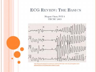
ECG Review The Basics
- Megan Chan, PGY-1
- UHCMC 2015
http//thepracticalpsychosomaticist.com/2013/04/01
/qtc-interval-prolongation-and-antipsychotics-by-e
lysha-elson-pharm-d-mph/
2
The Basics
http//flylib.com/books/en/2.569.1.27/1/
3
The ECG Unit
http//cal.vet.upenn.edu/projects/lgcardiac/ecg_tu
torial/printerval.htm
4
The Systematic Process
- Rate
- 300/( large boxes between RR interval)
- 300-150-100-75-60-50
- Rhythm
- Regular vs irregular
- Sinus rhythm?
- P before every QRS (easiest to see in leads II
and V1) - Positive p wave in I II negative p in aVR
- Axis
- Normal axis?
- Positive QRS sum in I and II (or aVF )
- Left deviation?
- Up in I, down in II
- Right deviation?
- Down in I, up/down in II
5
The Systematic Process Cont.
- Intervals
- PR interval normal 120-200ms (3-5 small boxes)
- Short PR interval WPW
- Long PR interval heart block
- QRS complex normal lt120ms ( 3 small boxes)
- Long QRS conduction delays, hyperkalemia,
ventricular rhythm - QT interval normal 430 in men, 450 in
females (less than RR/2) - Long QT MI, myocarditis, hypocalcemia,
hypothyroidism, subarachnoid hemorrhage,
drugssotolol, amiodarone, hereditary
6
The Systematic Process Cont.
- Conduction Abnormalities
- AV blocks
- RBBB
- LBBB
- IVCD (interventricular conduction delay)
- Left Anterior Fascicular Block
- Left Posterior Fascicular Block
7
http//healthybeatinghearts.blogspot.com/2011/01/f
irst-week-with-new-pacemaker.html
8
http//www.zuniv.net/physiology/book/images/11-13.
jpg
http//dualibra.com/wp-content/uploads/2012/04/037
8001/Part209.20Disorders20of20the20Cardiovas
cular20System/Section202.20Diagnosis20of20Car
diovascular20Disorders/221.htm
9
http//www.emedu.org/ecg/crapsanyallans.php
10
Hemi Blocks Left Fascicular Blocks
http//www.usfca.edu/fac-staff/ritter/Image74.gif
11
LAFB
LPFB
http//aliem.com/wp-content/uploads/2013/08/LAFB.p
ng
http//cdn.lifeinthefastlane.com/wp-content/upload
s/2011/02/avhisbb.jpg
http//aliem.com/wp-content/uploads/2013/08/LPFB.p
ng
12
Hypertrophy
http//dualibra.com/wp-content/uploads/2012/04/037
8001/Part209.20Disorders20of20the20Cardiovas
cular20System/Section202.20Diagnosis20of20Car
diovascular20Disorders/221.htm
13
The Systematic Process Cont.
- Chamber size
RAE LAE RVH LVH
Tall P gt 2.5 mm in lead II Large diphasic P with large initial phase in V1 Pgt 120ms Diphasic p with downward terminal phase gt 1mm wide and 1mm deep in V1 M-shaped P in I, II, or aVL R in aVR gt 5mm (or RgtQ) R in V1 gt 7mm qR in V1 R in V1 S in V5/V6 gt 10mm Deep S in V5/V6 gt 7mm R in aVL gt 11mm R in V5/V6 S in V1/V2 gt 35mm R in I S in III gt 25 mm R in aVF gt 20mm S in aVR gt 14mm
14
The Systematic Process Cont.
- Ischemia
- What ECG changes do you expect to see?
- Hyperacute T waves ? Inverted T waves ? ST
segment elevation ? Q waves - ST depressions ???
- Subendocardial ischemia
- ST elevations ???
- Transmural ischemia
- What are Pathologic Q waves?
- 1 small box wide and/or gt5mm or 1/3 of R wave
deep - Other changes
- Old septal infarct No R waves in V1-V3
- Old lateral infarct No R wave progression in
V4-V6 - RV infarct ST elevation in V4 V5 with right
sided EKG
15
The Systematic Process Cont.
- Everything Else
- Pericardial Effusion
- Low voltage (R waves lt 5mm in limb leads, lt10mm
in precordial leads) - Pericarditis
- Diffuse ST elevations and PR depressions
- Pulmonary Embolism
- S1Q3T3S wave in I, Q wave in III, T wave
inversion in III
16
Location Leads Occluded Vessel
Anterior V2-V4 LAD
Anteroseptal V1-V4 LAD
Anterolateral V1-V6, I, aVL LAD, diagonal
Lateral V5-V6, I, aVL Circumflex, diagonal
Inferior II, III, aVF RCA, circumflex
Posterior Tall R in V1-V3, ST depression in V1-V2 RCA
http//www.edoctoronline.com/media/19/photos_245a9
75b-66ad-4f7e-86d8-82d3ca7d0120.jpg
17
http//dotwordpressdotcom.wordpress.com/med-school
/clinical-skills/ecgs/
18
THE DR. ORTIZ METHOD
- 4 step method to interpreting 80 of ECGs in 1
minute - What are the most important ECG leads?
- II best axis, dx inferior wall MI, most studied
- V1best p wave, dx anterior wall MI RBBB
- V5dx lateral wall MI, LBBB, LVH
- What 2 leads are best for determining axis?
- I II
- 100 sensitive specific w/ zero false
- Normal axis is -30 to 90
- aVF was used gt 100 years ago
- Special thanks to Dr. Jose Ortiz!
19
THE DR. ORTIZ METHOD
- Step 1 Demographics
- Verifying pt name and calibration of ECG
- Step 2 Two second look at lead II
- Regularity of the tracing. Any funny beats?
- P waves
- Upright ? sinus
- M shape ? LAE
- Mountain peaks ? RAE
- Axis QRS positive ? 50 chance of normal axis
- Intervals
- Normal QRS lt3 boxes
- gt3 boxes BBB
- Q waves 75 risk for inferior MI
20
THE DR. ORTIZ METHOD
- Step 3 Study three things about the QRS
- Axis normal vs L deviation vs R deviation
- Confirm suspected axis by looking at lead I
- Width normal vs RBBB vs LBBB
- gt 3 boxes wide abnormal
- Look at V1 ? If RSR then RBBB If large S then
LBBB. - Height normal vs low voltage vs LVH
- Remember 14-12-35 for LVH
- Lead I R gt 14
- Lead aVL R gt 12
- S in V1 R in V5/V6 gt 35
21
THE DR. ORTIZ METHOD
- Step 4 Rate, ST segments, T waves, Infarcts
- Anterior/Septal infarct V1-V4
- Inferior infarct II, III, aVF
- Lateral infarct aVL, I, V5, V6
22
Draw A Normal ECG
http//www.lysosomalstorageresearch.ca/Fabry_eClin
ic/electrocardiography-ecg.html
23
How To Draw A Normal ECG
aVR
V4
I
V1
Same as aVR but T P waves can be or
Similar to V3 with smaller S, taller R
Same as II
Inverted II
aVL
V2
II
V5
Similar to V3 but less QRS voltage
Similar to V3 with larger S, smaller R
Similar to V4 with smaller S, taller R (R wave
progression)
aVF
III
V3
V6
Same as II
Same as II
Same as II
Biphasic QRS
24
REFERNCES
- Agabegi SS, Agabegi ED. Step up to Medicine, 3rd
ed. 2013. Lippincott Williams Wilkins.
Philadelphia, PA. - Gomella LG, Haist SA. Basic EKG reading. In
Clinicians Pocket Reference. McGraw-Hill 2007.
http//flylib.com/books/en/2.569.1.27/1/.
Accessed Nov 18, 2014. - Longo DL, Fauci AS, Kasper DL, et al.
Electrocardiography. In Harrisons Principles of
Internal Medicine, 18th ed. 2012. McGraw Hill.
New York, NY. - University of Illinois at Chicago. Online ICU
Guidebook. 2013. http//chicago.medicine.uic.edu/U
serFiles/Servers/Server_442934/Image/1.1/residentg
uides/final/icuguidebook.pdf. Accessed December
1, 2014.
Recommended
CrystalGraphics Presentations


