
Salivary Glands - PowerPoint PPT Presentation
Title:
Salivary Glands
Description:
Salivary Glands Major salivary glands: Lie at some distance from the mucosa and communicate with one or more ducts. Three paired masses of the parotid, submandibular ... – PowerPoint PPT presentation
Number of Views:365
Avg rating:3.0/5.0
Title: Salivary Glands
1
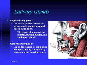
Salivary Glands
- Major salivary glands
- Lie at some distance from the mucosa and
communicate with one or more ducts. - Three paired masses of the parotid,
submandibular, and sublingual glands. - Minor Salivary glands
- Lie in the mucosa or submucosa and open directly
or indirectly via many short excretory ducts.
2
Salivary Glands
- Function
- Lubrication of food, assisting swallowing.
- Moistening of the buccal mucosa, essential for
speech. - Provision of an aqueous solvent, necessary for
taste. - Provision of a fluid seal, necessary for sucking.
- Secretion of digestive enzymes, amylase.
- Secretion of hormones, Glucagons-like protein and
? serotonin
3
Salivary Glands
The Parotid gland
- The Parotid gland
- The largest of the salivary and is predominantly
serous with few scattered mucous acini. - It lies below the external auditory meatus in a
deep hollow between the ramus, the mastoid and
the styloid process.
4
Salivary Glands
The Parotid gland
- It is roughly wedge shape, with an upper and
lower poles with the base above and the apex
below behind the angle of the mandible. - It is also wedge shaped in the horizontal plan
with three surfaces, lateral, antero-medial and
postero-medial, the base is the lateral and apex
lies against the pharyngeal wall
5
Salivary Glands
The Parotid gland
- The parotid sheath
- is a tough capsule, derived from the investing
layer of the deep cervical fascia - Superficial layer is attached to the zygomatic
arch - The deep part to the styloid process, the
mandible (stylo-mandibular ligament) and the
tympanic part of temporal bone
6
Salivary Glands
The Parotid gland
- The upper pole
- adhere to the cartilage of external auditory and
the TMJ capsule - The lower pole
- is rounded and lie below and behind the angle of
the mandible. - The facial nerve
- divide it into superficial and deep parts or lobes
7
Salivary Glands
The Parotid gland
- Lateral surface
- is flat and covered with skin and superficial
fascia containing great auricular nerve. - Lymph nodes lie within the substance or under
the sheath. - The duct and the five branches of the facial
nerve emerge from its anterior border
8
Salivary Glands
The Parotid gland
- Anteromedial surface
- U-shaped clasping the posterior border of the
ramus with masseter laterally and medial
pterygoid medially and inferiorly, separated by
the stylomandibular ligament from the
submandibular salivary gland
9
Salivary Glands
The Parotid gland
- The posteromedial surface
- Indented by the mastoid process and its muscles
- Lie against the styloid process with its three
muscles and two ligaments - The accessory gland
- Lies on the masseter between the duct and the
zygomatic arch
10
Salivary Glands
Parotid gland
- The Parotid duct
- Emerge from the convex anterior border of the
superficial part and pass anteriorly over the
masseter muscle. - At the anterior border of the muscle it turn
medial and pierce the buccal pad fat and
buccinator muscle, pass for a short distance
under the mucous membrane before it opens
opposite the upper second molar
11
Salivary Glands
Parotid gland
- Structures within the gland
- Facial nerve
- enter the gland from the deep surface and emerge
from behind the anterior border. - Retromandibular vein
- Deep to the branches of the nerve
12
Salivary Glands Parotid gland
- The capsule
- Derived from the deep cervical fascia which split
at the lower pole to encase the gland - The deep layer pass beneath the gland and ie
attached to the skull base - Part is inserted to styloid process and angle of
mandible is thickened, stylomandibular ligament
separating the gland from the submandibular one - The superficial layer pass anterior on the
masseter muscle and is attached to the lower
border of the zygomatic arch - Tight insertion will lead to sever pain on
inflammation and swelling of parotid
13
Salivary Glands
Parotid gland
- External carotid artery
- With its two terminal branches it is the deepest
structure - Periauricular lymph nodes
- Lies within the gland or under the capsule
14
Salivary Glands
Parotid gland
- Blood supply
- External carotid artery and retomandibular vein
- Nerve supply
- Secretomotor fibres
- from the inferior salivary nucleus to the
glossopharyngeal nerve, tympanic branch, tympanic
plexus, lesser superficial petrosal to otic
ganglion, to the Auriculotemporal nerve - Sympathetic fibres
- From the superior cervical plexus
- Vasoconstrictor
- Sensory fibres
- Gland, auriculotemporal
- Capsule, greater auricular (C2)
15
Salivary Glands Parotid gland
- The capsule send septa to the interior of the
gland separating it into lobules and making
surgical removal difficult. - Submandibular gland is loosely enveloped and can
be shelled out easily - Medially, the capsule is closely related to the
pharynx and the great vessels in the carotid
sheath - Break through by tumour or sharp instrument