
MALE%20REPRODUCTIVE%20SYSTEM - PowerPoint PPT Presentation
Title:
MALE%20REPRODUCTIVE%20SYSTEM
Description:
MALE REPRODUCTIVE SYSTEM It is a series of ducts and tubules. It has components in the abdomen, pelvis and perineum. The major components are: Testis. – PowerPoint PPT presentation
Number of Views:166
Avg rating:3.0/5.0
Title: MALE%20REPRODUCTIVE%20SYSTEM
1
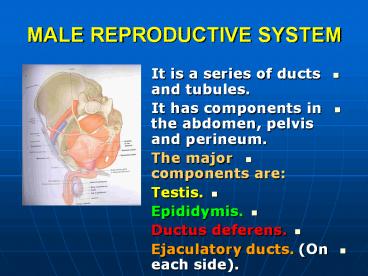
MALE REPRODUCTIVE SYSTEM
- It is a series of ducts and tubules.
- It has components in the abdomen, pelvis and
perineum. - The major components are
- Testis.
- Epididymis.
- Ductus deferens.
- Ejaculatory ducts. (On each side).
2
MALE GENITAL ORGANS
- Three types of accessory glands are associated
- Unpaired Prostate.
- A Pair of Seminal vesicles.
- A Pair of Bulbourethral glands.
3
VAS DEFERENS (PELVIC PART)
- It descends along the lateral pelvic wall.
- It crosses the ureter at the ischial spine.
4
VAS DEFERENS
- It runs downward and medially along the posterior
surface of the bladder. - Its terminal part is dilated to form the Ampulla
of the vas deferens. - The inferior end of the ampulla narrows and
shares in forming the ejaculatory ducts.
5
EJACULATORY DUCTS
- They are formed by the union of the lower end of
the vas deferens and the duct of the seminal
vesicle. - The ducts pierce the posterior surface of the
prostate.
6
EJACULATORY DUCTS
- They open into the prostatic urethra, close to
the margins of the prostatic utricle. - They drain the seminal fluid into the prostatic
urethra.
7
SEMINAL VESICLES
- Two lobulated organs (5) cm long.
- Lying on the posterior surface of the bladder.
- They are formed of a much coiled tube embedded in
a connective tissue.
8
SEMINAL VESICLES
- Upper ends
- Widely separated.
- Lower ends
- Close together.
- Medial
- Terminal part of the vas deferens.
9
SEMINAL VESICLES
- Posterior
- The rectum.
- Inferior
- They form the ejaculatory ducts.
10
SEMINAL VESICLES
- Function
- Their secretion contributes to increase the
volume of the semen. - It can nourish the spermatozoa.
11
PROSTATE
- It is unpaired accessory structure of the male
reproductive system. - It surrounds the urethra in the pelvic cavity.
12
SHAPE
- It is conical in shape.
- Its base lies against the neck of the bladder.
- Its apex lies against the urogenital diaphragm.
13
RELATIONS
- Superior
- Neck of the bladder.
- The urethra enters the center of the base of the
prostate. - The smooth muscle of the two organs is continuous.
14
RELATIONS
- Anterior
- Symphysis pubis.
- It is separated from it by retropubic pad of fat
(in cave of Retzius). - Its fibrous sheath is connected to the pubic
bones by puboprostatic ligaments.
15
RELATIONS
- Posterior
- Rectal ampulla.
- It is separated from it by rectovesical septum
(fascia of Denonviller). - It is formed in the fetal life by fusion of the
inferior extension of the rectovesical pouch.
16
PROSTATE
- The ejaculatory ducts pierce the upper part of
the posterior surface.
17
RELATIONS
- Lateral
- Anterior fibers of levator ani (levator prostate).
18
STRUCTURE
- It is surrounded by a fibrous capsule
(internally) and a fibrous sheath (from the
pelvic visceral fascia) externally.
19
STRUCTURE
- The glands are embedded in a mixture of smooth
muscle and connective tissue. - They open in the prostatic urethra.
20
LOBES
- It is incompletely divided into (5) lobes
(according to urethra). - Anterior
- In front of the urethra.
- It has no glandular tissue.
21
LOBES
- Median (middle).
- It is wedge in shape.
- It is between the urethra and the ejaculatory
ducts. - Its upper part forms the uvula vesicae in the
trigone. - It is rich in glands.
22
LOBES
- Posterior
- It is behind the urethra, below the ejaculatory
ducts. - It has glandular tissue.
23
LOBES
- Lateral (right and left)
- Lie on either side of the urethra.
- They are rich in glands.
- They are connected to each other by a vertical
groove on the posterior surface of the prostate.
24
FUNCTION
- It produces thin milky fluid that added to the
seminal fluid at the time of ejaculation. - It is alkaline secretion.
- It neutralizes the acidity of the vagina.
25
ARTERIAL SUPPLY
- Prostatic branches from
- Inferior vesical.
- Middle rectal.
26
VENOUS DRAINAGE
- Prostatic venous plexus
- It is between the capsule and the fibrous sheath.
- It receives
- Vesical veins.
- Deep dorsal vein of the penis.
- It drains into
- Internal iliac vein.
27
NERVE SUPPLY
- Inferior hypogastric plexus.
- The sympathetic fibers stimulate the smooth
muscle in the capsule and stroma to contract
during ejaculation.
28
PROSTATIC URETHRA
- It is the widest and most dilatable part of the
urethra. - It is (3cm) long.
- It begins at the neck of the bladder.
- It passes through the prostate from the base to
the apex. - It continues with the membranous part of the
urethra.
29
PROSTATIC URETHRA
- On the posterior wall
- 1. Urethral crest
- A longitudinal ridge.
- 2. Prostatic sinus
- It is a groove on each side of the crest.
- The prostatic glands open in the sinuses.
30
POSTERIOR WALL
- 3. Prostatic utricle
- It is a depression on the summit of the urethral
crest. - It is analog to the uterus and vagina in female.
- The ejaculatory ducts open on the edge of the
mouth of the utricle.
Recommended
CrystalGraphics Presentations


