
Plasma Membrane Composition - PowerPoint PPT Presentation
Title:
Plasma Membrane Composition
Description:
Plasma Membrane Composition Phospholipid Bilayer-phospholipid molecules in a double-layered sheet-hydrophilic (water loving) head facing out-hydrophobic (water ... – PowerPoint PPT presentation
Number of Views:205
Avg rating:3.0/5.0
Title: Plasma Membrane Composition
1
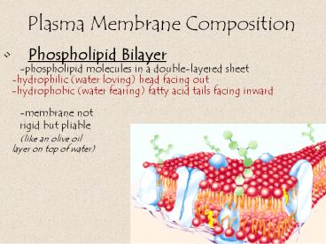
Plasma Membrane Composition
- Phospholipid Bilayer
- -phospholipid molecules in a double-layered
sheet - -hydrophilic (water loving) head facing out
- -hydrophobic (water fearing) fatty acid tails
facing inward - -membrane not
- rigid but pliable
- (like an olive oil
- layer on top of water)
2
Plasma Membrane Composition
- 1. Phospholipid Bilayer
- -membrane not rigid but pliable
- fluid mosaic model molecules of
- membrane flow and move about each other
- in constant
- motion
- sliding past
- one another
3
Plasma Membrane Composition
- 2. Proteins
- - found on surface of the membrane and in the
membrane among the phospholipid molecules - - identify the cell
- - form channels through which other molecules
can be transported - - act as receptors
4
Plasma Membrane Composition
2. Proteins
5
Plasma Membrane Composition
- Cholesterol
- -plays role in stabilizing the membrane
- -helps keep the membrane flexible
6
Movement across a membrane. . .
- Plasma membranes are selectively permeable
- selectively permeable membrane a membrane that
allows some things to pass through it and other
things not
7
- Diffusion - the tendency of molecules to move
from a higher concentration to a lower
concentration until equilibrium is reached.
8
Movement across a membrane. . .
- Diffusion
- movement of molecules across a membrane from area
of higher percent concentration to area of lower
percent concentration until they are equally
distributed - no energy required
9
Passive Transport
- Diffusion across a membrane.
- Cell does not use any energy for diffusion.
- Selectively permeable membrane.
- Examples water, O2 and CO2 gas exchange
10
- Osmosis - passive transport of water across a
semi-permeable membrane. - The water is the substance that moves across
membrane not the solute.
11
Movement across a membrane. . .
- Osmosis
- isotonic solution environment equal
concentrations of substances (solutes) and water
(solvent) in cell and in environment around cell - Most bodily fluids will be isotonic to inside of
cells (ex. plasma and RBCs)
12
Hypertonic
- Solution with a higher concentration of solute
and a lower concentration of water. - Hyper above
13
Movement across a membrane. . .
- Osmosis
- hypertonic solution environment
- lower concentration of water, higher
concentration of solute - water leaves cell it shrinks
14
Hypotonic
- Solution with a lower solute concentration and a
higher water concentration. - Hypo below
15
Movement across a membrane. . .
- Osmosis
- hypotonic solution environment
- higher concentration of water, lower
concentration of solute - water enters the cell and it swells / bursts
16
Isotonic
- Solutions of equal solute concentration.
- Isos equal
17
(No Transcript)
18
Effect on living animal cells
- Osmoregulation control of water balance.
- Animals must use this when exposed to hypertonic
and hypotonic environments for survival. - Example fresh water fish live in hypotonic
environment use kidneys and gills to prevent
excess water buildup in body.
19
Effect on living plant cells
- Most plants thrive in a hypotonic environment
when cell wall is turgid and vacuole is full. - Plants become wilted in isotonic environment.
- Plasmolysis plant in hypertonic environment
causes cell water loss, cell shrivels and the
cell membrane pulls away from the cell wall and
can kill the cell.
20
Other Ways to Get in. . . .
- Facilitated diffusion
- -involves protein carriers that
- combine w/ the molecule to
- move them across the membrane
- -the diffusion is ALWAYS
- from high to low concentration
- (down the concentration gradient)
- -no energy required
21
Other Ways to Get in. . . .
- Facilitated diffusion
- -use carrier proteins
- -from high to low concentration
- -no energy required
22
Other Ways to Get in. . . .
- active transport
- uses carrier proteins to carry molecules
against the concentration gradient, from low to
high concentrations - -requires ATP energy
- (backwards from diffusion - - not passive)
23
Other Ways to Get in. . . .
- Endocytosis
- process cells use to wrap membrane around a
particle (usually food) and engulf it - ex. leukocytes (white blood cells) use this to
surround invading bacteria, viruses, and other
foreign materials - Exocytosis
- the opposite
- of endocytosis
- particles are
- released
exocytisis
endocytosis
24
Types of Microscopes
- Light Microscope - the models found in most
schools, use compound lenses to magnify objects.
The lenses bend or refract light to make the
object beneath them appear closer.
Common magnifications 40x, 100x, 400x Oil
Immersion lenses can improve quality of focus and
magnification
25
Imaging technologies provide new views of life.
- Light microscopes (LM)
- Light and lenses used to magnify specimens
- limited magnification
- can be used to study living specimens
- shows a two-dimensional image of a specimen.
- Shows actual color of the specimen.
LM magnification 400X
26
(No Transcript)
27
Stereoscope- dissecting microscope
- This microscope allows for binocular (two eyes)
viewing of larger specimens. - Usually magnifies 10x to 20x
- Can be used for thicker specimens
- Creates a 3D view of specimen
28
SEM
- Scanning Electron
- microscope
29
- Scanning electron microscopes (SEM)
- Deflection of electrons used to magnify
specimens - provides high magnification and a
- three-dimensional black-and-white image that can
be - colored by computer
- cannot be used to study living specimen
SEM magnification 1500X
30
31
Transmission Electron Microscope (TEM)
32
- Transmission electron microscopes (TEM)
- Electrons passing through a specimen
- used to magnify specimen
- provides high magnification
- two-dimensional black-and-white image
- that can be colored by computer
- cannot be used to study living
- specimens
33
TEM of a cell, notice you see the inside of the
cell and not the surface.
34
(No Transcript)
35
The Light Microscope Guidelines for Use
- Always carry with 2 hands
- Only use lens paper for cleaning
- Do not force knobs
- Always store covered
- Keep objects clear of desk and cords
36
MagnificationYour microscope has 3
magnifications Scanning, Low and High. Each
objective will have written the magnification. In
addition to this, the ocular lens (eyepiece) has
a magnification. The total magnification is the
ocular x objective
37
- General Procedures
- Make sure all backpacks and junk are out of the
aisles and off the tops of desks. - 2. Plug your microscope in to the extension
cords. - 3. Store with cord wrapped around microscope and
the scanning objective clicked into place. - 4. Carry by the base and arm with both hands.
38
Focusing Specimens 1. Always start with the
scanning objective. Odds are, you will be able
to see something on this setting. Use the Coarse
Knob to focus, image may be small at this
magnification, but you won't be able to find it
on the higher powers without this first
step. Do not use stage clips, try moving the
slide around until you find something.
39
- 2. Once you've focused on Scanning, switch to Low
Power. Use the FINE Knob ONLY to refocus. Again,
if you haven't focused on this level, you will
not be able to move to the next level. - 3. Now switch to High Power. (If you have a thick
slide, or a slide without a cover, do NOT use the
high power objective). Again, ONLY use the Fine
Adjustment Knob to focus specimens. - Recap
- 1. Scanning --gt use coarse knob
- 2. Low power --gt use fine knob
- 3. High power --gt use fine knob
DO NOT SKIP STEPS!!!!
40
- Your slide MUST be focused on low power before
attempting this step - Click the nosepiece to the longest objective
- Do NOT use the Coarse Focusing Knob, this could
crack the slide or the lens - Use the Fine Focus Knob to bring the slide
41
- Drawing Specimens
- 1. Use pencil - you can erase and shade areas
- 2. All drawings should include clear and proper
labels (and be large enough to view details).
Drawings should be labeled with the specimen name
and magnification. - 3. Labels should be written on the outside of the
circle. The circle indicates the viewing field as
seen through the eyepiece, specimens should be
drawn to scale - ie..if your specimen takes up
the whole viewing field, make sure your drawing
reflects that.
42
- Making a Wet Mount
- 1. Gather a thin slice/piece of whatever your
specimen is. If your specimen is too thick, then
the coverslip will wobble on top of the sample
like a see-saw, and you will not be able to view
it under High Power. - 2. Place ONE drop of water directly over the
specimen. If you put too much water, then the
coverslip will float on top of the water, making
it hard to draw the specimen, because they might
actually float away. (Plus too much water is
messy) - 3. Place the cover slip at a 45 degree angle
(approximately) with one edge touching the water
drop and then gently let go. Performed correctly
the coverslip will perfectly fall over the
specimen.
Do not drop vertically, set one edge down and let
the other side drop.
43
- How to Stain a Slide
- 1. Place one drop of stain (iodine, methylene
blue..there are many kinds) on the edge of the
coverslip. - 2. Place the flat edge of a piece of paper towel
on the opposite side of the coverslip. The paper
towel will draw the water out from under the
coverslip, and the cohesion of water will draw
the stain under the slide. - 3. As soon as the stain has covered the area
containing the specimen, you are finished. The
stain does not need to be under the entire
coverslip. If the stain does not cover as needed,
get a new piece of paper towel and add more stain
until it does. - 4. Be sure to wipe off the excess stain with a
paper towel.
44
- Cleanup
- Store microscopes with the scanning objective in
place. - 2. Wrap cords and cover microscopes.
- Double check to make sure
you didn't leave a slide - 3. Wash slides in the sinks and dry them, placing
them back in the slide boxes to be used later. - 4. Throw coverslips away. (these are not
reusable) - Be careful not to drop these in the sink,
they can clog drain. - 5. Place microscopes in their designated location
(probably a cabinet)
45
Troubleshooting
- Occasionally you may have trouble with working
your microscope. Here are some common problems
and solutions. - 1. Image is too dark!
- Adjust the diaphragm, make sure your light is on.
- 2. There's a spot in my viewing field, even when
I move the slide the spot stays in the same
place! - Your lens is dirty. Use lens paper, and only lens
paper to carefully clean the objective and ocular
lens. The ocular lens can be removed to clean the
inside. The spot is probably a spec of dust. - 3. I can't see anything under high power!
- Remember the steps, if you can't focus under
scanning and then low power, you won't be able to
focus anything under high power. Start at
scanning and walk through the steps again. - 4. Only half of my viewing field is lit, it looks
like there's a half-moon in there! - You probably don't have your objective fully
clicked into place..
46
Practice Labeling the Parts
47
Quiz Over the Microscope
- 1. When focusing a specimen, you should always
start with the ___________________ objective. - 2. When using the high power objective, only the
________ ___________ knob should be used. - 3. The type of microscope used in most science
classes is the _________________ microscope - 4. Stains can be drawn under the slide (and over
a specimen) by using a _____________________ - 5. What part of the microscope can adjust the
amount of light that hits the slide?
______________________________
48
- 6. You should carry the microscope by the
________ and the __________. - 7. The objectives are attached to what part of
the microscope (it can be rotated to click the
lenses into place) _______________
________________ - 8. You should always store you microscope with
the ________________ objective in place. - 9. A microscope has an ocular objective of 10x
and a high power objective of 50x. What is this
microscope's total magnification? ____________ - 10. SEM is an abbreviation for ____________
____________ ________________
Recommended
CrystalGraphics Presentations


