
The Respiratory System: Breath deeply the air of knowledge - PowerPoint PPT Presentation
1 / 31
Title:
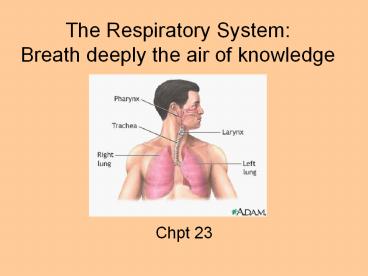
The Respiratory System: Breath deeply the air of knowledge
Description:
The Respiratory System: Breath deeply the air of knowledge Chpt 23 Respiratory System Function: supply body with oxygen and dispose of carbon dioxide Requirements ... – PowerPoint PPT presentation
Number of Views:216
Avg rating:3.0/5.0
Title: The Respiratory System: Breath deeply the air of knowledge
1
The Respiratory SystemBreath deeply the air of
knowledge
- Chpt 23
2
Respiratory System
- Function supply body with oxygen and dispose of
carbon dioxide
3
Requirements
- pulmonary ventilation (movement of air into and
out of lungs) - external respiration (gas exchange between blood
and lungs) - transport of respiratory gases (via blood)
- internal respiration (gas exchange between blood
and cells)
4
5 Functions of the Respiratory System
- Provides extensive gas exchange surface area
between air and circulating blood - Moves air to and from exchange surfaces of lungs
- Protects respiratory surfaces from outside
environment - Produces sounds-speaking, singing
- Helps maintain pH
5
Anatomy of the Respiratory System
Figure 231
6
Anatomy Nose
- Functions
- provides airway for respiration
- moistens and warms entering air
- filters air
- serves as a resonating chamber for speech
- houses olfactory receptors
7
Mucus
8
Nasal Structures
- External nose bridge, root, dorsum nasi, apex,
external nares, alae - Nasal cavity nasal septum, internal nares (aka
posterior nares or choanae), hard palate, soft
palate, vestibule (with sebaceous and sweat
glands and vibrissae) - Mucous membranes of the nasal cavity
- olfactory mucosa (with smell receptors)
- respiratory mucosa- secrete sticky mucus with
antibacterial substances - Conchae- superior, middle, and inferior help to
trap particles
9
Sinuses
- Paranasal sinuses
- Frontal
- Sphenoid
- Ethmoid
- Maxillary
- Function to lighten skull warm and moisten the
air
10
The Nasopharynx
- only an air passageway
- during swallowing, closed off by the soft palate
and uvula to prevent food from entering the nasal
cavity - pseudostratified ciliated columnar
- Pharyngeal tonsil (adenoids)
11
The Oropharynx
- swallowed food and inhaled air
- palatine lingual tonsils
- stratified squamous
12
The Laryngopharynx
- Inferior portion of the pharynx
- stratified squamous
- where resp. and dig. pathways diverge
13
Larynx aka voice box
- Functions
- open airway
- switching mechanism to route air and food into
proper channels - voice production via vocal cords
14
Larynx aka voice box
- Anatomy
- 9 cartilages connected by membranes and ligaments
(8 are hyaline) - 9th cartilage is the epiglottis- elastic closes
over trachea during swallowing - Vocal folds aka true vocal cords- avascular
vibrate as air passes through them to produce
sound - Medial opening between vocal folds is the glottis
15
(No Transcript)
16
(No Transcript)
17
Anatomy of the Trachea
Figure 236
18
Trachea aka windpipe10-12 cm long 2.5 cm diam
- Layers of tracheal wall (internal to external)
- Mucosa- pseudostr. ciliated columnar
- submucosa
- adventitia- with 16-20 rings of C-shaped hyaline
cartilage, allowing flexibility
19
Trachea aka windpipe
- 1520 tracheal cartilages
- strengthen and protect airway
- discontinuous where trachea contacts esophagus
- Ends of each tracheal cartilage are connected by
- an elastic ligament and trachealis muscle
- The carina (keel) marks the point where the
trachea splits into the 2 primary bronchi (approx
T7).
20
(No Transcript)
21
The Bronchial Tree
- R and L primary bronchus
- Secondary (lobar) bronchi- 3 R, 2 L
- About 20 smaller branches
- Bronchioles- less than 1 mm in diameter
22
- The Respiratory Zone thin-walled alveoli
(clustered into the alveolar sacs) where gas
exchange occurs
23
(No Transcript)
24
Lungs
- apex- narrow, superior tip
- base- concave, inferior surface on diaphragm
- L lung 2 lobes- upper and lower separated by
oblique fissure - R lung 3 lobes- upper, middle, and lower
separated by the horizontal and oblique fissures - Each lung lobe is divided into 10
bronchopulmonary segments
25
Lung lobes
26
Relationship between Lungs and Heart
Figure 238
27
Pleural Cavities and Pleural Membranes
Figure 238
28
Pleural Cavities and Pleural Membranes
- 2 pleural cavities
- are separated by the mediastinum
- Each pleural cavity
- holds a lung
- is lined with a serous membrane (the pleura)
29
The Pleura
- Consists of 2 layers
- parietal pleura
- visceral pleura
- Pleural fluid
- lubricates space between 2 layers
30
Pleurae (membrane around the lungs)
Parts of the parietal pleura. (parietal pleura
in blue visceral pleura in purple)
31
Grab a copy of the article Struggling to Inhale
- ANSWER THE FOLLOWING QUESTIONS
- There are 2 different words for croup. List them
and write what they each mean. - Explain how the virus that causes croup causes
infection. - What is the treatment for croup?
- What is a cricothyrotomy? Explain how doctors
perform these. - What type of infection did the older patient
have?