
Chapter 16 The Molecular Basis of Inheritance - PowerPoint PPT Presentation
1 / 33
Title:
Chapter 16 The Molecular Basis of Inheritance
Description:
Chapter 16 The Molecular Basis of Inheritance In April 1953, James Watson and Francis Crick shook the scientific world with an elegant double-helical model for the ... – PowerPoint PPT presentation
Number of Views:192
Avg rating:3.0/5.0
Title: Chapter 16 The Molecular Basis of Inheritance
1
Chapter 16The Molecular Basis of Inheritance
2
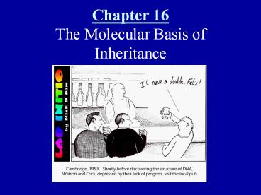
- In April 1953, James Watson and Francis Crick
shook the scientific world with an elegant
double-helical model for the structure of
deoxyribonucleic acid, or DNA.
3
(No Transcript)
4
- A. DNA as the Genetic Material
- 1. The search for genetic material led to DNA.
- Until the 1940s, the great heterogeneity and
specificity of function of proteins seemed to
indicate that proteins were the genetic material. - The discovery of the genetic role of DNA began
with research by Frederick Griffith in 1928. - He studied Streptococcus pneumoniae, a bacterium
that causes pneumonia in mammals. - One strain, the R strain, was harmless.
- The other strain, the S strain, was pathogenic.
- Griffith mixed heat-killed S strain with live R
strain bacteria and injected this into a mouse. - The mouse died, and he recovered the pathogenic
strain from the mouses blood. - Griffith called this phenomenon transformation, a
phenomenon now defined as a change in genotype
and phenotype due to the assimilation of foreign
DNA by a cell.
5
(No Transcript)
6
Avery experiment
7
- Finally in 1944, Oswald Avery, Maclyn McCarty,
and Colin MacLeod announced that the transforming
substance was DNA. - Viruses consist of DNA (or sometimes RNA)
enclosed by a protective coat of protein. - Viruses that specifically attack bacteria are
called bacteriophages or just phages. - In 1952, Alfred Hershey and Martha Chase showed
that DNA was the genetic material of the phage
T2. - To determine the source of genetic material in
the phage, Hershey and Chase designed an
experiment in which they could label protein or
DNA and then track which entered the E. coli cell
during infection. - They grew one batch of T2 phage in the presence
of radioactive sulfur, marking the proteins but
not DNA. - They grew another batch in the presence of
radioactive phosphorus, marking the DNA but not
proteins. - They allowed each batch to infect separate E.
coli cultures. - Hershey and Chase found that when the bacteria
had been infected with T2 phages that contained
radiolabeled proteins, most of the radioactivity
was in the supernatant that contained phage
particles, not in the pellet with the bacteria. - When they examined the bacterial cultures with
T2 phage that had radiolabeled DNA, most of the
radioactivity was in the pellet with the
bacteria. - Hershey and Chase concluded that the injected
DNA of the phage provides the genetic information
that makes the infected cells produce new viral
DNA and proteins to assemble into new viruses.
8
(No Transcript)
9
(No Transcript)
10
- By 1947, Erwin Chargaff had developed a series
of rules based on a survey of DNA composition in
organisms. - The bases could be adenine (A), thymine (T),
guanine (G), or cytosine (C). - Chargaff noted that the DNA composition varies
from species to species. - In any one species, the four bases are found in
characteristic, but not necessarily equal,
ratios. - Chargaffs rules.
- In all organisms, the number of adenines was
approximately equal to the number of thymines (T
A). - The number of guanines was approximately equal
to the number of cytosines (G C). - Human DNA is 30.9 adenine, 29.4 thymine, 19.9
guanine, and 19.8 cytosine.
11
(No Transcript)
12
Chargaff ratios
13
(No Transcript)
14
(No Transcript)
15
- 2. Watson and Crick discovered the double helix
by building models to conform to X-ray data.. - Among the scientists working on the problem were
Linus Pauling in California and Maurice Wilkins
and Rosalind Franklin in London. - The sugar-phosphate chains of each strand are
like the side ropes of a rope ladder. - Pairs of nitrogenous bases, one from each
strand, form rungs. - The ladder forms a twist every ten bases.
- The nitrogenous bases are paired in specific
combinations adenine with thymine and guanine
with cytosine. - Only a pyrimidine-purine pairing produces the
2-nm diameter indicated by the X-ray data. - In April 1953, Watson and Crick published a
succinct, one-page paper in Nature reporting
their double helix model of DNA.
16
The Dark Lady
17
xray
18
- B. DNA Replication and Repair
- 1. During DNA replication, base pairing enables
existing DNA strands to serve as templates for
new complementary strands. - When a cell copies a DNA molecule, each strand
serves as a template for ordering nucleotides
into a new complementary strand. - One at a time, nucleotides line up along the
template strand according to the base-pairing
rules. - The nucleotides are linked to form new strands.
- Watson and Cricks model, semiconservative
replication, predicts that when a double helix
replicates, each of the daughter molecules will
have one old strand and one newly made strand.
19
(No Transcript)
20
(No Transcript)
21
Meselson and Stahl Experiment
22
- 2. A large team of enzymes and other proteins
carries out DNA replication. - It takes E. coli 25 minutes to copy each of the
5 million base pairs in its single chromosome and
divide to form two identical daughter cells. - A human cell can copy its 6 billion base pairs
and divide into daughter cells in only a few
hours. - This process is remarkably accurate, with only
one error per ten billion nucleotides. - The replication of a DNA molecule begins at
special sites, origins of replication. - Replication proceeds in both directions until
the entire molecule is copied. - In eukaryotes, there may be hundreds or
thousands of origin sites per chromosome. - At the origin sites, the DNA strands separate,
forming a replication bubble with replication
forks at each end. - DNA polymerases catalyze the elongation of new
DNA at a replication fork. - In eukaryotes, at least 11 different DNA
polymerases have been identified so far. - The strands in the double helix are
antiparallel. - The sugar-phosphate backbones run in opposite
directions. - The 5 ? 3 direction of one strand runs counter
to the 3 ? 5 direction of the other strand. - DNA polymerases can only add nucleotides to the
free 3 end of a growing DNA strand. - A new DNA strand can only elongate in the 5 ?
3 direction. - The DNA strand made by this mechanism is called
the leading strand.
23
(No Transcript)
24
Replication
25
- The other parental strand (5 ? 3 into the
fork), the lagging strand, is copied away from
the fork. - Unlike the leading strand, which elongates
continuously, the lagging stand is synthesized as
a series of short segments called Okazaki
fragments. - Another enzyme, DNA ligase, eventually joins the
sugar-phosphate backbones of the Okazaki
fragments to form a single DNA strand. - DNA polymerases cannot initiate synthesis of a
polynucleotide. - They can only add nucleotides to the 3 end of
an existing chain that is base-paired with the
template strand. - The initial nucleotide chain is called a primer.
- In the initiation of the replication of cellular
DNA, the primer is a short stretch of RNA with an
available 3 end. - Primase, an RNA polymerase, links
ribonucleotides that are complementary to the DNA
template into the primer. - For synthesis of the lagging strand, each
Okazaki fragment must be primed separately. - Another DNA polymerase, DNA polymerase I,
replaces the RNA nucleotides of the primers with
DNA versions, adding them one by one onto the 3
end of the adjacent Okazaki fragment. - Helicase untwists the double helix and separates
the template DNA strands at the replication fork. - Single-strand binding proteins keep the unpaired
template strands apart during replication. - The lagging strand is copied away from the fork
in short segments, each requiring a new primer.
26
(No Transcript)
27
(No Transcript)
28
- 3. Enzymes proofread DNA during its replication
and repair damage in existing DNA. - Mistakes during the initial pairing of template
nucleotides and complementary nucleotides occur
at a rate of one error per 100,000 base pairs. - DNA polymerase proofreads each new nucleotide
against the template nucleotide as soon as it is
added. - If there is an incorrect pairing, the enzyme
removes the wrong nucleotide and then resumes
synthesis. - The final error rate is only one per ten billion
nucleotides. - In mismatch repair, special enzymes fix
incorrectly paired nucleotides. - A hereditary defect in one of these enzymes is
associated with a form of colon cancer. - In nucleotide excision repair, a nuclease cuts
out a segment of a damaged strand. - DNA polymerase and ligase fill in the gap.
- The importance of the proper functioning of
repair enzymes is clear from the inherited
disorder xeroderma pigmentosum. - These individuals are hypersensitive to
sunlight. - In individuals with this disorder, mutations in
their skin cells are left uncorrected and cause
skin cancer.
29
(No Transcript)
30
(No Transcript)
31
DNA Replication overview
- More replication
32
- 4. The ends of DNA molecules are replicated by a
special mechanism. - The usual replication machinery provides no way
to complete the 5 ends of daughter DNA strands. - Repeated rounds of replication produce shorter
and shorter DNA molecules. - The ends of eukaryotic chromosomal DNA
molecules, the telomeres, have special nucleotide
sequences. - Eukaryotic cells have evolved a mechanism to
restore shortened telomeres in germ cells, which
give rise to gametes. - An enzyme called telomerase catalyzes the
lengthening of telomeres in eukaryotic germ
cells, restoring their original length. - There is now room for primase and DNA polymerase
to extend the 5 end. - It does not repair the 3-end overhang, but it
does lengthen the telomere. - Telomerase is not present in most cells of
multicellular organisms. - Normal shortening of telomeres may protect
organisms from cancer by limiting the number of
divisions that somatic cells can undergo. - Cells from large tumors often have unusually
short telomeres, because they have gone through
many cell divisions. - Active telomerase has been found in some
cancerous somatic cells.
33
(No Transcript)