Spine - PowerPoint PPT Presentation
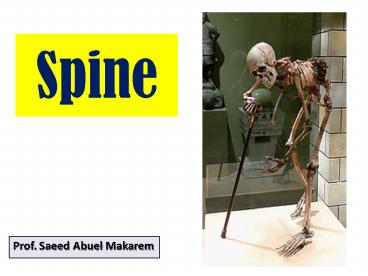
Title: Spine
1
Spine
- Prof. Saeed Abuel Makarem
2
Spinal fractures
- Spinal fractures are different than a broken arm
or leg. - A fracture or dislocation of a vertebra can cause
bone fragments that pinch or damage the spinal
nerves or even the spinal cord. - Most spinal fractures occur due to
- Car accidents,
- Falls,
- Gunshot, or
- Sports.
- Injuries can range from mild ligament or muscle
strains, to fractures and dislocations of the
vertebrae, and debilitating spinal cord damage.
3
Spinal fractures
- Depending on how severe your injury is, you may
experience - Pain,
- Difficulty walking,
- Unable to move your arms or legs (paralysis).
- Many fractures heal with conservative treatment.
- However severe fractures may require surgery.
- To understand spinal fractures, it is helpful to
understand how your spine are formed and works.
4
SPINAL COLUMN
- The vertebral column is a complex construct that
includes a variety of - Bones,
- Joints,
- Tendons,
- Nerves,
- Ligaments,
- Muscles and
- Vessels
- All woven together.
- The spines extends from the base of the skull to
the pelvis. - Ligaments, joints, muscles and tendons connect
the bones together and keep them aligned.
5
SPINAL COLUMN
- It is consists of 24 single vertebrae and 2
bones, - Sacrum and
- Coccyx, which made from fused vertebrae.
- Of the 24 single bones,
- 7 vertebrae in the neck (cervical),
- 12 vertebrae in the chest (thoracic), and
- 5 supporting the lower back and are called
(lumbar).
6
- Body or Centrum
- Anterior discoid, weight-bearing part of the
vertebra. - And
- Vertebral arch
- 2 pedicles and
- 2 Laminae.
- They join together to complete the arch.
- Vertebral foramen Between body and the arch and
contains the spinal cord, ligament, fat and
blood vessels. - The arch carries 7 processes.
- 2 Transverse processes two lateral projections
from the vertebral arch.
TYPICAL VERTEBRA
- One spinous process single posterior projection
arising from the vertebral arch. - 2 Superior and 2 inferior articular processes
paired projections allowing a vertebra to
articulate with adjacent vertebrae.
7
C E R V I C A L VERTEBRA
8
The vertebrae in each region have unique features
that help them to perform their main functions.
ATLAS AXIS
- The 7 cervical vertebrae (identified as C1 to
C7). - The first two (atlas axis) are different
because they perform functions not shared by the
other cervical vertebrae. - The neck has the greatest range of motion
because of these 2 specialized vertebrae.
The atlas has no body. The superior surfaces of
its 2 lateral masses contain large kidney- shaped
facet that articulate with the occipital
condyles. This joint allows you to nod- that is
to say "yes." Atlanto-occipital joint. The axis
acts as a pivot for the rotation of the atlas
(and the skull above). It has a large upright
process, called the odontoid process, or dens,
which acts as the pivot joint. The atlantoaxial
joint allows you to rotate your head that is to
say "no."
9
TYPICAL CERVICAL VERTEBRAE
- The "typical" cervical vertebrae (C3 to C7) are
the smallest, lightest vertebrae. - Their spinous processes are often short and
bifid, except the 7th. - Its transverse processes contain foramina through
which the vertebral vessels pass. - The main function of the cervical spine is to
support the weight of the head (about 10 pounds). - Also, it is the most movable region of the spine.
10
THORACIC VERTEBRAE
- The 12 thoracic vertebrae (T1 toT12) are larger
than the cervical vertebrae. - The body is somewhat heart-shaped and has two
costal demifacets on each side, which receive the
heads of the ribs. - The spinous process is long and hooks sharply
downward. - The range of motion in the thoracic spine is
limited.
11
LUMBAR VERTEBRAE
- The 5 lumbar vertebrae (L1-- L5) have massive,
block like bodies. - They have short, hatchet-shaped spinous
processes. - They are the most solid of all vertebrae.
- Their main function is to bear the weight of the
body. - They also have a moderate range of movements.
12
SACRUM
- The sacrum is formed of 5 fused vertebrae.
- It articulates with L5 above.
- And below with the coccyx.
- It also articulate with the hip bones at the
alae, to form the sacroiliac joints. - The sacrum forms the posterior wall of the
pelvis. - Its dorsal midline surface is roughened by the
median sacral crest, (fused dorsal spines). - The median sacral crest is flanked laterally by
the dorsal sacral foramina. - The sacral canal is the continuation of the
vertebral canal.
13
COCCYX
- The coccyx is formed of 3 to 5 (usually 4) tiny,
irregularly shaped vertebrae. - Also called the tailbone it provides attachment
for ligaments and muscles of the pelvic floor.
14
Intervertebral Discs
- The vertebral bodies are separated by pads of
flexible fibrocartilage called intervertebral
discs. - The discs looks like a jelly doughnut
- There are a total of 23 discs in the spinal
column. - There are no discs between the Atlas Axis and
Sacrum Coccyx. - The intervertebral discs forms about one fourth
of the whole length of the spinal column. - Its primary function
- It act as a shock absorber between 2 adjacent
vertebrae. - It also acts as a cartilaginous joints that allow
for slight mobility. - It also acts as ligaments that hold the vertebrae
together. - Intervertebral discs are avascular and receive
its nutrition from the vertebral end plates.
15
Intervertebral Discs
- The intervertebral discs are formed of an outer
annulus fibrosus and an inner soft, jelly-like
material (nucleus pulposus). - The annulus fibrosus is a strong radial tirelike
structure made up of concentric lamellae of
collagen fibers connected to the vertebral end
plates. - The nucleus pulposus contains a mucoprotein
gellike material that is sealed by the annulus
fibrosus. - The nucleus pulposus needs to be well-hydrated in
order to maintain its strength and softness. - It serve as the major carrier of the bodys axial
load that resists compression.
16
Intervertebral Discs
- Both the annulus fibrosus and nucleus pulposus
are composed of - Water,
- Collagen, and
- Proteoglycans (PGs).
- The amount of fluid (water PGs) is greatest in
the nucleus pulposus. - PG molecules are important as they can attract
and retain water in the discs. - The amount of water in the nucleus varies
throughout the day depending on the body
activity. - Unfortunately, the amount of water becomes less
by old age.
17
Intervertebral Discs
- The vertebral discs in the spine is an
interesting and unique structure. - The nucleus acts like a ball-bearing when you
move, allowing the vertebral bodies to roll over
the incompressible gel. - The gel-filled nucleus is composed mostly of
fluid. - This fluid absorbed during the night as you lie
down and is pushed out during the day as you move
upright.
18
HERNIATED DISC
- With age, our discs increasingly lose the ability
to reabsorb fluid and become brittle and flatter. - This is why we get shorter as we grow older.
- Also diseases, such as osteoarthritis
osteoporosis, can cause bone spurs (osteophytes)
to grow. - Injury and strain can cause the nucleus to
herniate, out of the annulus and compresses the
nerve roots causing back pain.
19
HERNIATED DISC
- The herniated disc is usually prevented to
herniate posteriorly because of the presence of
the posterior longitudinal ligament,(PLL). - Herniation is mostly posterolateral.
- So disk herniation impinge on a spinal nerves
rather than on the spinal cord itself!
20
Curvatures
- The S-shaped curves of the vertebral column work
together with the discs to prevent shock to the
head when we walk or run. - They also make the body trunk flexible.
- Curves act like a coiled spring to absorb shock,
maintain balance, and allow range of motion
throughout the spinal column. - The spinal curves in the thoracic and sacral
regions are referred to as primary curves as they
are present when we are born. - Later, the secondary curves develop.
- The cervical curve appears by the 6th month, when
the baby begins to set and hold his head. - While the lumbar curve develops by the end of the
1st year, when the baby begins to walk.
21
Muscles and Posture
- Muscles and correct posture maintain the natural
spinal curves. - Good posture involves training your body
- To stand up,
- To walk,
- To sit,
- To lie down, and
- To carry weight.
- So that the least amount of strain are placed on
the spine during movement or weight-bearing
activities. - Excess body weight, big abdominal belly, weak
muscles, and other factors can affect the spinal
alignment.
22
Spine
- It is important to know that
- Strong Bones
- Strong Muscles,
- Flexible Tendons,
- Flexible Ligaments
- Sensitive Nerves.
- All Contribute to a healthy spine.
- Keeping your spine healthy is vital if you want
to live an active life without back pain.
23
Abnormal Spinal Curves
- An icreased curvature of the thoracic spine is
called kyphosis, or hunch back. - An abnormal curve of the lumbar spine is called
lordosis, or sway back. - An abnormal curve from side-to-side is called
scoliosis.
Kyphosis
Lordosis
Scoliosis
24
Muscles
- Flexor are in the front and include the abdominal
muscles. - These muscles enable us to flex, or bend forward,
and are important in lifting and controlling the
arch in the lower back.
- Two main muscle groups that affect the spine are
extensors and flexors. - Extensor muscles enable us to stand up lift
objects. - Extensors are attached to the back of the spine.
25
MUSCLES OF THE BACK
- Most of the body weight lies anterior to the
spinal column. - So the axis of gravity descends anterior to the
vertebral column, knee and ankle. - The deep muscles of the back are important in
maintaining the spinal alignment and the normal
postural curves of the spinal column in the
standing position. - There are 3 groups of muscles in the back
- Superficial muscles associated with the shoulder
girdle. - Intermediate muscles involved in respiration.
- Deep muscles belonging to the spinal column.
26
SUPERFICIAL MUSCLES
- The superficial muscles belong to the upper limb.
- These are
- Trapezius
- Latissimus dorsi
- Levator scapulae
- Rhomboids minor
- Rhomboids major
27
INTERMEDIATE MUSCLES
- The intermediate muscles are associated with
respiration. - These are
- Serratus posterior superior.
- Serratus posterior inferior.
- Levatores costarum.
28
DEEP MUSCLES
- The deep muscles of the back form a deep, broad
muscular column, which occupies the hollow on
each side of the spinous processes. - They extend from the sacrum to the skull.
- This muscle mass is composed from many small
muscles of different length. - Each individual muscle causes one or several
vertebrae to be extended or rotated on the
vertebra below.
29
CLASSIFICATION OF THE DEEP MUSCLES OF THE BACK
- Superficial vertically running muscles
- Erector spinae
- Iliocostalis
- Longissimus
- Spinalis
- Intermediate oblique running muscles
- Transversospinalis
- Semispinalis
- Multifidus
- Rotatores
- Deepest muscles
- Interspinalis
- Intertransversarii
30
BLOOD AND NERVE SUPPLY
- Arterial supply Dorsal branches of the posterior
intercostal arteries. - Venous drainage Posterior intercostal veins.
- Nerve supply Posterior rami of the spinal nerves
31
Misalignment
- Back muscles stabilize your spine.
- Poor muscle tone or a large belly can pull your
entire body out of alignment. - Misalignment puts incredible strain on the spine.
32
Facet joints
- The facet joints of the spine allow spinal
motion. - Each vertebra has four facet joints,
- One pair that connects to the vertebra above
(superior facets) - One pair that connects to the vertebra below
(inferior facets).
33
Ligaments
- The ligaments are strong fibrous bands that hold
the vertebrae together, stabilize the spine, and
protect the discs. - The three major ligaments of the spine are
- Anterior longitudinal ligament (ALL),
- Posterior longitudinal ligament (PLL).
- The ALL and PLL are continuous bands that run
from the top to the bottom of the spinal column
along the vertebral bodies. - They prevent excessive movement of the vertebrae,
and hold the vertebral bodies and discs together. - Ligamentum flavum,
- The ligamentum flavum attaches between the lamina
of all vertebrae.
34
What Types of Vertebral Injuries May Occur?
- The two main types of injuries to the spinal
bones (vertebrae) are fractures and dislocations.
- A fracture is a break to any part of the
vertebra. - A dislocation is when the vertebrae do not line
up correctly or are out of place. - These injuries may cause damage to the spinal
nerves or the spinal cord. - There are several types of fractures and
dislocations that can occur.
35
Compression fracture
- This usually results from a hyperflexion (front
to back) injury where part of the vertebral
column is forced forward and downward.
36
Burst Fracture
- A burst fracture is a very serious form of
compression fracture. - In this type the bone is shattered from the
injury. - Bone fragments may pierce the spinal cord.
- The injury usually occurs from a downward or
upward force along the spine. - It is often result in serious spinal cord injury.
37
Subluxation
- In subluxation, the joints in the back of the
vertebrae are weakened by abnormal movement of
the bones. - It is a partial dislocation of the vertebrae.
- It happens if the muscles and ligaments in the
spine are injured. - It may also cause injury to the spinal cord.
38
Dislocation
- A dislocation also may occur when ligaments are
badly stretched from the injury. - This allows too much movement of the vertebrae.
- The vertebrae may "lock" over each other on one
or both sides. - A spinal cord injury may occur, depending on how
much extra movement is allowed by the torn
ligaments. - The vertebrae that are not lined up correctly are
returned to a normal position by a "reduction".
Traction or surgery is often required for a
reduction. - A brace, vest, or surgery to fuse the vertebrae
is sometimes needed to keep the vertebrae lined
up correctly.
39
Fracture- Dislocation
- This occurs when there is a fracture and a
dislocation of the vertebrae. - There is usually serious ligament and soft tissue
injury and this may also cause injury to the
spinal cord
40
(No Transcript)


