
Muscle Overview - PowerPoint PPT Presentation
1 / 57
Title:
Muscle Overview
Description:
... Muscle fiber shortening occurs as myosin pulls on actin in a repetitive ratcheting fashion Thin filaments move toward the center of the sarcomere Activation ... – PowerPoint PPT presentation
Number of Views:108
Avg rating:3.0/5.0
Title: Muscle Overview
1
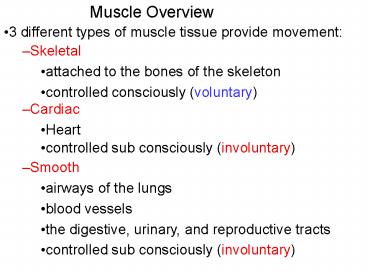
Muscle Overview
- 3 different types of muscle tissue provide
movement
- Skeletal
- attached to the bones of the skeleton
- controlled consciously (voluntary)
- Cardiac
- Heart
- controlled sub consciously (involuntary)
- Smooth
- airways of the lungs
- blood vessels
- the digestive, urinary, and reproductive tracts
- controlled sub consciously (involuntary)
2
Characteristics of Muscle Tissue
- Excitability, or irritability
- the ability to receive and respond to stimuli
- Conductivity
- the ability to create and conduct an action
potential along the cell membrane
- Contractility
- the ability to shorten forcibly
- Extensibility
- the ability to be stretched or extended
- Elasticity
- the ability to recoil after being stretched
3
Muscle Terminology
- Prefixes
- sarco- flesh
- Sarcolemma muscle plasma membrane
- Sarcoplasm cytoplasm of a muscle fiber (cell)
- my- muscle
- Myocyte muscle fiber
- Epimysium the sheath of connective tissue that
surrounds a skeletal muscle
4
Skeletal Muscle Gross Anatomy
5
Skeletal Muscle Gross Anatomy
- Three connective tissue sheaths surround a muscle
and holds the cells together
- Epimysium
- connective tissue on the outside of the muscle
- Perimysium
- connective tissue that surrounds a group of
muscle fibers - organizes muscle fibers into fascicles
- Endomysium
- connective tissue inside the muscle that wraps
around an individual muscle fiber
6
Motor Unit The Nerve-Muscle Functional Unit
- A skeletal fiber will contract only after it is
excited - stimulated to generate an action potential
-A skeletal fiber stimulated by the exocytosis of
neurotransmitters from a motor neuron at a
synapse called the neuromuscular junction (NMJ)
-generates an action potential in the
skeletal fiber which triggers contraction
-A single motor neuron is capable of stimulating
multiple skeletal muscle fibers
-one axon branches creating multiple axon
termini
-the anatomical relationship between a
motor neuron and all skeletal fibers that it
causes to contract is called a motor unit
7
Motor Unit The Nerve-Muscle Functional Unit
8
- The number of muscle fibers per motor unit can
range - few (small motor unit)
-control fine movements (fingers,
eyes)
- several hundred (large motor unit)
- large weight-bearing muscles (back)
- control gross movements (arms, legs)
9
Neuromuscular Junction
- The axon termini have synaptic vesicles that
contain the neurotransmitter acetylcholine (ACh)
-ACh receptors (ligand-gated Na channels) are
localized to a portion of the sarcolemma called
the motor end plate
10
Neuromuscular Junction
11
Neuromuscular Junction
12
Muscle Twitch
-The contraction followed by the relaxation of a
muscle fiber to a single, brief threshold
stimulus by a motor neuron is called a twitch
-There are three phases of a muscle twitch
- Latent (lag) period
- time between the stimulation by a motor neuron
and the beginning of contraction (few
milliseconds)
- Contractile period
- contractile proteins within the fiber hydrolyze
ATP causing the fiber to shorten resulting in an
increase in tension (force)
13
- Relaxation period
- fiber lengthens causing tension to decrease
14
Muscle Twitch
15
Contraction of Skeletal Muscle
-The two types of muscle contractions are
- Isometric contraction same length
- muscle contracts and produces tension, but does
not shorten - trying to lift a car
- Isotonic contraction same tension
- muscle contracts and produces tension
- shortens as it contracts
- lifting a pencil
16
Isometric Contractions
- Tension increases to the muscles capacity, but
the muscle neither shortens nor lengthens - Occurs if the load is greater than the tension
the muscle is able to develop
17
Isotonic Contractions
- In isotonic contractions, the muscle changes in
length and moves the load
18
Variety of Muscle Responses
-Variations in the force of muscle contraction is
required for proper control of skeletal movement
-moving a pencil vs. a textbook with your
hand uses the same muscles, but requires a
different amount of force
- Skeletal muscle contractions are varied by
- altering the frequency of muscle stimulation
-determined by the frequency of
action potentials traveling down a motor neuron
arriving at a fiber
- altering the number of muscle fibers that will
contract
- determined by the number of motor units that are
propagating action potentials to a muscle
19
Muscle Response Stimulation Frequency
- A single stimulus results in a single muscle
twitch producing a constant amount of tension
20
Muscle Response Stimulation Frequency
- More rapidly delivered stimuli result in the
summation of muscle twitches resulting in an
incomplete tetanus - muscle tension does not return to baseline
- If stimuli are given quickly enough, complete
tetanus is observed where the contractile force
reaches a maximum, but individual twitches
blended together
21
Muscle Response Stimulation Strength
- The number of muscle fibers actively contracting
determines the force that muscle produces - directly correlated to the number of active motor
units
- The first observable muscle contraction occurs
following a threshold stimulus - activates one motor unit
- As stimulus strength is increased more motor
units are activated - recruitment
- The maximum force that a muscle is capable of
generating is reached when all motor units are
activated - an increase in stimulus intensity results in no
further increase in force generated
22
Types of Skeletal Muscle Fibers
- There are 3 different types skeletal muscle
fibers based on the duration of a twitch and the
method of ATP production
- slow oxidative fibers
- fast oxidative fibers
- fast glycolytic fibers
- Skeletal muscles of your body contain a
combination of all three fiber types, but their
ratio determines the overall function of that
muscle
23
Oxidative vs. Glycolytic fibers
-Oxidative fibers contain greater amounts of
mitochondria compared to glycolytic fibers
- Oxidative fibers contain an oxygen-binding
protein called myoglobin to maintain a high
concentration of oxygen within the fiber for
aerobic respiration
- similar in structure to the blood protein
hemoglobin - provides a red color to oxidative fibers
- a lack of myoglobin in glycolytic fibers results
in a white color
24
Characteristics of Skeletal Muscle Fiber Types
- Slow oxidative fibers
- have a slow twitch (use ATP slowly)
- fatigue resistant
- muscle fibers used to maintain posture
- Fast oxidative fibers
- have a fast twitch (use ATP quickly)
- moderate resistance to fatigue
- muscle fibers used for non-exertive movement
(walking)
- Fast glycolytic fibers
- have a fast twitch (use ATP quickly)
- easily fatigued
- muscle fibers used for powerful movements
(jumping and sprinting)
25
Microscopic Anatomy of a Skeletal Muscle Fiber
-Each fiber is long (up to 30 cm) and cylindrical
with multiple nuclei just beneath the sarcolemma
- the sarcolemma contains both voltage-gated Na
and K capable of generating an action potential
- portions of the sarcolemma called transverse (t)
-tubules fold inward toward the center of the
fiber - propagate APs to the center of the muscle cell
- Muscle fibers contain an elaborate, smooth
sarcoplasmic (endoplasmic) reticulum (SR)
- physically associated with the t-tubules
- storage site of intracellular calcium (Ca2)
26
- An action potential in the t-tubules causes the
release of Ca2 from the SR into the
sarcoplasm which increases the cytoplasmic level
of Ca2 - triggers the contraction of a muscle fiber
27
Skeletal Muscle
- Has repeating pattern of contractile proteins
- (striations)
- Easily fatigable (tired)
28
Microscopic Anatomy of a Skeletal Muscle Fiber
29
Contractile Proteins
-The contractile proteins (myofilaments) are
arranged within the sarcoplasm in long bundles
called myofibrils
- composed of 2 types of myofilaments that overlap
and slide past one another during contraction and
relaxation - thin
- thick
- The arrangement of contractile proteins within
myofibrils creates a repeating pattern of
striations called sarcomeres
- the basic/repeating contractile unit of a muscle
- thousands of sarcomeres per myofibril
30
Myofilaments Banding Pattern
-The overlapping arrangement of thin and thick
filaments in a sarcomere creates an ordered
banding pattern within a single sarcomere
- Z disc
- constitutes one end of a sarcomere
- anchors the thin filaments
- A band
- the length of the thick filaments
- I band
- the length of thin filaments within a sarcomere
that is not overlapping with the thick filaments
- H (bare) zone
- the length of thick filaments within in a
sarcomere that is not overlapping with the thin
filaments
31
Microscopic Anatomy of a Skeletal Muscle Fiber
32
Sarcomeres
33
Arrangement of the Filaments in a Sarcomere
34
Sliding Filament Model of Contraction
-In the relaxed state, thin and thick filaments
overlap only slightly
- Upon stimulation, the thick filaments pull the
thin filaments toward the center of the sarcomere
- filaments overlap to a greater degree
- shortening the sarcomere
- As all of the sarcomeres in a muscle shortens,
the entire muscle shortens
35
Sliding Filament Model of Contraction
36
Structure of Thin Filaments
- Thin filaments are composed of 3 proteins
- Actin is a helical polymer of protein subunits
- each subunit contains a binding site for the
myosin head
- Tropomyosin blocks the interaction between actin
and myosin - prevents an unstimulated muscle from contracting
- Troponin C is attached to tropomyosin
- binds to Ca2 in the sarcoplasm during
contraction
37
Structure of Thin Filaments
38
Structure of Thick Filaments
- Thick filaments are composed of many molecules of
the protein myosin
-Each myosin protein has a rodlike tail and two
heads
- Myosin heads (also known as cross bridges)
- hydrolyze a molecule of ATP
- uses the chemical energy to contract
- Temporarily bind to actin
- pull on actin causing the shortening sarcomere
39
Structure of Thick Filaments
40
Skeletal Muscle Contraction
-In order to contract, a skeletal muscle must be
stimulated by a motor neuron
- generates an action potential in the muscle fiber
- causes an increase in the amount of cytoplasmic
Ca2
- causes the muscle fiber to contract
-Linking the action potential to the contraction
of a muscle fiber is called excitation-contraction
coupling
41
Excitation-Contraction Coupling
42
Excitation-Contraction Coupling
- Binding of ACh to its receptors opens the channel
and allows both Na and K to diffuse - diffusion of more Na than K causes the membrane
potential to depolarize (endplate potential)
43
Excitation-Contraction Coupling
- The endplate potential brings the membrane
potential to threshold - opens voltage-gated Na and K channels to
generate an action potential in the sarcolemma
44
Excitation-Contraction Coupling
- Action potentials propagate along the sarcolemma
into the t-tubules - action potentials in the t-tubules cause the
release of Ca2 from the SR into the sarcoplasm
45
Excitation-Contraction Coupling
- Ca2 in the sarcoplasm binds to troponin C
- changes the position of troponin C
- moves tropomyosin away from the myosin binding
site on actin
46
Events of Contraction (Cross bridge cycling)
- Muscle fiber shortening occurs as myosin pulls on
actin in a repetitive ratcheting fashion
- Thin filaments move toward the center of the
sarcomere
- Activation of the myosin head
- a molecule of ATP is hydrolyzed and the energy is
used by the myosin head to change the shape of
myosin into the high-energy state
- Cross bridge formation
- myosin cross bridge attaches to actin filament
- Power stroke
- myosin head pivots and pulls thin filament over
thick filament
- Cross bridge detachment
- The binding of a molecule of ATP to the myosin
head causes it to detach from actin
47
Events of Contraction (Cross bridge cycling)
48
Muscle Fiber Relaxation
- The motor neuron stops the exocytosis of ACh
- The remaining ACh is hydrolyzed into acetate and
choline by the enzyme Acetylcholine esterase
located in the synaptic cleft of the NMJ - ACh receptors close
- membrane potential returns to resting value
49
Muscle Fiber Relaxation
- Ca2 is pumped back into the SR by Ca2-ATPase in
the SR membrane - decreases Ca2 in the sarcoplasm
- troponin C moves back to resting position
- Tropomyosin covers the binding site for myosin on
G actin
50
Skeletal Muscle Tissue
- Packaged in skeletal muscles that attach to and
cover the bony skeleton - Has obvious stripes called striations
- Is controlled voluntarily (i.e., by conscious
control) - Contracts rapidly but tires easily
- Is responsible for overall body motility
- Is extremely adaptable and can exert forces
ranging from a fraction of an ounce to over 70
pounds
51
Skeletal Muscle
- -Striated
- -Multinucleated
- -Cells not defined
52
Cardiac Muscle Tissue
- Occurs only in the heart
- Is striated like skeletal muscle but is not
voluntary (involuntary) - Contracts at a fairly steady rate set by the
hearts pacemaker - Neural controls allow the heart to respond to
changes in bodily needs
53
Cardiac Muscle
- -Striated
- -Defined cells with single nucleus
- only rests between beats
54
Smooth Muscle Tissue
- Found in the walls of hollow visceral organs,
such as the stomach, urinary bladder, and
respiratory passages - Forces food and other substances through internal
body channels - It is not striated and is involuntary
55
Skeletal Muscle Nerve and Blood Supply
- Each muscle is served by one nerve, an artery,
and one or more veins - Each skeletal muscle fiber is supplied with a
nerve ending that controls contraction - Contracting fibers require continuous delivery of
oxygen and nutrients via arteries - Wastes must be removed via veins
56
Skeletal Muscle Attachments
- Most skeletal muscles span joints and are
attached to bone in at least two places - When muscles contract on the movable bone, the
muscles insertion moves toward the immovable
bone, the muscles origin
57
Skeletal Muscle Attachments
- Muscles attach
- Directly epimysium of the muscle is fused to
the periosteum of a bone - Indirectly connective tissue wrappings extend
beyond the muscle as a tendon or aponeurosis
Recommended
CrystalGraphics Presentations


