
Inception SMX-225CT - PowerPoint PPT Presentation
Title: Inception SMX-225CT
1
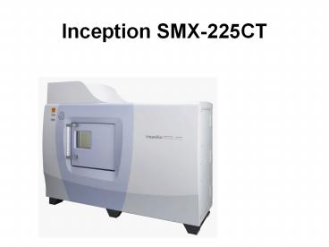
Inception SMX-225CT
2
inspeXio sm25C
- First in the Industry CT Scan Auto-Positioning
via Exterior Camera True 3D Non-Destructive
Testing Machine - Featuring a proprietary microfocus X-ray tube and
high-sensitivity ima - inspeXio SMX-225CT is Shimadzu's top-of-the-line
microfocus X-ray CT - system. An intuitive user interface ensures the
easy acquisition of clear CT - images of a sample's internal structure.
Previously impossible with earlier - inspection machines, the SMX-225CT's wide sample
stage allows easy - inspection of large samples and covers a variety
of applications, from - miniaturized electronic devices to aluminum
die-casting for automobiles. - Equipped with a wide range of useful functions,
inspeXio SMX-225CT is the - highest grade of CT inspection machine.
3
- Shimadzu's microfocus X-ray tube provides
high-power output to penetrate samples, while the
collimated X-ray focus acquires clear images,
even at high magnification. - Improved image intensifier lenses enhance image
quality, and Shimadzu's continuous improvements
to major system components ensure beautiful,
clear images that satisfy every customer's
highest expectations.
- Conventional instruments typically demanded both
complex operation and a high degree of operator
skill. - Now, our calibration-free operation eliminates
the need for laborious calibration work. - And determining the imaging position couldn't be
easier. Positioning based on the exterior camera
image and Easy Zoom functions, ensure imaging
operations are simple.
- A range of convenient functions significantly
reduce the time required prior to imaging. An
ultrafast CT image-processing engine provides
astonishingly fast processing, while a 3D
cone-beam CT quickly creates 3D views of internal
structures in a matter of moments.
4
Feature
Exterior Camera for Intuitive Manipulation
(Patent Pending)
Exterior camera image lets you easily decide the
CT imaging position. No need for complex
operations or special operator skills.
? 300mm
H 300mm
Step 1
Load the sample on the stage. (Max. sample size
300 mm dia. x 300 mm H)
Min.field-of-view
Use the mouse to specify the CT scan area on the
exterior camera image.
Curent.field-of-view
Max.field-of-view
Click
Set the CT parameters and start the scan.
CT scan is completed
RESULT
5
Result Tomographic image is displayed.
Exterior Camera
Detector
X-Ray Tube
CT Stage
6
- Easy Zoom (Patent Pending)
- After the initial CT, just a few simple steps
produce CT scan of your area of interest.
- Align the CT scan area with the region of
interest. - (This synchronizes stage movement.)
- Display the current CT scan area on the MPR
screen.
- Result Enlarged tomographic image of the region
of interest is displayed.
- Close the MPR screen. Set the CT parameters and
start the CT scan.
7
Software
Filled with Useful Software
- MPR Display Function
- Obtained by placing CT data at equal intervals
along the Z-axis in virtual space, the
"Multi-Planar Reconstruction (MPR) function
renders virtual 3D data as cross-sections
(oblique or double-oblique images) for any
horizontal or vertical position.
Oblique image (1) formed by cross-section on
plane B
CT image -D-line shows position of CT image in
top-left frame
Oblique image (2) formed by Double-oblique image
formed by cross-section on plane A
Double-oblique image formed by cross-section on
plane C
8
- VGStudio
- This software reconstructs slice images taken by
the CT unit to create a 3D representation. It
fully exploits the hardware performance to
visualize and analyze images using high-speed
volume rendering, even creating animations that
rotate the visualized 3D image.
- X-Ray Image Viewer
- 2D image analysis software that offers
sophisticated image processing.
Brightness profile display
Image display
View of arbitrary image plane
Angle/distance measurements from image
9
Safety Measures
Door Interlock Circuit (X-Ray) This
double-interlock circuit is installed in the
sliding door to prevent X-ray emission while the
door is open. Door Interlock Circuit (CT
Stage) Allows stage operation to be stopped while
the sliding door is open. Collision
Sensor Installed near the X-ray source unit, this
collision senso?prevents collisions between the
CT stage and the X-ray generator.
- Sliding-Door Safety Mechanism Prevents Injuries
This safety mechanism protects against pinched
fingers in the sliding door. To close the door,
turn the door safety lock counterclockwise with
the left hand, grip the door handle with the
right hand and gently slide it to the left.
This safety mechanism protects against pinched
fingers in the sliding door. To close the door,
turn the door safety lock counterclockwise with
the left hand, grip the door handle with the
right hand and gently slide it to the left.
Object
Movement Limit Function Use this convenient
software function to enter the size of the sample
and prevent collisions. Limit Area
Human
Human
Human
Human
Human
Human
10
- Applications
Electronic Components
Digital camera
11
Electronic Components
Cylindrical lithium cell
Prismatic lithium cell Electrolytic
capacitor
12
Electronic Components
13
- Applications
Automobile Parts
14
Other Applications
15
Specifications
Model inspeXio SMX-225CT inspeXio
SMX-90CT(Benchtop Type) P/N 362-69100-02 362-65000
-52 X-ray generator Non-enclosure tube type,
Maximum tube voltage 225 kV, Maximum tube
current 1000 A, Maximum output 135 W Sealed
tube type, Maximum tube voltage 90 kV, Maximum
tube current 250 A, Maximum output 10 W X-ray
detector Image intensifier Digital flat panel
detector Maximum stroke of SOD axis (1) 670
mm SOD axis (1) 200 mm CT stage SID axis(2)
600 (400 to 1000)Z axis 300 mm Z axis 50 mm
max. Maximum sample size 300 mm (dia.) H300 mm, 9
kg 160 mm (dia.) H100 mm, 4 kg CT
field-of-view(FOV) 200 mm (dia.) 50 mm
(dia.) Scanning methods Normal Scan, Half Scan,
Offset Scan, FS Scan (3), 2DCT (4), CBCT
(5) Offset CBCT CT Data acquisition time Any
value from 10 seconds to 30 minutes. Any value
from 40 seconds to 30 minutes. CT reconstruction
matrix 512 x 512,1024 x 1024, 2048 x 2048, 4096 x
4096 512x512 1024x 1024 Size and Weight of shield
box W 2,170 x D 1,350 x H 1,860 mm, 3,100 kg W
830 x D 601 x H 587 mm, 250 kg Size and Weight of
specialized desk W 1,200 x D 700 x H 1,270 mm, 50
kg W 1,500 x D 750 x H 740 mm, 60 kg Power
supply Main unit AC 200 V 10 , 50/60 Hz, 3
kVA Computer AC 100 V 10 , 50/60 Hz, 1 kVA AC
100 V 10 50/60 Hz, 1 kVA X-ray leakage rate 1
uSv max. 1 uSv/h max.
(1) Abbreviation for Source to Object Distance.
It is the distance from X-ray focus point to a
sample. (2) Abbreviation for Source to Image
Distance. It is the distance from X-ray focus
point to a detector. (3) Abbreviation for
Fan-Shaped Scan. It is a scanning method that
obtains CT images at rotation angles of 60, 90,
and 120 degrees. (4) Abbreviation for
2-Dimensions Computed Tomography. It is the
scanning method which obtains CT image of one
slice or three slices with one scan. (5)
Abbreviation for Cone Beam Computed Tomography.
It is the scanning method which obtains CT images
of hundreds of slices with one scan.
16
Special large CT scan area model
Comply with all local regulations when installing
an SMX system.
Accommodates samples up to 500 mm dia. x 500 mm
height Max. CT field-of-view 200 mm dia.
Recommended
CrystalGraphics Presentations


