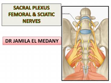
SACRAL PLEXUS FEMORAL - PowerPoint PPT Presentation
1 / 29
Title:
SACRAL PLEXUS FEMORAL
Description:
... To iliacus (flexor of hip joint). In lower limb: ... Sciatic nerve: to lower limb. SCIATIC NERVE ... FOOT DROP It is a peripheral nerve injury that affects a ... – PowerPoint PPT presentation
Number of Views:150
Avg rating:3.0/5.0
Title: SACRAL PLEXUS FEMORAL
1
- SACRAL PLEXUS FEMORAL SCIATIC NERVES
DR JAMILA EL MEDANY
2
OBJECTIVES
- By the end of the lecture, students should be
able to - Describe the formation of sacral plexus (site
root value). - List the main branches of sacral plexus.
- Describe the course of the femoral the sciatic
nerves - List the motor and sensory distribution of
femoral sciatic nerves. - Describe the effects of lesion of the femoral
the sciatic nerves (motor sensory).
3
LUMBAR PLEXUS
- Formation
- Ventral (anterior) rami of the upper 4 lumbar
spinal nerves (L1,2,3 and L4). - Site Within the substance of the psoas major
muscle. - Main branches
- Iliohypogastric ilioinguinal to anterior
abdominal wall. - Obturator to medial (adductor) group of the
thigh. - Femoral to anterior group of the thigh.
4
FEMORAL NERVE
Femoral N
- Origin
- from lumbar plexus (L2,3,4).
- Course
- Descends lateral to psoas major enters the
thigh behind the inguinal ligament. - Passes lateral to femoral artery divides into
anterior posterior divisions.
5
MUSCULAR BRANCHES OF FEMORAL NERVE
- In abdomen
- To iliacus (flexor of hip joint).
- In lower limb
- To anterior compartment of the thigh
- Flexors of hip joint
- sartorius pectineus
- Extensors of knee joint
- quadriceps femoris.
P
S
6
CUTANEOUS BRANCHES OF FEMORAL NERVE
- To antero-medial aspect of the thigh.
- To medial side of knee, leg and foot (saphenous
nerve).
7
INJURY OF THE FEMORAL NERVE
- MOTOR EFFECT
Iliacus
Pectinus
Paralysis of Movement affected
Iliacus Flexion of the hip
Sartorius Flexion and abduction of the hip
Pectineus Flexion and adduction of the hip
Quadriceps femoris Extension of the knee
sartorius
Quadriceps
- SENSORY EFFECT
- Loss of sensation of the areas supplied by
femoral nerve.
8
FEMORAL NERVE INJURY
- MOTOR MANIFESTATION
- Wasting of quadriceps femoris.
- Loss of extension of knee.
- Weak flexion of hip (psoas major is intact).
- SENSORY MANIFESTATION
- loss of sensation over areas supplied
(antero-medial) aspect of thigh medial side of
leg foot.
9
SACRAL PLEXUS
- Formation
- By the ventral (anterior) rami of a part of
L4 whole L5 (lumbosacral trunk) S1,2,3 and
most of S 4. - Site
- in front of the piriformis muscle.
10
SACRAL PLEXUS
- Main branches
- Pelvic splanchnic nerves are the sacral part of
the parasympathetic system and arise from the
second, third, and fourth sacral nerves. - They are distributed to the pelvic viscera.
- Pudendal nerve to perineum.
- Sciatic nerve to lower limb.
11
SCIATIC NERVE
- It is the largest nerve of the body.
- Origin
- Sacral plexus (L4,5, S1, 2,3).
- Course
- Leaves the pelvis through greater sciatic
foramen, below piriformis passes in the gluteal
region (between ischial tuberosity greater
trochanter) then to posterior compartment of
thigh. - Termination
- Divides into tibial common peroneal (fibular)
nerves in the middle of the back of the thigh
12
TIBIAL NERVE
- Course
- Descends through popliteal fossa to the posterior
compartment of leg, accompanied with posterior
tibial vessels. - Passes deep to flexor retinaculum (behind the
medial malleolus) to reach the sole of foot where
it divides into 2 terminal branches, (Medial
Lateral planter nerves.
13
COMMON PERONEAL (FIBULAR) NERVE
- Course
- Leaves popliteal fossa close to the lateral
aspect of neck of the fibula. - Then divides into
- Superficial peroneal descends into lateral
compartment of leg. - Deep peroneal descends into anterior compartment
of leg.
14
MUSCULAR BRANCHES OF THE SCIATIC NERVE
- To Hamstrings (flexors of knee extensors of
hip). - To all muscles in the leg foot through
- Common peroneal
- TO Muscles of anterior lateral
compartments of leg (Dorsiflexors of ankle,
Extensors of toes, Evertors of foot). - 2. Tibial
- TO Muscles of posterior compartment of leg
intrinsic muscles of sole (Planterflexors of
ankle, Flexors of toes, Invertors of foot).
15
Cutaneous BRANCHES OF SCIATIC NERVE
- To all leg foot
- EXCEPT
- areas supplied by saphenous nerve (blue), branch
of femoral nerve.
16
CAUSES OF SCIATIC NERVE INJURY
- The sciatic nerve is most frequently injured by?
- I- Badly placed intramuscular injections in the
gluteal region. - To avoid this, injections into the gluteus
maximus or medius should be made into the upper
outer quadrant of the buttock. - Most nerve lesions are incomplete, and in 90 of
injuries, the common peroneal (part of the nerve)
is the most affected. Why? - - The common peroneal nerve fibers lie
superficial in the sciatic nerve.
II-Posterior dislocation of the hip joint
17
- The following clinical features are present
- Motor
- The hamstring muscles are paralyzed, but weak
flexion of the knee is possible. Why? - - because of the action of the sartorius
(femoral nerve) and gracilis (obturator nerve). - All the muscles below the knee are paralyzed, and
the weight of the foot causes it to assume the
plantar-flexed position, or Foot Drop.
18
FOOT DROP
- It is a peripheral nerve injury that affects a
patients ability to lift the foot at the ankle.
While foot drop injury is a neuromuscular
disorder, it can also be a symptom of a more
serious injury, such as a nerve compression or
herniated disc. - Symptoms of foot drop
- Inability to point toes toward the body (dorsi
flexion) - Pain
- Weakness
- Numbness (on the shin or top of the foot)
- Loss of function of foot
- High-stepping walk (called Steppage
gait or Footdrop Gait)
19
- SENSORY MANIFESTATION
- Sensation is lost below the knee, except for a
narrow area down the medial side of the lower
part of the leg and along the medial border of
the foot as far as the ball of the big toe, which
is supplied by the saphenous nerve (femoral
nerve).
20
SCIATICA
- Sciatica describes the condition in which
patients have pain along the sensory distribution
of the sciatic nerve. - Thus the pain is experienced in the posterior
aspect of the thigh, the posterior and lateral
sides of the leg, and the lateral part of the
foot.
21
- Sciatica can be caused by
- Prolapse of an intervertebral disc, with pressure
on one or more roots of the lower lumbar and
sacral spinal nerves, - Pressure on the sacral plexus or sciatic nerve by
an intrapelvic tumor, or - Inflammation of the sciatic nerve or its terminal
branches.
22
Common Peroneal Nerve Injury
- The common peroneal nerve is in an exposed
position as it leaves the popliteal fossa it
winds around neck of the fibula to enter peroneus
longus muscle, (Dangerous Position).
The common peroneal nerve is commonly injured In
Fractures of the neck of the fibula and By
pressure from casts or splints.
23
Common Peroneal Nerve Injury
- The following clinical features are present
- Motor
- The muscles of the anterior and lateral
compartments of the leg are paralyzed, - As a result, the opposing muscles, the plantar
flexors of the ankle joint and the invertors of
the subtalar joints, cause the foot to be Plantar
Flexed (Foot Drop) and Inverted, an attitude
referred to as Equinovarus.
24
Tibial Nerve Injury
Complete division results in the following
clinical features Motor All the muscles in the
back of the leg and the sole of the foot are
paralyzed. The opposing muscles Dorsiflex the
foot at the ankle joint and Evert the foot at
the subtalar joint, an attitude referred to as
Calcaneovalgus.
- The tibial nerve leaves the popliteal fossa by
passing deep to the gastrocnemius soleus. - Because of its deep and protected position, it is
rarely injured.
25
SUMMARY
- The lumbar plexus is formed by ventral (anterior)
rami of L1,2,3 and most of L4, in the substance
of psoas major muscle. - The sacral plexus is formed by ventral (anterior)
rami of a part of L4 whole L5 (lumbosacral
trunk) S1,2,3 and most of S4, in front of
piriformis msucle. - The femoral nerve, a branch of lumbar plexus
(L2,3,4). Its injury leads to weak flexion of hip
loss of extension of knee as well as loss of
sensation of skin of antero-medial aspects of the
thigh, medial side of knee, leg and foot.
26
SUMMARY
- The sciatic nerve is a branch of sacral plexus
(L4,5, S1,2,3). Its injury leads to affection of
Flexion of knee, Extension of hip, all movements
of leg foot, as well as loss of sensation of
skin of leg foot (Except areas supplied by
saphenous branch of femoral nerve).
27
SCIATIC NERVE INJURY
- MOTOR EFFECT
- Marked wasting of the muscles below the knee.
- Weak flexion of the knee (sartorius gracilis
are intact). - Weak extension of hip (gluteus maximus is
intact). - The foot assumes the position of Foot Drop
(planter flexed position) by its weight. - SENSORY EFFECT
- Loss of sensation below knee (EXCEPT medial side
of leg foot).
28
Test your knowledge!
- Which of the following is supplied by the femoral
nerve ? - Extensors of hip.
- Skin of dorsum of foot.
- Hamstrings.
- Extensors of knee.
- Injury of common peroneal nerve leads to
- Loss of dorsiflexion of ankle.
- Loss of inversion of foot.
- Loss of extension of knee.
- Loss of flexion of toes.
29
THANK YOU


