
Nerve activates contraction - PowerPoint PPT Presentation
1 / 45
Title:
Nerve activates contraction
Description:
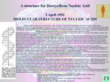
A structure for Deoxyribose Nucleic Acid 2 April 1953 MOLECULAR STRUCTURE OF NUCLEIC ACIDS We wish to suggest a structure for the salt of deoxyribose nucleic acid (D ... – PowerPoint PPT presentation
Number of Views:37
Avg rating:3.0/5.0
Title: Nerve activates contraction
1
A structure for Deoxyribose Nucleic Acid
2 April 1953MOLECULAR STRUCTURE OF NUCLEIC ACIDS
We wish to suggest a structure for the salt of
deoxyribose nucleic acid (D.N.A.). This structure
has novel features which are of considerable
biological interest. A structure for nucleic
acid has already been proposed by Pauling and
Corey (1). They kindly made their manuscript
available to us in advance of publication. Their
model consists of three intertwined chains, with
the phosphates near the fibre axis, and the bases
on the outside. In our opinion, this structure is
unsatisfactory for two reasons (1) We believe
that the material which gives the X-ray diagrams
is the salt, not the free acid. Without the
acidic hydrogen atoms it is not clear what forces
would hold the structure together, especially as
the negatively charged phosphates near the axis
will repel each other. (2) Some of the van der
Waals distances appear to be too small. Another
three-chain structure has also been suggested by
Fraser (in the press). In his model the
phosphates are on the outside and the bases on
the inside, linked together by hydrogen bonds.
This structure as described is rather
ill-defined, and for this reason we shall not
comment on it. We wish to put forward a radically
different structure for the salt of deoxyribose
nucleic acid. This structure has two helical
chains each coiled round the same axis (see
diagram). We have made the usual chemical
assumptions, namely, that each chain consists of
phosphate diester groups joining
ß-D-deoxyribofuranose residues with 3',5'
linkages. The two chains (but not their bases)
are related by a dyad perpendicular to the fibre
axis. Both chains follow right- handed helices,
but owing to the dyad the sequences of the atoms
in the two chains run in opposite directions.
Each chain loosely resembles Furberg's2 model No.
1 that is, the bases are on the inside of the
helix and the phosphates on the outside. The
configuration of the sugar and the atoms near it
is close to Furberg's 'standard configuration',
the sugar being roughly perpendicular to the
attached base. There is a residue on each every
3.4 A. in the z-direction. We have assumed an
angle of 36 between adjacent residues in the
same chain, so that the structure repeats after
10 residues on each chain, that is, after 34 A.
The distance of a phosphorus atom from the fibre
axis is 10 A. As the phosphates are on the
outside, cations have easy access to them. The
structure is an open one, and its water content
is rather high. At lower water contents we would
expect the bases to tilt so that the structure
could become more compact. The novel feature of
the structure is the manner in which the two
chains are held together by the purine and
pyrimidine bases. The planes of the bases are
perpendicular to the fibre axis. The are joined
together in pairs, a single base from the other
chain, so that the two lie side by side with
identical z-co-ordinates. One of the pair must be
a purine and the other a pyrimidine for bonding
to occur. The hydrogen bonds are made as follows
purine position 1 to pyrimidine position 1
purine position 6 to pyrimidine position 6. If it
is assumed that the bases only occur in the
structure in the most plausible tautomeric forms
(that is, with the keto rather than the enol
configurations) it is found that only specific
pairs of bases can bond together. These pairs are
adenine (purine) with thymine (pyrimidine), and
guanine (purine) with cytosine (pyrimidine). In
other words, if an adenine forms one member of a
pair, on either chain, then on these assumptions
the other member must be thymine similarly for
guanine and cytosine. The sequence of bases on a
single chain does not appear to be restricted in
any way. However, if only specific pairs of bases
can be formed, it follows that if the sequence of
bases on one chain is given, then the sequence on
the other chain is automatically determined. It
has been found experimentally (3,4) that the
ratio of the amounts of adenine to thymine, and
the ration of guanine to cytosine, are always
bery close to unity for deoxyribose nucleic
acid. It is probably impossible to build this
structure with a ribose sugar in place of the
deoxyribose, as the extra oxygen atom would make
too close a van der Waals contact. The previously
published X-ray data (5,6) on deoxyribose nucleic
acid are insufficient for a rigorous test of our
structure. So far as we can tell, it is roughly
compatible with the experimental data, but it
must be regarded as unproved until it has been
checked against more exact results. Some of these
are given in the following communications. We
were not aware of the details of the results
presented there when we devised our structure,
which rests mainly though not entirely on
published experimental data and stereochemical
arguments. It has not escaped our notice that the
specific pairing we have postulated immediately
suggests a possible copying mechanism for the
genetic material. Full details of the structure,
including the conditions assumed in building it,
together with a set of co-ordinates for the
atoms, will be published elsewhere. We are much
indebted to Dr. Jerry Donohue for constant advice
and criticism, especially on interatomic
distances. We have also been stimulated by a
knowledge of the general nature of the
unpublished experimental results and ideas of Dr.
M. H. F. Wilkins, Dr. R. E. Franklin and their
co-workers at King's College, London. One of us
(J. D. W.) has been aided by a fellowship from
the National Foundation for Infantile Paralysis.
J. D. WATSON F. H. C. CRICK Medical Research
Council Unit for the Study of Molecular Structure
of Biological Systems, Cavendish Laboratory,
Cambridge. April 2. 1. Pauling, L., and Corey, R.
B., Nature, 171, 346 (1953) Proc. U.S. Nat.
Acad. Sci., 39, 84 (1953). 2. Furberg, S., Acta
Chem. Scand., 6, 634 (1952). 3. Chargaff, E.,
for references see Zamenhof, S., Brawerman, G.,
and Chargaff, E., Biochim. et Biophys. Acta, 9,
402 (1952). 4. Wyatt, G. R., J. Gen. Physiol.,
36, 201 (1952). 5. Astbury, W. T., Symp. Soc.
Exp. Biol. 1, Nucleic Acid, 66 (Camb. Univ.
Press, 1947). 6. Wilkins, M. H. F., and Randall,
J. T., Biochim. et Biophys. Acta, 10, 192 (1953).
2
Rosalind Franklin
We wish to suggest a structure for the salt of
deoxyribose nucleic acid (D.N.A.). This structure
has novel features which are of considerable
biological interest."-- James Watson and
Francis Crick, in a brief letter to the journal
Nature, April 2, 1953
3
The search for genetic material lead to DNA
Genetic Material DNA or Protein?????
4
- Frederick Griffith (1928)
- Transformation- a change in genotype and
phenotype due to the assimilation of a foreign
substance (now known to be DNA) by a cell.
5
- Oswald Avery discovers DNA as the transforming
substance (1944)
6
- Hershey and Chase 1952
- Bacteriophage virus that infects bacteria
7
(No Transcript)
8
(No Transcript)
9
Known so far
- DNA is polymer made of Nucleotides
- Nucleotides have sugar-phosphate-and a nitrogen
base - Nitrogen bases can be Adenine, Guanine, Cytosine,
or Thymine - Not known - how the monomers connect to make a
molecule that can carry genetic material
10
Relative Proportions () of Bases in
DNA Organism A T G C Human 30.9 29.41
9.91 9.8 Chicken 28.8 29.2 20.5 21.5
Grasshopper 29.3 29.3 20.5 20.7 Sea
Urchin 32.8 32.1 17.7 17.3 Wheat 27.3 27.1 22.
7 22.8 Yeast 31.3 32.9 18.7 17.1 E.
Coli 24.7 23.6 26.0 25.7
11
Chargaffs Rule (1947)
- DNA composition varies in different species
- In any one species, all 4 bases are not equal in
number - of A of T (AT)
- of G of C (G C)
- Nucleotide
- Sugar (deoxyribose)
- Phosphate
- Nitrogenous base (Adenine, Guanine, Cytosine, or
Thymine)
12
- DNA a double helix
13
Hydrogen Bonding
- Between purine and pyrimidine
- Purines are Adenine and Guanine
- Pyrimidines are Thymine and Cytosine
- A and T
- G and C
14
Sugar Phosphate Backbone
- The phosphate group of one nucleotide is attached
to the sugar of the next nucleotide in line. - The result is a backbone of alternating
phosphates and sugars, from which the bases
project.
Fig. 16.3
15
Sugar Phosphate Backbone
- The 2 strands are antiparallel (5 to 3 in one
strand and 3 to 5 in the other) - 5 end has a P
- 3 end has a -OH
Sugar Phosphate Backbone
16
Double Helix
- 10 bases per turn of helix
- Major and minor grooves sites of protein
interaction - Base complementarity easy replication
17
(No Transcript)
18
(No Transcript)
19
DNA Replication
When does this occur???
Template strands
20
- Semiconservative replication
- - when a double helix replicates, each of the
daughter molecules will have one old strand and
one newly made strand.
21
- Meselson and Stahl (1958)
22
A large team of enzymes and other proteins
carries out DNA replication
- It takes E. coli less than an hour to copy each
of the 5 million base pairs in its single
chromosome and divide to form two identical
daughter cells. - A human cell can copy its 6 billion base pairs
and divide into daughter cells in only a few
hours. - This process is remarkably accurate, with only
one error per billion nucleotides. - More than a dozen enzymes and other proteins
participate in DNA replication.
23
- The replication of a DNA molecule begins at
special sites, origins of replication.
- ORI SITE special DNA sequence recognized by
proteins - 1 ORI Site in bacteria (circular chromosome)
- Many ORI Sites in eukaryotes (replication bubbles
and forks)
24
- 1) Helicase unwinds DNA.
- 5 end has a P
- 3 end has a -OH
- A new DNA strand can only elongate in the 5-gt3
direction.
25
- 2) Leading and Lagging strands are synthesized
by 2 different mechanisms
- Leading strand is synthesized continuously in
5-gt 3 direction towards the replication fork - Lagging strand uses Okazaki fragments short
segments copied away from the fork
26
3)DNA polymerase adds nucleotides to 3 end of
growing strand
27
- DNA polymerases catalyze the elongation of new
DNA at a replication fork using nucleoside
triphosphates
28
So has DNA Polymerase!!
- DNA polymerases cannot initiate synthesis of a
polynucleotide because they can only add
nucleotides to the end of an existing chain that
is base-paired with the template strand.
29
4) Solution use Primase to make a short Primer
- To start a new chain requires a primer, a short
segment of RNA. - The RNA primer is about 10 nucleotides long in
eukaryotes.
30
- Another DNA polymerase later replaces the
primer ribonucleotides with deoxyribonucleotides
complimentary to the template.
31
- Leading strand requires the formation of only a
single primer as the replication fork continues
to separate. - The lagging strand requires formation of a new
primer as the replication fork progresses.
32
5) DNA Ligase
- DNA ligase joins the fragments together.
33
(No Transcript)
34
(No Transcript)
35
Oops, its a wrong base!!!
- One error per 10,000 base pairs.
- DNA polymerase proofreads each new nucleotide
- If there is an incorrect pairing, the enzyme
removes the wrong nucleotide and then resumes
synthesis. - The final error rate is only one per billion
nucleotides.
36
X Rays
- Can cause DNA mutations
37
Cosmic Rays
- Can cause DNA mutations
38
UV Rays
- Can cause DNA mutations
- produces thymine dimers between adjacent thymine
nucleotides.
39
- Each cell continually monitors and repairs its
genetic material, with over 130 repair enzymes
identified in humans.
- In nucleotide excision repair, a nuclease cuts
out a segment of a damaged strand. - The gap is filled in by DNA polymerase and ligase.
40
- In mismatch repair, special enzymes fix
incorrectly paired nucleotides.
41
The ends of DNA molecules are replicated by a
special mechanism
- Repeated rounds of replication produce shorter
and shorter DNA molecules.
This animation is WRONG!
42
Fig. 16.18
43
- The ends of eukaryotic chromosomal DNA molecules,
the telomeres, have special nucleotide sequences. - In human telomeres, this sequence is typically
TTAGGG, repeated between 100 and 1,000 times. - Telomeres protect genes from being eroded through
multiple rounds of DNA replication.
44
(No Transcript)
45
- Telomerase is not present in most cells of
multicellular organisms. - Therefore, the DNA of dividing somatic cells and
cultured cells does tend to become shorter. - Thus, telomere length may be a limiting factor in
the life span of certain tissues and the
organism. - Telomerase is present in germ-line cells,
ensuring that zygotes have long telomeres. - Active telomerase is also found in cancerous
somatic cells. - This overcomes the progressive shortening that
would eventually lead to self-destruction of the
cancer.