
A New Option for Keratoconus - PowerPoint PPT Presentation
1 / 49
Title:
A New Option for Keratoconus
Description:
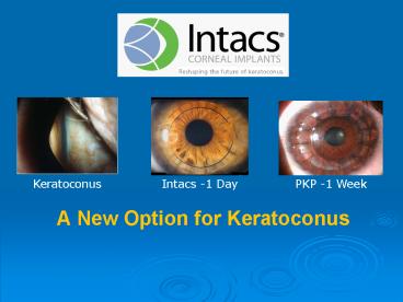
Keratoconus Intacs -1 Day PKP -1 Week A New Option for Keratoconus Objective - Bridge the gap between frustration and (PKP) the point of no return Contact Lens ... – PowerPoint PPT presentation
Number of Views:109
Avg rating:3.0/5.0
Title: A New Option for Keratoconus
1
A New Option for Keratoconus
Keratoconus
Intacs -1 Day
PKP -1 Week
2
Objective - Bridge the gap between frustration
and (PKP) the point of no return
- Contact Lens Intolerant Keratoconus
- Steep K s, 45 to 60
- Changing refractions, eyes irritated, frequent
visits/re-fits - Lenses not providing functional vision
- Outright failure
- Shortened wearing time
- Inability to achieve 20/40
- keratoconus personality exacerbated
- Apprehensive about transplant
- Active, younger or risk averse
3
Reshape the Cornea for CL Success
4
INTACS HistoryConcept for Corneal Reshaping
- Oklahoma optometrist first conceptualized the
idea in 1978 - One of the early medical champions of contact
lenses in the U.S. - Developed CorneaScope in late 1960s - led to
todays topography
Gene Reynolds, O.D. 1921 - 1994
5
How does it work?
Arc-Shortening Model for Treating Myopia
Preoperative Representation of the
Cornea
6
How does it work?
Arc-Shortening Model for Treating Myopia
Representation of the Cornea After
Placement of INTACS Inserts
7
History
1984
Adjustable Ring
As conceived by Dr. Reynolds
8
Milestones
- 1978 A.E. Reynolds, O.D. conceives of
- Intrastromal Corneal Ring (ICR)
- 1985 - First pre-clinical studies on Dr.
Reynolds' product - 1991 - First human clinical trials begun - Brazil
- 1996 - U.S. myopia clinical trial begun, 150º
ICR - - CE Mark approval of ICR in Europe, -1.00 to
-4.50 D - 1997 - Joseph Colin, MD inserts first ICR for
Keratoconus
9
INTACS Design Features
- Precision manufactured to 0.01mm
- 150 arcs PMMA
- Unique hexagonal cross-section design with 7mm
wide optical zone - Positioning holes for manipulation
- Inserts placement
- In peripheral cornea
- Between stromal layers
6.9 mm
8.1 mm
10
How INTACS Work
- Inserts placed at 75 corneal depth
- Inserts separate corneal lamellae
- Separation shortens corneal arc length
- Central cornea flattens
- Increased flattening achieved with thicker
segments
11
Milestones contd
- 1999 - FDA approval for myopia, -1.00 to -3.00
D - 2001 - Addition Technology purchased INTACS
technology to pursue keratoconus indication - 2003 - CE approval granted for keratoconus in
Europe - 2004 - FDA approval for keratoconus under
Humanitarian Device Exemption (HDE) - 2005 Over 5000 INTACS corneal implants
procedures for keratoconus performed
worldwide
12
Keratoconus
- Non-Inflammatory Ectasia
- Stromal Thinning
- Disruption of Bowmans Membrane
- Corneal Ectasia
- Myopia
- Irregular Astigmatism
- Optical Correction
- Spectacles early
- Contact Lenses later
13
Keratoconus
- Demographics
- Estimates vary from 50 to 170 per 100,000
- Obscure Etiology
- Heredity
- Allergies, Eye Rubbing
14
Why Does the Cornea Bulge in Keratoconus?
- Corneal tissue is abnormal
- Too elastic?
- Abnormal cross-linking of collagen?
- Loss of structural integrity of Bowmans Layer?
- Keratocyte apoptosis
- Trauma (eye rubbing)
- Corneal tissue bulges because it is too thin?
15
Current Surgical Options - Keratoconus
- 10 to 20 of Keratoconus Patients Ultimately
Require Surgery - Lamellar Keratoplasty
- Interface haze limits visual result
- Penetrating Keratoplasty
- Most frequent procedure 4,771 cases in 2004
(US) - 80-90 successful
- Issues
- Graft rejection rate 17.9
- Continued astigmatism
- Endothelial cell loss (limited longevity of
graft) - Recurrence of Keratoconus
16
INTACS a New Surgical Option
- Goal is to restore functional vision
- Effective functional refraction with soft,
soft-toric, or rigid contact lenses - Create cornea more receptive to contact lenses
17
Watch the Pre-op and Post-op mire INTACS
Normalize Corneal Shape
The INTACS Procedure
Courtesy David Schanzlin, MD Shiley Eye Inst. UCSD
18
Pre-Op
Procedure Outcome
Post-Op (Day 1)
- UCVA 20/200
- MR
- -4.75 5.25 X 005 20/40
- RGP intolerant
- UCVA 20/50
- MR
- -1.00 2.75 X 150 20/20
- Soft Toric
Courtesy David Schanzlin, MD Shiley Eye Inst. UCSD
19
Fitting CLs on keratoconus patients who have
INTACS is feasible and has a role in augmenting
their vision Nepomuceno, Boxer Wachler,
Weissman, CLAE 2003 175-180
- pre-op BCVA post-op BCVA post-op BCLVA Lens
- 31 F 20/32 20/25 20/16 soft toric
- 44 M 20/125 20/50 20/25 cust. RGP
- 34 M 20/63 20/40 20/20 cust. RGP
- All were CF UCVA pre-op and 20/200 or better
post-op
20
INTACS Case Files
Case 1
Pre-Op
Anterior
Posterior
- UCVA CF
- BCVA 20/50
- MR -7.00 -6.00 _at_ 60
- Max K 46.60 _at_ 175
- Custom RGP Intolerant
21
INTACS Case Files
Case 1
Post-Op
Anterior
Posterior
- UCVA 20/80
- BCVA 20/30
- MR -2.00 -2.75 _at_ 60
- Max K 43.40 _at_ 14
- Soft Toric
22
Architecture Modification
23
Architecture Modification
Pentacam Images
24
INTACS Case Files
Case 2
OD Pre-OP
- UCVA CF
- BCVA 20/50
- MR -4.75 5.00 _at_ 20
- Max K 55.78 _at_ 90
- Custom RGP Intolerant
25
INTACS Case Files
Case 2
OD Post-OP
- UCVA 20/40
- BCVA 20/25
- MR -2.00
- Max K 51.69 _at_ 89
- RGP Tolerant
26
INTACS Optics
- Maintains prolate cornea
- Enhances structural integrity (second limbus)
- Additive Removable - Replaceable
- Large, clear central optical zone
27
INTACS The Prolate Cornea
In vivo Hartman-Shack analysis
28
Peer Reviewed Literature INTACS for Keratoconus
Primary Auth. Title Eyes
Levinger Keratoconus Managed with Intacs, Arch Ophthal, Oct 05 53
Uusitalo Treating Keratoconus with Intacs, JRS May 05 50
Alio One or Two Intacs for correction of Keratoconus, JCRS May 05 26
Colin Current Surgical Options for Keratoconus, JCRS Feb 03 0
Tunc Intacs for Asymetrical Astigmatism of Keratoconus, Journal of French Ophthal. Oct 03 9
Boxer Wachler Intacs for Keratoconus, Ophthalmology May 03 74
Colin Intacs and Refractive IOL to Correct Keratoconus, JCRS Apr 03 1
Siganos Management of Keratoconus With Intacs, AJO Jan 03 33
Colin Intacs for Treating Keratoconus, Ophthalmology Aug 01 10
Colin Utilization of Refractive Technology in Keratoconus and Transplants, Cur Opin Ophthal 2002 0
Alio Changes in Keratoconic Corneas after Intacs Expantation and Reimplantation, Opthalmology Apr 04 5
Lemp Intacs Safety in Keratoconic Eyes, Invest Ophthalmol Vis Sci ARVO 04 164
Colin Correcting Keratoconus with Intracorneal Rings, JCRS Aug 00 10
Guell Are Intacs Usefull in Refractive Surgery, Curr Opinion Ophthal. 2005 222
Weissman Feasibility of Contact Lens Fitting on Keratoconus Patients with Intacs, CLAE 2003 3
Total Eyes Summarized 660
Unique Eyes Summarized 338
29
INTACS Clinical Overview
- First case 1997 Joseph Colin, MD
- Decentered Cone
- Segment Placement
- Superior thin segment 0.25 mm
- Inferior thick segment 0.45 mm
- Very encouraging results
- Patient scheduled for immediate PKP,
- Transplant has been deferred 7 years with
acceptable BSCVA - Reduction in myopia and astigmatism
- Results stable over time
30
Combined Studies 1997- 2003
- Colin (2001) 10 eyes
- Ophthalmology 2001 1081409-1414.
- Siganos (2003) 33 eyes
- American Journal of Ophthalmology 2003
135164-70. - Boxer-Wachler (2003) 74 Eyes
- Ophthalmology. 2003 1101031-1040.
- European Clinical (2003) 34 eyes
- Accepted for Publication Ophthalmology
31
(No Transcript)
32
(No Transcript)
33
Combined Studies 1997 - 2003
- Follow-up shows stable and lasting effect
- Very Few Surgical Complications Observed
- Postoperative Complications
- Superficial placement
- Segment migration
- Visual symptoms
- Lack of effect
- Manageable with INTACS Removal
- 14/174 eyes (8)
- Majority of patients returned to preoperative
refraction upon removal - Several have gone on to have successful corneal
transplantation
34
European Keratoconus Study Results Summary
- Dr. Joseph Colin (France) pioneered the use of
INTACS in Keratoconus - First case in 1997
- 7 years follow up with stable results
- Very few INTACS patients have required corneal
transplants in 7 years - In the few cases where PKP was performed, no
problems were reported
35
European Keratoconus Study
- Change in MRSE
- Mean - 3.1 Diopters Corrected
- Range -1.6 to 8.7 Diopters
- Change in Cylinder
- Mean - 2.9 Diopters Corrected
- Range - 0 to 7.5 Diopters
- Stability of refraction achieved at 3 to 6
months - 75 within 1 Diopter
- 50 within 0.5 Diopter
36
European Keratoconus Study2 year data - Joseph
Colin, MD
- 96 of 100 eyes, initially referred for PKP,
successfully implanted with INTACS and remain
stable after 24 months - 100 became contact lens tolerant, some patients
became correctable with spectacles and a subset
required no correction - 80 have improved UCVA and 68 improved BCVA at
year 2 - Manifest refraction, cylinder, MRSE and
pachymetry continued to improve at year 2 over
year 1 and preoperative exams
Accepted for Publication JCRS
37
INTACS PKP Comparison
Transplant
Intacs
8.00 (.)-2.00 X 180
-0.75
38
INTACS - PKP Comparison
- PKP
- Irreversible Procedure
- Time 1 Hour
- Rehab Time 12-18 Months
- Intraocular Procedure
- Lifetime Follow-up required
- Complications
- Cataract
- Glaucoma
- Endophthalmitis
- Rejection
- Expulsive hemorrhage
- Corneal ulcer
- Neovascularization
- Induced astigmatism
- Disease recurrence
- Risk of viral transference
- INTACS
- Reversible Out-Patient Procedure
- Time 20-30 Minutes
- Rehab Time 1-2 Weeks
- (Visual Function Immediate)
- Corneal Lamellar Procedure
- Periodic Follow-up
- Complications
- Unsatisfactory ring placement
- Segment extrusion
- (All easily managed with segment removal)
39
INTACS - PKP Comparison
- PKP
INTACS
- Significant loss of endothelial cells
- Permanently weakened cornea with risk of
additional trauma - Outcomes unpredictable, often unstable
- Endothelial cell loss, not clinically
significant1 - Provides structural integrity, PKP still an
option without complication - Outcomes predictable, case dependent
- 1Two-Year Endothelial Cell Assessment following
INTACS implantation, Azar et al, J Refract Surg.
2001 Sept-Oct
40
Conclusions INTACS Intervention is Superior to
Transplant
- Goal of INTACS is to restore functional vision
- Effective functional refraction with soft,
soft-toric, or rigid contact lenses is likely - Creates cornea more receptive to contact lenses
- INTACS implantation reduces corneal coning
- Central cone is flattened
- Asymmetrical cones are repositioned centrally
- Post-surgical recovery
- Visual improvement can be immediate
- Vision stabilizes in months rather than a year or
longer - High potential to defer transplant
41
INTACS Case Files
Case 3
OS Pre-Op
- UCVA CF
- BCVA 20/45
- MR -6.25 -4.75 _at_ 175
- Max K 54.43 _at_ 79
- Custom RGP Intolerant
42
INTACS Case Files
Case 3
OS Post-Op
- UCVA 20/80
- BCVA 20/30
- MR -.50 -3.00 _at_ 135
- Max K 51.69 _at_ 89
- RGP Tolerant
43
INTACS Removal Replacement Summary
- Easy to remove
- In FDA study, no complications post-removal
- Preliminary data indicates that the patients
return to their preoperative refractive error in
most cases - Patients are able to return to their original
mode of correction or to pursue an alternative
refractive procedure
44
Keratoconus Treatment Flow The Old Paradigm
Work-Up, PKP Surgery, Post-Op 1 to 3 Months
Patient Recovery Surgeon
Disease Identification Management Spectacles,
Contacts, Custom Lenses Optometric Physician
Identification of Surgical Need Contact Lens
Intolerance or Central Scarring Optometric
Physician
Long-Term Follow-Up Specialty CL Fitting,
Regular Monitoring (Re-Graft 17.9) Surgeon/Opto
metric Physician (Specialist)
PKP Post-Op Care 12 to 24 Months Surgeon
Post PKP Fitting Specialty Custom
Lenses Surgeon/Optometric Physician (Specialist)
45
Keratoconus Treatment Flow The New Paradigm
Work-Up, INTACS Surgery, 1-Day 3-Month
Post-Op 1-2 Days Patient Recovery Surgeon
Disease Identification Management Spectacles,
Contacts, Custom Lenses Optometric Physician
Identification of Surgical Need Contact Lens
Intolerance or Risk of Scarring Optometric
Physician
Post-Op Management Outcome Analysis Re-Referral
if Complications or Atypical Outcomes
Optometric Physician
Long-Term Follow-Up Include CL Fitting,
Periodic Monitoring (Defer PKP) Optometric
Physician
Ongoing Follow-Up Include Initial CL
Fit Optometric Physician
46
Why recommend INTACS ?
- Contact lens intolerant keratoconus
- Improve contact lens success, UCVA, BCVA
- Defer PKP and associated risks
- Keep on the conservative side of leading edge
patient care technology - Retain patient loyalty and follow-up care
47
Ideal INTACS Patients
- Contact Lens Intolerant Keratoconus
- K readings 45 to 60
- Contact lenses not providing functional vision
- Outright failure
- Shortened wearing time
- Inability to achieve 20/40
- Desire to forestall central scarring
- Apprehensive about transplant
- Or, if Surgical Intervention is Medically
Necessary
48
INTACS a refractive option for
- Those who strongly desire refractive surgery, but
work-up exhibits concerning signs - Posterior anomaly
- Forme fruste keratoconus or pellucid-like
topography - Those who desire refractive surgery, but fear
no-return of laser ablation - Wish to retain options for future conditions or
technologies - Advanced, Additive, Removable
- Up to -3.00D sphere and 1.00D astigmatism
49
Thank you !
Recommended
CrystalGraphics Presentations


