Anemia - PowerPoint PPT Presentation
Title: Anemia
1
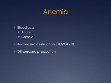
Anemia
- Blood Loss
- Acute
- Chronic
- IN-creased destruction (HEMOLYTIC)
- DE-creased production
2
Hypoproliferative
Marrow damage gt Infiltration fibrosis gt
Aplasia gt Myelodysplasia gt Drug or radiation
injury Iron deficiency B12 deficiency Folate
deficiency Stimulus gt Inflammation gt
Endocrine defect gt Renal disease Hypersplenism
Clues from morphology
microcytic, normocytic, or macrocytic poikilocyto
sis anisocytosis nucleated red cells target
cells Howell-Jolly bodies hypersegmented polys
3
Retics normal or increased
Hemorrhage and Hemolysis
Clues from morphology
Blood loss Hemolysis gt Antibody-mediated gt
Membrane defect gt Metabolic defect gt Red cell
fragmentation Hemoglobinopathy
microcytic, normocytic, or macrocytic red cell
fragmentation red cell clumping nucleated red
cells target cells
4
Features of All Anemias
- Pallor, where?
- Tiredness
- Weakness
- Dyspnea, why?
- Palpitations
- Heart Failure (high output), why?
5
Blood Loss
- Acute
- Trauma
- Chronic
- Lesions of gastrointestinal tract, gynecologic
disturbances. The features of chronic blood loss
anemia are the same as iron deficiency anemia,
and is defined as a situation in which the
production cannot keep up with the loss.
6
Hemolytic
- Hereditary
- MEMBRANE disorders e.g., spherocytosis
- ENZYME disorders e.g., G6PD deficciency
- HGB disorders (hemoglobinopathies)
- Acquired
- MEMBRANE disorders (PNH)
- ANTIBODY MEDIATED, transfusion or autoantibodies
- MECHANICAL TRAUMA
- INFECTIONS
- DRUGS, TOXINS
- HYPERSPLENISM
7
Impaired Production
- Disturbance of proliferation and differentiation
of stem cells aplastic anemias, pure RBC
aplasia, renal failure - Disturbance of proliferation and maturation of
erythroblasts - Defective DNA synthesis (Megaloblastic)
- Defective heme synthesis (Fe)
- Deficient globin synthesis (Thalassemias)
8
Modifiers
- MCV, microcytosis, macrocytosis
- MCHC, hypochromic
- RDW, anisocytosis
9
Hemolytic Anemias
- Life span LESS than 120 days
- Marrow hyperplasia (ME), EPO
- Increased catabolic products, e.g., bilirubin,
serum HGB, hemosiderin
10
Hemolysis
- INTRA-vascular (vessels)
- EXTRA-vascular (spleen)
11
ME Ratio normally 31
12
HEREDITARY SPHEROCYTOSIS
Genetic defects affecting ankyrin, spectrin,
usually autosomal dominant Children,
adults Anemia, hemolysis, jaundice,
splenomegaly, gallstones
13
Glucose-6-Phosphate Dehydrogenase (G6PD)
Deficiency
A- and Mediterranean are most significant types
14
Features of G6PD Defic
- Genetic Recessive, X-linked
- Can be triggered by foods (fava beans), oxidant
substances drugs (primaquine, chloroquine), or
infections - HGB can precipitate as HEINZ bodies
- Acute intravascular hemolysis can occur
- Hemoglobinuria
- Hemoglobinemia
- Anemia
15
Sickle Cell Disease
- Classic hemoglobinopathy
- Normal HGB is a2 ß2 ß-chain defects
(Val-gtGlu) - Reduced hemoglobin sickles in homozygous
- 8 of American blacks are heterozygous
16
Clinical features of HGB-S disease
- Severe anemia
- Jaundice
- PAIN (pain CRISIS)
- Vaso-occlusive disease EVEREWHERE, but
clinically significant bone, spleen
(autosplenectomy) - Infections Pneumococcus, Hem. Influ., Salmonella
osteomyelitis
17
(No Transcript)
18
(No Transcript)
19
THALASSEMIAS
- A WIDE VARIETY of diseases involving GLOBIN
synthesis, COMPLEX genetics - Alpha or beta chains deficient synthesis involved
- Often termed MAJOR or MINOR, depending on
severity, silent carriers and traits are seen - HEMOLYSIS is uniformly a feature, a microcytic
anemia - A crew cut skull x-ray appearance may be seen
20
(No Transcript)
21
Hemoglobin H Disease
- Deletion of THREE alpha chain genes
- HGB-H is primarilly Asian
- HGB-H has a HIGH affinity for oxygen
- HGB-H is unstable and therefore has classical
hemolytic behavior
22
Hydrops Fetalis
- FOUR alpha chain genes are deleted, so this is
the MOST SEVERE form of thalassemia - Many/most never make it to term
- Children born will have a SEVERE hemolytic anemia
as in the erythroblastosis fetalis of Rh disease - Pallor (as in all anemias)
- Edema (hence the name hydrops)
- Massive hepatosplenomegaly (hemolysis)
23
Paroxysmal Nocturnal Hemoglobinuria (PNH)
GlycosylphosPhatidylInositol
- ACQUIRED, NOT INHERITED like all the previous
hemolytic anemias were - ACQUIRED mutations in phosphatidylinositol glycan
A (PIGA) - It is P and N only 25 of the time
24
Immunohemolytic Anemia
- All of these have the presence of antibodies
and/or compliment present on RBC surfaces - NOT all are AUTOimmune, some are caused by drugs
25
IMMUNOHEMOLYTIC ANEMIAS
- WARM (IgG), will NOT hemolyze at room temp
- Primary Idiopathic (most common)
- Secondary (Tumors, especially leuk/lymph, drugs)
- COLD AGGLUTININS (IgM), WILL hemolyze at room
temp - Mycoplasma pneumoniae, HIV, mononucleosis
- COLD HEMOLYSINS (IgG) Cold Paroxysmal
Hemoglobinuria, hemo-LYSIS in body, ALSO often
follows mycoplasma pneumoniae
26
Coombs Test
- DIRECT Patients CELLS are tested for surface
Abs - INDIRECT Patients SERUM is tested for Abs.
27
Direct anti-globulin test
28
HEMOLYSIS/HEMOLYTIC ANEMIAS DUE TO RBC TRAUMA
- Mechanical heart valves breaking RBCs
- MICROANGIOPATHIES
- TTP
- Hemolytic Uremic Syndrome
29
NON-Hemolytic Anemiasi.e., DE-creased Production
- Megaloblastic Anemias
- B12 Deficiency (Pernicious Anemia)
- Folate Deficiency
- Iron Deficiency
- Anemia of Chronic Disease
- Aplastic Anemia
- Pure Red Cell Aplasia
- OTHER forms of Marrow Failure
30
MEGALOBLASTIC ANEMIAS
- Differentiating megaloblasts (marrow) from
macrocytes (peripheral smear, MCVgt94) - Impaired DNA synthesis
- For all practical purposes, also called the
anemias of B12 and FOLATE deficiency - Often VERY hyperplastic/hypercellular marrow
31
- Decreased intake
- Inadequate diet, vegetarianism
- Impaired absorption
- Intrinsic factor deficiency
- Pernicious anemia
- Gastrectomy
- Malabsorption states
- Diffuse intestinal disease, e.g., lymphoma,
systemic sclerosis
32
- Ileal resection, ileitis
- Competitive parasitic uptake
- Fish tapeworm in
- Fish tapeworm infestation
- Bacterial overgrowth in blind loops and
diverticula of bowel - Increased requirement
- Pregnancy, hyperthyroidism, disseminated cancer
33
Vit-B12 Physiology
- Oral ingestion
- Combines with INTRINSIC FACTOR in the gastric
mucosa - Absorbed in the terminal ileum
- DEFECTS at ANY of these sites can produce a
MEGALOBLASTIC anemia
34
Please remember that ALL megaloblastic anemias
are also MACROCYTIC (MCVgt94 or MCV100), and
that not only are the RBCs BIG and
hyperplastic/hypercellular, but so are the
neutrophils, and neutrophilic precursors in the
bone marrow too, and even more so,
HYPERSEGMENTED!!!
35
PERNICIOUS ANEMIA
- MEGALOBLASTIC anemia
- LEUKOPENIA and HYPERSEGS
- JAUNDICE
- NEUROLOGIC posterolateral spinal tracts
- ACHLORHYDRIA
- Cant absorb B12
- LOW serum B12
- Flunk Schilling test, i.e., cant absorb B12,
using a radioactive tracer
36
FOLATE DEFICIENCY MEGALOBLASTIC AMEMIAS
- Decreased Intake diet, etoh-ism, infancy
- Impaired Absorption intestinal disease
- DRUGS anticonvulsants, BCPs, CHEMO
- Increased Loss Hemodialysis
- Increased Requirement Pregnancy, infancy
- Impaired Usage
37
APLASTIC ANEMIAS
- ALMOST ALWAYS involve platelet and WBC
suppression as well - Some are idiopathic, but MOST are related to
drugs, radiation - FANCONIs ANEMIA is the only one that is
inherited, and NOT acquired - Act at STEM CELL level, except for pure red
cell aplasia
38
APLASTIC ANEMIAS
39
APLASTIC ANEMIAS
- CHLORAMPHENICOL
- OTHER ANTIBIOTICS
- CHEMO
- INSECTICIDES
- VIRUSES
- EBV
- HEPATITIS
- VZ
40
MYELOPHTHISIC ANEMIAS
- Are anemias caused by metastatic tumor cells
replacing the bone marrow extensively
41
(No Transcript)
42
Fe Deficiency Anemia
- Due to increased loss or decreased ingestion,
almost always, in USA, nowadays, increased loss
is the reason - Microcytic (low MCV), Hypochromic (low MCHC)
- THE ONLY WAY WE CAN LOSE IRON IS BY LOSING BLOOD,
because FE is recycled!
43
Fe Transferrin Ferritin (GREAT test) Hemosiderin
44
Regulation of iron absorption
Gut lumen
Heme Fe
DMT1
Enterocyte
Ferritin
Fe
Fe
MTP1
Enterocyte precursor
Plasma transferrin
Transferrin Receptor
HFE
Hepcidin
45
Gastrointestinal absorption 1 mg/day
Functional iron Blood, marrow, myoglobin 2 grams
Storage iron Liver, RES 1 gram
Plasma transferrin 2 mg
Daily physiologic loss 1 mg
46
Clinical Fe-Defic-Anemia
- Adult men GI Blood Loss
- PRE menopausal women menorrhagia
- POST menopausal women GI Blood Loss
47
2 BEST lab tests
- Serum Ferritin
- Prussian blue hemosiderin stain of marrow (also
called an iron stain)
48
(No Transcript)
49
Iron stores
Erythron iron
Marrow iron stores 1 - 3 0 - 1 0 0
Ferritin 50 - 200 lt20 lt15 0
TIBC 300 - 360 gt360 gt380 gt400
Serum iron 50 - 150 50 - 150 lt50 lt30
Red cells normal normal normal microcytic, hypochromic
50
Serum transferrin receptor
Storage iron 107 mg
Storage iron 335 mg
Storage iron 1,102 mg
Serial measurement of sTfr during phlebotomy in 3
individuals
Goodnough, Skikne, Brugnara. Blood, 2000 96 823
- 833
51
Ratio of serum transferrin receptor to ferritin
as a measure of total body iron
Cook, Flowers, Skikne. Blood 2003 101 3359 - 64
52
Serum ferritin and total body iron
Kaltwasser, Gottschalk. Kidney Int. 1999
55(suppl) S49 - S56
53
Treatment of iron def anemia
- Oral iron is the preferred initial treatment
- Recommended daily dose is 150-200mg/day of
elemental iron - 325 mg of ferrous sulfate contains 65 mg of
elemental iron - One table three times a day
- Administer iron on an empty stomach with half a
glass of OJ or 250mg ascorbic acid
54
Serum iron after oral iron in patients with iron
deficiency
80
60
Serum iron
40
20
1
2
3
4
Hours
WH Crosby, Arch Int Med circa 1970
55
Safety of intravenous iron
Sodium ferric gluconate in sucrose
(Ferrlecit) Available in Europe gt 30 years 2.7 x
106 doses/year in Germany Italy in 1995 Iron
dextran (Imferon until 1992, InFed since 1992) 3
x 106 doses/year in US in 1996
Faich, Strobos. Am J Kidney Dis 1999 33(3)464-70
56
Safety of intravenous iron
Reported severe adverse reactions (1976 -
1996) SFGS 3.3 severe allergic reactions/106
doses, no fatalities ID 8.7 severe allergic
reactions/106 doses, 31 fatalities
Faich, Strobos. Am J Kidney Dis 1999 33(3)464-70
57
Safety of intravenous iron
Other theoretical risks iron overload sepsis a
cceleration of atherosclerosis
Faich, Strobos. Am J Kidney Dis 1999 33(3)464-70
58
Medicare warning (
Recombinant human erythropoietin is approved only
for treatment of anemia caused by renal failure
or by cancer treatment and for certain
hematologic malignancies. Sodium ferric
gluconate in sucrose is approved only for
treatment of anemia in patients on hemodialysis
and for patients who have had a severe reaction
to iron dextran.
59
Anemia of Chronic Disease
- CHRONIC INFECTIONS
- CHRONIC IMMUNE DISORDERS
- NEOPLASMS
- LIVER, KIDNEY failure
Please remember these patients may very very
much look like iron deficiency anemia, BUT, they
have ABUNDANT STAINABLE HEMOSIDERIN in the marrow!
60
Anemia of chronic disease
Typical lab findings Serum iron lt 50 TIBC lt
150 Normochromic or hypochromic red
cells Normal ferritin Normal serum transferrin
receptor
61
Anemia of chronic disease
Mechanisms blunted erythropoietin
response diminished response of erythroid
precursors to erythropoietin decreased delivery
of iron from RES, increased intracellular
ferritin in macrophages decreased
gastrointestinal iron absorption
62
Anemia of chronic disease
Mediators IL-1 IL-6 g-interferon TNF-a
63
Anemia of chronic disease
Inflammation Tissue necrosis Infection Neoplasia C
ongestive heart failure Acute myocardial
infarction
64
(No Transcript)
65
(No Transcript)
66
(No Transcript)
67
(No Transcript)

