
The Human Body: An Orientation - PowerPoint PPT Presentation
Title:
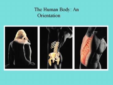
The Human Body: An Orientation
Description:
Title: PowerPoint Presentation Author: Terri Bicknell Last modified by: tbicknell Created Date: 12/19/2004 4:40:19 PM Document presentation format – PowerPoint PPT presentation
Number of Views:521
Avg rating:3.0/5.0
Title: The Human Body: An Orientation
1
The Human Body An Orientation
2
The Human Body An Orientation
- Anatomy the study of structure
- Physiology the study of function
3
Levels of Structural Organization
Smooth muscle cell
Molecules
Cellular levelCells are made up of molecules
2
Atoms
Chemical levelAtoms combine to form molecules
1
Smooth muscle tissue
Heart
Tissue levelTissues consist of similar types of
cells
3
Cardiovascular system
Blood vessels
Epithelial tissue
Smooth muscle tissue
Blood vessel (organ)
Organismal levelThe human organism is made up of
many organ systems
6
Connective tissue
Organ levelOrgans are made up of different types
of tissues
4
Organ system levelOrgan systems consist of
different organs that work together closely
5
4
4 Types of Organic Molecules
- 1. Carbohydrates
- Sugars
- Empirical formula CH2O
- Three forms Monosaccharide, Disaccharide,
Polysaccharide - Quick Energy in body
- Glucose, Sucrose, Starch, Glycogen
5
- 2. Lipids
- Monomers - fatty acids and glycerol
- Hydrophobic
- Make waterproof coverings (part of cell membrane)
6
- 3. Proteins
- Monomers - amino acids (20 naturally occurring)
- Bonded by peptide bonds
- Includes enzymes (lock and key model)
7
- 4. Nucleic Acids
- Monomers-Nucleotides
- DNA and RNA
- Genetic Expression and protein synthesis
8
Life Functions of Humans
- Maintaining boundaries
- Keeping internal and external environments
separate - Cells use the cell membrane
- Humans use integumentary system
- Movement
- Moving muscles, bones, blood, urineetc
- Responsiveness
- Also called irritability
- Sensing stimuli and responding to them
9
- Digestion
- Breaking down polymers to use as energy
- Hydrolysis
- Metabolism
- All chemical reactions that happen in the body
- Excretion
- Moving wastes out of body
- Includes digestive system and respiratory system
10
- Reproduction
- Making a new organism or new cells
(mitosis/meiosis)
- Growth
- Increase in body size
11
Homeostasis
- Maintaining a constant internal environment
- Every cell, tissue and organ in the human body
contributes toward total body homeostasis
12
Ex Blood cells carry wastes from various tissues
toward kidneys which filters blood and then sends
waste materials toward excretory system
13
Homeostatic Control Mechanisms
- Control mechanisms are the way that the body
maintains homeostasis - Variable The event that is being regulated
- Receptor A sensor that monitors the environment
- Stimuli Changes in environment
14
- Control Centers Analyze the stimuli and
determines the course of action - Effector Provides the control centers response
to the stimuli (also called output)
15
Feedback Systems
- Negative feedback
- Output turns off or reduces stimulus
- Variables are changed opposite to stimulus
16
Ex Body is cold, you begin to shiver to raise
body temperature Ex Your body needs oxygen,
your respiration rate increases to meet this O2
need
17
- Positive Feedback
- Response to stimulus enhances so the output is
raised - Usually control infrequent events
- Ex Labor contractions and blood clotting
18
- Homeostatic Imbalance
- Negative feedback is overwhelmed and harmful
positive feedback takes over. - Ex If heart muscles fail to contract and
negative feedback can not control the muscles,
positive feedback takes over and heart failure
can occur
19
Language of Anatomy
Body erect, feet slightly apart, palms facing
forward, thumbs point away from body
Anatomical Position
20
- Body Region Terms
- Axial - Makes up the main axis of the
body-head, neck and trunk
- Appendicular - Appendages that are attached to
the axis
21
Directional Terms
- Superior toward the head
- Inferior away from the head
22
- Anterior toward the front of the body
- Posterior back of the body
23
- Medial toward the midline
- Lateral away from the midline
24
- Proximal closer to the origin of the body
- Distal farther from the origin of the body
25
- Superficial toward or at the body surface
- deep away from the body surface more internal
26
- Human Body Planes
- Sectioned along a flat surface into the following
divisions - Sagittal Vertical plane, divided into left and
right sides
27
- Frontal Plane Vertical plane that divides body
into anterior and posterior sections. - Transverse or horizontal plane Divides body into
superior and inferior parts. Also called cross
section
28
- Body Cavities
- 1. Dorsal Body Cavity - Protects nervous system
organs - Includes cranial cavity that encases brain
29
- Ventral Body Cavity - Largest body cavity, holds
the internal organs - Thoracic Cavity - Superior portion of cavity
- Pleural cavity - Encloses each lung
- Pericardial cavity - Encloses heart
30
- Abdominopelvic Cavity - Inferior portion of
cavity separated from thoracic cavity by
diaphragm - Abdominal cavity stomach, intestines, spleen,
liveretc
- Pelvic cavity bladder, reproductive organs,
rectum
31
Four Types of Tissues
- Epithelial tissue Covering tissue
- Connective tissue Support tissue
- Muscle tissue Movement tissue
- Nervous tissue Control tissue
32
Epithelium
- Covers body surface or lines body cavities
- Outer layer of skin, open cavities,
cardiovascular system (veins/arteries), digestive
system, respiratory system, covers walls of
organs, forms glands
33
Functions of epithelial tissue
- Protection
- Absorption
- Filtration
- Excretion
- Secretion
- Sensory Reception
34
- Special characteristics of epithelial tissue
- 1. Supported by connective tissues
- Epithelial tissue rests upon connective tissue
that forms a basement membrane
35
- 2. Avascular
- Epithelial tissue has no blood vessels (only
capillaries)
- Epithelial tissues does have nerves
- 3. Regeneration
- Epithelial tissue goes through mitosis
quickly-this tissue is replaced quickly
36
- How do we differentiate epithelial tissue?
- 1. Layers
- Simple Thin layer usually used for absorption
- Stratified Thick layer of 2 or more cells used
for protection (skin)
37
(No Transcript)
38
- 2. Shape of cells
- All polyhedral shape, allows for close
connection of cells
- Cell nuclei conform to shape
- 3 shapes
- 1. Squamous cells - flat and scale like
39
2. Cuboidal cells - box-like or blunt pyramids
3. Columnar cells - tall and column shaped
40
- Simple Squamous Epithelium
- Cells flattened laterally, not much cytoplasm
- Found where filtration and the exchange of
substances by diffusion is needed - Kidneys and lungs
41
- Simple Cuboidal Epithelium
- Secretion and absorption cells
- Walls of small ducts of glands
- Kidney tubules
- Looks like strings of beads when viewed
microscopically
42
- Simple Columnar Epithelium
- Layers of tall cells with closely packed
membranes - Lines digestive tract from the stomach to the
rectum - Absorption and secretion cells
43
- Modifications of columnar
- Microvilli Projections that increase absorption
- Goblet cells Secrete protective mucus
44
- Pseudostratified Columnar Epithelium
- Layers of different height cells that rest on a
basement membrane (appears layered) - Secretes or absorbs
- Ciliated version are goblet cells that line the
respiratory tract
45
- Stratified Epithelial Tissue
- Stratified Squamous Tissue
- Most widespread type (areas of wear and tear)
- Skin
- Stratified Cuboidal Tissue
- Rare in body, usually found in glands (mammary
and sweat) - Stratified Columnar Tissue
- Pharynx, male urethra
46
Connective Tissue
- Found everywhere, most abundant of primary
tissues
- Skin is mostly connective tissue
- 4 classes of connective tissue
- 1. Connective tissue proper
- 2. Cartilage
- 3. Bone Tissue
- 4. Blood
47
- 3 elements make up connective tissue
- 1. Ground Substance
- Material that fills in space between cells, also
called matrix - 2. Fibers
- Provide support
- Collagen Fibers Very strong, high tensile
strength, also called white fibers - Elastic Fibers Stretchy fibers made
- of elastin, found in lungs, blood vessels, also
called yellow fibers
48
- Reticular Fibers Fine network of collagen found
around capillaries
49
- 3. Cells
- Blast forming (baby or developing cells)
- Cyte cell (adult cells)
- Fibroblast/Fibrocyte Cells in connective tissue
proper - Chondroblast/Chondrocyte Cartilage cells
- Osteoblast/Osteocyte Bone cells
- Hematopoietic Stem Cells Blood developing cells
50
- Types of Connective Tissue
1. Connective Tissue Proper loose connective
tissues
a. Areolar Connective Tissue
- Supports and joins fibers of other tissues
universal packing material of tissues
- Holds body fluids
51
- Defends against infection- when an area is
inflamed, this tissue soaks up extra body
fluids-called edema
- Stores nutrients in fat
- Most widespread connective tissue
52
- b. Adipose Tissue
- Fat storage tissue
- Adipocytes (fat cells) 90 of tissue mass, large,
rounded, with large fat droplets, faint outlines
- High amount of veins, arteries, metabolic
activity, nutrient storage ability
53
- Acts as a shock absorber, insulator and energy
storage site
- 18 of an average persons body weight is this
type of tissue
- Can be deposited around organs such as the heart,
lymph nodes and various muscles
54
c. White Fibrous Connective Tissue - Dense
Regular Connective Tissue
- Tendons and ligaments
- Matrix of regular arranged collagenous fibers
55
- 2. Cartilage
- Resists tension and compression
- Tough, flexible tissue
- Avascular, no nerve fibers
- Receives nutrients from blood vessels located in
the connective tissues that surround
it
56
- Ground substance is collagen
- Chondroblasts in growing cartilage until
maturity-becomes chondrocyte
57
- Types of Cartilage
- 1. Hyaline
- gristle
- Most abundant cartilage in body
- Matrix appears bluish-white
- Trachea, lungs, nose, rib attachment to sternum,
growth plate (epiphyseal plate), embryonic
skeleton
58
2. Elastic
- Elastin fibers
- External ear, epiglottis
59
3. Fibrocartilage
- Compressible, resists tension
- Support and pressure
- Intervertebral discs, cartilage of knee
60
- 3. Bone Tissue
- Support and protect body
- Provide cavities for fat storage and the
synthesis of blood cells
- Matrix includes calcium salts
Cells Osteoblasts (develop bones)
Osteocytes (mature bones)
61
- 4. Blood Tissue
- Consists of red blood cells, white blood cells,
lymphocytes, monocytes, eosinophils, basophiles
- Is considered connective tissue, but it doesnt
connect anything
62
Muscle Tissue
- Responsible for body movement
- Three types
skeletal muscle,
cardiac muscle,
smooth muscle
63
Nervous Tissue
- Main component of nervous system (brain, spinal
cord, nerves)
- Cells Neurons
64
Tissue Repair
- Process
- 1. Inflammation
- Tissue trauma causes inflammation chemicals to be
released
- Is a negative feedback system-causes capillaries
to dilate to accommodate WBCs and clotting
factors
- Clot forms, the part of the clot that is exposed
to air becomes a scab
65
2. Organization
- Clot is replaced by granulation tissue which
becomes scar tissue
3. Regeneration and fibrosis
- Regeneration
- New tissue (same type) takes the place of the
damaged tissue
66
- Fibrosis
- Fibrosis connective tissue (scar tissue) takes
the place of the damaged tissue
67
Tissue Development
- Tissues develop from three germ layers
- Ectoderm
- Mesoderm
- Endoderm
68
By second month of development, all tissues have
appeared and all organs are formed
69
Tissue cells (except neural cells) remain mitotic
until adulthood is reached
- As adults only blood and skin cells are highly
mitotic
- Skin cells can go through mitosis once a day