
Mass Spectrometry - PowerPoint PPT Presentation
Title:
Mass Spectrometry
Description:
Title: PowerPoint Presentation Author: DSP Last modified by: chickosj Created Date: 11/16/2004 11:26:06 PM Document presentation format: On-screen Show (4:3) – PowerPoint PPT presentation
Number of Views:175
Avg rating:3.0/5.0
Title: Mass Spectrometry
1
Mass Spectrometry
2
Exact Mass Measurements What is exact
mass? m mass of a proton 1.672623 10-24
g mass of a neutron 1.674927 10-24 g mass of
a deuteron 3.3427 10-24g Avogadros Number
(AN) 6.0254 1023 Molar mass of 2D AN mD
6.02541023 3.3427 10-24 g 2.0141 g
mol-1 Carbon 6 2D 62.0141
12.0846 Carbon 6(P N) 6(1.6726231.674927)
0.60254 12.1022 Mass of carbon 12.0 Why the
discrepancy?
3
E m C2 Where E is the energy given off from
a mass discrepancy of m and C is the speed of
light. E 0.0846 g (31010 cm sec-1)2
4
5
Suppose you determined the exact mass of an ion
by mass spectrometry to be 56.0377. Nominal mass
56
Using the rule of 13, the hydrocarbon formula is
C4H8 Other possible molecular formulas are C4H8
- CH4 C3H4O C4H8 - CH2 C3H6N X C4H8 -
2CH4 C2O2 C4H8 - 2CH4 C2S C4H8 -
2CH2 C2H4N2 C4H8 - CH4, CH2
C2H2NO X C4H8 - 3CH2 CH2N3 X C4H8 - C
C3H20 X C4H8 - CH4, 2CH2 CN2O C4H8 -
4CH2 N4
6
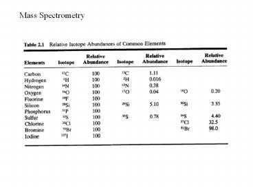
Element Exact Mass 12C 12.0000 1H
1.00783 14N 14.0031 16O 15.9949 19F 18.9984 28Si 2
7.9769 31P 30.9738 32S 31.9721 35Cl 34.9689 79Br 7
8.9183 127I 126.9045
7
The exact mass of an ion by mass spectrometry was
determined to be 56.0377 amu Nominal mass
56 exact mass N4 414.0031 56.0124
CN2O 12.00214.0031 15.9949
56.0011 CH2N3 56.0249 C2O2 55.9898 C2H
2NO 56.0136 C2H4N2 56.0375 C3H4O 56.0
262 C3H6N 56.0501 C4H8 56.0626
8
(No Transcript)
9
What is the origin of the peak at 141 called the
P1 peak For a molecular formula of C9H16O,
whats the probability of having 1
13C? Probability is (XY)n where X and Y is the
probability of having isotope 12C and 13C,
respectively and n is the number of C (12C
13C)9 1 n 0 1 1 n
1 1 2 1 n 2 1 3
3 1 n 3 1 4
6 4 1 n 4 1 5
10 10 5 1 n 5
1 6 15 20 15 6
1 n 6 1 7 21 35 35 n
7 1 8 28 56
56 n 8 1 9 36
84 n 9 (12C)9 9(12C)8(13C)
36(12C)7(13C)2 All 12C 1 13C 2 13C (0.989)9
0.905 9(0.989)8(0.011) 0.091
36(0.989)7(0.011)2 0.004
10
On the basis of the molecule with only 12C
100 Then (0.989)9 100(0.905/0.905) 100
9(0.989)8(0.011) 100(0.091/0.905) 10.0
36(0.989)7(0.011)2 0.004/.905 0.45
Including 1oxygen 17O 0.04 18O
0.2 P 100 P1 10.04 P2 0.65 The
contribution of 2H is pretty small
11
What about other elements?
12
Electron impact mass spectrum of CCl4
13
Single focusing instrument
14
(No Transcript)
15
(No Transcript)
16
(No Transcript)
17
- -
The quadrupole mass spectrometer consists of four
precisely straight and parallel rods so arranged
that the beam of ions from the ionization source
of the spectrometer is directed axially between
them. A voltage comprising a
a DC component and a radio frequency
electric field is applied between adjacent rods,
reinforcing and then overwhelming the DC field.
Once inside the quadrupole, the ions will
oscillate normal to the field as a result of the
high frequency electric field. The oscillations
are only stable for a certain function of
frequency and the DC voltage otherwise the ions
will strike the rods and become dissipated. The
mass range of the oscillating ions is scanned by
changing the DC voltage and the frequency,
keeping the ratio of the DC voltage to the
frequency constant. Typical operating parameters
include rf voltages of several thousand volts,
frequencies in the 106 range and DC voltages of
several hundred volts. Unlike a magnetic sector
instrument, the mass is linear as the DC and
frequency are scanned.
18
An ion trap is a combination of electric or
magnetic fields that captures ions in a region of
a vacuum system or tube. A quadrupole ion trap
exists in both linear and 3D varieties and refers
to an ion trap that uses constant DC and
radiofrequency (RF) oscillating AC electric
fields to trap ions.
19
The motion of an ion is complex but it is clear
that specific frequencies are involved. The
frequencies can be used to manipulate the ion
population in a mass selective fashion.
20
Time of Flight MS A variety of ways can be used
to create ions. Ions are not suitable for
analysis for a time of flight mass spectrometer
unless they are all ejected from the ion source
with the same starting time. This is easy to do
with a pulsed laser This results in a group of
ions which can be turned on and off during time
t rapidly so that it only creates ions only
during time t. The ions once formed are
accelerated by a negative grid of known
potential. Once accelerated, all ions have the
same kinetic energy but different velocities
(1/2mv2). They reach the detector at different
times.
21
(No Transcript)
22
(No Transcript)
23
Formation of Ions
24
(No Transcript)
25
CH5
P 143
P H
PC3H6
26
CH5
P 69
PH
PC3H5
27
P 390
Different energetics associated with different
ionization methods
28
Single focusing instrument and metastable ions
Some ions are relatively unstable and fall apart
shortly after being formed. If they survive long
enough to be accelerated as m1 but then fragment
shortly in the field free region to m2before
encountering the magnetic field, then
Metastable Ions
29
Metastable ions accelerated as mp but analyzed
as md where mp gt md , then a peak often
broadened as a result of energy release
accompanying decomposition, can be found
at (md)2/mp The usefulness of metastable is
that they permit you to identify connectivity of
fragmentation (i.e. which parent ion gave rise to
which daughter ion) Metastables are lost in
instruments that use a quadruple mass filter such
as in most GCMS instruments.
30
(No Transcript)
31
Metastables observed at m/e 136.2
1602/188 131.4 1452/160 108.9
1322/160 103.7 1172/132 94.4
1172/145 67.7 892/117
32
- Fragmentation Patterns in EI MS
- Electrons with 70 eV are used to bombard the
sample. In addition to a molecular ion formed by
loss of an electron, the resulting ions
frequently have sufficient energy to fragment
into daughter ions. - The easiest way to interpret fragmentation
patterns is to focus on the molecular ion formed.
The electron with the lowest ionization potential
is lost first. Secondary reactions focus around
this center. - Electrons in C-C bonds have lower ionization
energies than C-H bonds. - Electrons in ? bonds are easier to lose than
sigma bonds. - Non-bonded electrons on heteroatoms are lost the
easiest. - Conventions used in mass spectrometry means
movement of 2 electrons
- ? means movement of one electron
33
m/e 121 P-CH3
m/e 93 P C3H7
68
m/e 68 P- C4H8
93
C10H16
136
34
CH2CH-CH2-CH3
m/e 41 P CH3
P
35
57
m/e 57 P C4H9
m/e 114 parent
m/e 99 P-CH3
36
m/e 91 P - H
m/e 92 parent
37
m/e 91 P C2H5
m/e 120 parent
38
m/e 45 P C2H5 CHO
m/e 74 Parent
m/e 59 P CH3
39
m/e 77 P CHO C2H5
m/e 106 parent
m/e 105 P - H
40
43
m/e 58 P C3H6 P- C2H2O
m/e 43 P- C4H9 P- C3H5O
58
m/e 100 parent
m/e 85 P CH3
85
41
Mw 88
m/e 60 P C2H4 P - CO
73
42
Loss of neutral molecules is frequently observed
43
m/e 83
m/e 69
MW 140
125 P-CH3
44
45
46
(No Transcript)
47
m/e 43
P-C4H9O P-C3H5O2
m/e 87
P-C2H4O
m/e 116
P-C2H5
m/e 56
P CH3
m/e 101
48
(No Transcript)