
Archaeal cell walls - PowerPoint PPT Presentation
1 / 31
Title:
Archaeal cell walls
Description:
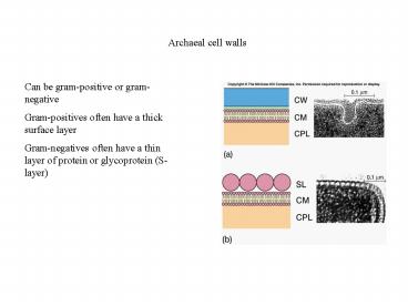
Archaeal cell walls Can be gram-positive or gram-negative Gram-positives often have a thick surface layer Gram-negatives often have a thin layer of protein or ... – PowerPoint PPT presentation
Number of Views:656
Avg rating:3.0/5.0
Title: Archaeal cell walls
1
Archaeal cell walls
Can be gram-positive or gram-negative Gram-positiv
es often have a thick surface layer Gram-negatives
often have a thin layer of protein or
glycoprotein (S-layer)
2
Pseudomurein
Often found in gram-positive archaea Similar to
peptidoglycan N-acetylalosaminuronic acid
replaces NAM Cross-bridges do not contain D-amino
acids
3
Plasma membrane Composed of lipids and proteins
4
Membrane lipids
Amphipathic molecules Hydrophilic heads and
hydrophobic tails
5
Membrane lipids
Amphipathic molecules Hydrophilic heads and
hydrophobic tails Allows lipids to interact with
water on one end and each other on the other
end Formation of lipid bilayers
6
Archaeal lipids
Contain branched chain hydrocarbons attached to
glycerol via ether links Other cells have fatty
acids attached to glycerol via ester links
(bacteria and eukaryotes)
7
Archaeal lipids
Two glycerol groups can be linked to form a
tetraether Tetraether chains are usually 40
carbons long Diether chains are usually 20
carbons long Length of tetraethers can be
adjusted by cyclizing the chain to form
pentacyclic rings
8
Archaeal lipids
Various combinations of lipids can result in
differences in rigidity and thickness of membrane
9
Sterols and hopanoids
Eukaryotic cell membranes often contain
sterols Also found in the membrane of some
bacteria that lack a cell wall Stabilize the
membrane and add rigidity
10
Sterols and hopanoids
Hopanoids are sterol-like molecules that are
found in bacterial membranes Play similar role as
sterols
11
Plasma membrane Composed of two layers of lipids
with hydrophobic ends in the interior of the
membrane Proteins can be peripheral or integral
12
Fluid mosaic model Most widely accepted model
for membrane structure Lipid composition varies
with temperature to maintain fluidity
13
Internal membrane systems
Mesosomes Invaginations of membrane Often in the
form of vesicles, tubules or lamellae Some
believe they are artifacts generated during
chemical fixation
14
Internal membrane systems
Photosynthetic prokaryotes Often have extensive
infoldings of the plasma membrane In the form of
flattened or spherical vesicles or tubules May
serve to provide larger surface area for
metabolic processes
15
Cytoplasmic matrix
16
Cytoplasmic matrix
Area between the plasma membrane and the
nucleoid Composed largely of water Specific
proteins positioned at particular sites (e.g.
poles or septum)
17
Inclusion bodies
Organic inclusion bodies usually contain glycogen
or poly-?-hydroxybutyrate Inorganic inclusion
bodies can store phosphate or sulfur
18
Inclusion bodies
Magnetosomes Iron containing inclusion bodies
used to orient cell in the Earths magnetic field
19
Inclusion bodies
Gas vacuoles Used by bacteria to regulate
buoyancy Composed of a collection of collapsible
gas vesicles
20
Ribosomes
Can be free in the cytoplasmic matrix or loosely
attached to the plasma membrane Membrane-associate
d ribosomes synthesize proteins that are
transported to the outside
21
Ribosomes
Are 70S vs. 80S Are composed of a 50S and a 30S
subunit
22
The nucleoid
The region of the cell where the chromosome is
located Irregularly-shaped Often appears to be
attached to plasma membrane Can rarely be bound
by a membrane
23
The nucleoid
Most prokaryotes have a single circular
chromosome Some bacteria have linear
chromosomes Some bacteria have two
chromosomes DNA-binding proteins associated with
chromosome
24
Endospores
Dormant structures that are resistant to
environmental stresses Can remain viable for
100,000 years Can survive boiling (must be
autoclaved)
25
Endospores
True endospores are only found in gram positive
bacteria
26
Endospores
Location of endospore in cell can aid in
identification Mother cell is called the
sporangium
27
Endospores
Are complex structures Covered by
exosporium Next layer is the spore coat
(responsible for resistance to chemicals)
28
Endospores
Cortex is beneath the spore coat and contains
peptidoglycan Spore cell wall surrounds the core
29
Resistance of endospores
Large amounts of dipicolinic acid is complexed
with calcium ions in the core May aid in
resistance DNA-binding proteins, dehydration of
core and DNA repair systems all contribute to
resistance
Dipicolinic acid
30
Sporogenesis/sporulation
31
Transformation into vegetative cells
Occurs in three stages
- 1. Activation
- 2. Germination
- 3. Outgrowth
Recommended
CrystalGraphics Presentations


