
Chap. 5 Molecular Genetic Techniques (Part A) - PowerPoint PPT Presentation
Title:
Chap. 5 Molecular Genetic Techniques (Part A)
Description:
A cDNA library (complementary DNA), is a collection of cloned DNA fragments corresponding to all mRNAs transcribed in a certain tissue or organism. – PowerPoint PPT presentation
Number of Views:76
Avg rating:3.0/5.0
Title: Chap. 5 Molecular Genetic Techniques (Part A)
1
Chap. 5 Molecular Genetic Techniques (Part A)
- Topics
- Genetic Analysis of Mutations to Identify and
Study Genes - DNA Cloning and Characterization
Goals Learn about genetic and recombinant DNA
methods for isolating genes and characterizing
the functions of the proteins they encode.
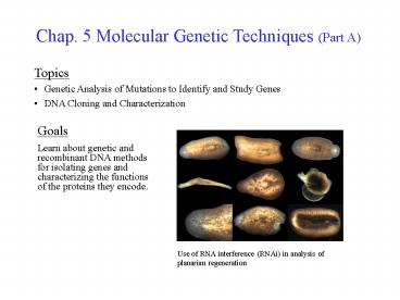
Use of RNA interference (RNAi) in analysis of
planarian regeneration
2
Leptin Receptor Knockout Mice
db/db DB/DB
3
Importance of Mutations in Gene Analysis
One of the most important ways in which the
function of a gene can be learned is by the study
of a mutant in which the gene has been
inactivated. Currently, mutants can be generated
by classical forward genetic methods, and by
more modern reverse genetic approaches (Fig.
5.1). In forward genetic analyses one generates a
mutant organism and then uses molecular
biological techniques to isolate the mutant gene
and characterize the protein responsible for the
phenotype of the mutant. In reverse genetic
approaches, a gene is inactivated and the
function of the gene is learned by study of the
properties of the mutant organism.
4
Genetics Terms
Alleles-Different versions (sequences) of a
gene. Mutant-Newly created allele made by
mutagenesis. Genotype-The complete set of alleles
for all genes carried by an individual. Wild
type-Standard reference genotype. Most common
allele for a certain trait. Phenotype-Observable
trait specified by the genotype. Point mutation-A
change in a single base pair (e.g., a G.C to A.T
transition). Silent mutation-A point mutation in
a codon that does not change the specified amino
acid. Missense mutation-A point mutation that
changes the encoded amino acid. Nonsense
mutation-A point mutation that introduces a
premature stop codon into the coding sequence of
a gene. Recessive dominant mutant alleles-(next
slide)
5
Recessive and Dominant Mutant Alleles
Diploid organisms have two copies of each gene
haploid organisms (e.g., some unicellular
organisms) contain only one. A recessive mutant
allele must be present in two copies (be
homozygous) to cause a phenotype in a diploid
organism (Fig. 5.2). Only one copy of a recessive
allele must be present for the phenotype to be
observable in a haploid organism. In contrast, a
dominant mutant allele needs to be present in
only one copy (heterozygous) in a diploid
organism for the phenotype to be observable. Most
recessive alleles cause gene inactivation and
phenotypic loss of function. Some dominant
alleles change or increase activity causing a
gain of function. However, a dominant affect can
be caused by gene inactivation if two copies of
the gene are needed for proper function
(haplo-insufficiency). Lastly, a dominant
negative mutation refers to a situation where the
product of the mutant gene inactivates the
product of the wild-type gene. This can occur if
a gene encodes one subunit of an oligomeric
protein.
6
Review of Mitosis
Mating experiments provide important information
about gene function. These experiments demand a
thorough knowledge of meiosis and production of
gametes (sperm egg cells in higher eukaryotes).
In Fig. 5.3, mitosis is described to contrast it
with meiosis. In mitosis, one round of DNA
replication in a diploid somatic cell is followed
by one cell division. The paternal and maternal
homologous chromosomes (homologs) first are
duplicated. The sister chromatids then are
separated by a cell division. The daughter cells
end up with one copy of each paternal and
maternal chromosome and are diploid (2n).
7
Review of Meiosis
In meiosis, one round of DNA replication in a
diploid germ cell is followed by two cell
divisions, resulting in four haploid gametes
(Fig. 5.3). Paternal and maternal homologous
chromosomes first are copied as in mitosis.
However, after alignment (synapsis) and crossing
over (recombination) of homologous chromosomes,
paternal and maternal chromosomes are randomly
segregated between the daughter cells formed in
the first cell division. Subsequently, the sister
chromatids of each chromosome are separated in a
second cell division, which produces the gametes
(1n). The two sets of gametes each contain a
random assortment of the paternal and maternal
chromosomes.
8
Identification of Dominant Mutations
Dominant mutations can be identified by mating
strains that each are homozygous for two alleles
of a given gene. Because all gametes from each
parent are of one type (Fig. 5.4a), all members
of the first filial generation from the cross
(F1) necessarily will be heterozygotes. If the
mutation is dominant, all F1 offspring will
display the mutant phenotype. On self crossing of
F1 cells, 3/4 of the second filial generation
(F2) will display the mutant phenotype, if it is
dominant.
9
Identification of Recessive Mutations
Recessive mutations also can be identified by
mating strains that are homozygous for two
alleles of a given gene. Again, because all
gametes from each parent are of one type (Fig.
5.4b), all members of the F1 generation from the
cross necessarily will be heterozygotes. None of
the F1 offspring will display the mutant
phenotype if it is recessive. On self crossing of
F1 cells, only 1/4 of the F2 generation will
display the phenotype, if it is recessive.
10
Analysis of Mutant Alleles in Yeast
The yeast Saccharomyces cerevisiae is an ideal
experimental organism for analysis of dominant
and recessive alleles. First, cells can exist in
either a haploid or diploid state. Second,
haploid cells occur in two mating types (a and a)
that are useful for performing crosses. The
diploid cells resulting from matings can be
examined to determine if a mutation is dominant
or recessive (Fig. 5.5). Finally, haploid cells
can be regenerated by meiotic sporulation of
diploid cells grown under starvation conditions.
11
Use of Conditional Mutations to Study Essential
Genes
The study of essential genes (needed for life)
requires special genetic screening techniques. In
diploid organisms, such as the fruit fly
Drosophila, lethal mutations in essential genes
can be maintained in the diploid state and
identified by inbreeding experiments. In haploid
organisms, such as haploid yeast (Fig. 5.6),
defects in essential genes can be isolated and
maintained through the use of
conditional mutations. Very often, conditional
mutations that display temperature-sensitive (ts)
phenotypes are used. ts mutations often result
from substitution mutations that cause an
essential protein to be unstable and inactive at
high (nonpermissive), but not low (permissive)
temperatures. A number of yeast cell-division
cycle (cdc) mutants have been isolated via this
technique (Fig. 5.6).
12
Complementation Analysis of Recessive Mutations
Many processes, including cell division involve
the combined actions of multiple genes. Thus, a
genetic screen for mutations affecting such
processes will turn up a collection of genes.
Through mating haploid yeast containing the
defective genes, one can establish in the diploid
cells whether the mutations fall in the same or
separate genes. As shown in Fig. 5.7, diploid
cells will grow under nonpermissive conditions if
the mutations reside in different genes (the wild
type genes complement the defective ones).
However, diploids with two defective copies of
the same gene will not survive.
13
Double Mutant Analysis of Biosynthetic Pathways
Genetic experiments can be used to determine the
order in which gene products act in carrying out
a process. Double mutant analysis can be applied
to order the enzyme-catalyzed steps in a
metabolic pathway. As shown in Fig. 5.8a, the
accumulation of intermediate 1 in the double
mutant strain indicates enzyme A operates prior
to enzyme B.
14
Suppressor Mutations
Suppressor mutation analysis is a powerful tool
for identifying proteins that interact with one
another in the performance of a certain cellular
process. In genetic suppression, a loss of
function mutation in Protein A is corrected by a
compensating mutation in Protein B. The resulting
gain of function phenotype of the double mutant
results from the recreation of interaction sites
between two proteins that are disrupted by each
individual amino acid substitution mutation (Fig.
5.9a).
15
Synthetic Lethal Mutations
The analysis of synthetic lethal mutations also
is an important tool for identifying proteins
that must interact to carry out a cellular
process (Fig. 5.9b). It also is a powerful method
to identify proteins that function in redundant
pathways needed for the production of an
essential cell component (Fig. 5.9c). Unlike
suppressor mutations, synthetic lethal double
mutants display a loss of function phenotype.
16
Intro to DNA Cloning by Recombinant DNA Methods
To study a gene, one must first prepare and
purify its DNA in relatively large amounts. This
is accomplished via the recombinant DNA (rDNA)
technology method known as DNA cloning. In
cloning, a DNA molecule of interest is spliced
into a vector such as a bacterial plasmid or
virus forming a rDNA molecule which can be
propagated in bacterial cells such as E. coli.
After replication and amplification of the rDNA
in the bacterium, it is purified for sequencing
and other manipulations used in gene
characterization.
17
DNA Cleavage by Restriction Enzymes
Restriction enzymes are nucleases that are very
important in rDNA technology. These enzymes make
double-stranded cuts in DNA molecules at specific
4-8 bp palindromic (two-fold symmetrical)
sequences called restriction sites. Many
restriction enzymes make staggered cuts in DNA
molecules resulting in single-stranded
complementary sticky ends (Fig. 5.11).
Sticky-ended fragments can be readily joined
together to synthesize rDNA molecules (Fig.
5.12). In many cases, cleavage at the restriction
site is blocked by methylation of bases in the
site.
18
(No Transcript)
19
Joining of DNA Molecules by Ligation
Plasmid vectors containing a DNA of interest
(e.g., genomic DNA) can be readily constructed by
ligating restriction fragments to vector DNA that
has been digested with the same restriction
enzyme (Fig. 5.12). Base-pairing between the
complementary sequences of the sticky ends aligns
the fragments for covalent linkage by a DNA
ligase, typically T4 DNA ligase. This enzyme uses
2 ATP to provide energy for joining the
3'-hydroxyl and 5'-phosphate groups of the
base-paired fragments together in 2 new 3'-5'
phosphodiester bonds. Note, all restriction
enzymes produce a 5'-phosphate and 3'-hydroxyl
group at the cut site.
20
E. coli Plasmid Cloning Vectors
Plasmids are autonomously replicating circular
DNAs found in bacterial cells. Naturally
occurring plasmids contain an origin of
replication (ori) for propagation in the host
cell and one or more genes that specify a trait
that may be useful to the host. Cloning vectors
are plasmids that have been genetically
engineered to reduce unneeded DNA and to
introduce selectable markers such as antibiotic
resistance genes (e.g., ampr) that are used to
force cells to maintain the plasmid. Polylinker
sequences that encode several unique restriction
sites for cloning purposes also are engineered
into these vectors (Fig. 5.13).
21
Cloning of DNA in Plasmid Vectors
An overview of the steps required for DNA cloning
in a plasmid vector is presented in Fig. 5.14. In
Step 1, the DNA of interest is ligated into a
plasmid cloning vector. In Step 2, the
recombinant plasmid is introduced into E. coli
host cells by transformation. In Step 3, cells
that have taken up the plasmid are selected on
antibiotic (ampicillin) agar. In Step 4, the
transformed cells replicate their chromosomal and
plasmid DNA and multiply to form a colony. Cells
in the colony contain the cloned DNA and are
themselves clones. The rDNA plasmid then is
harvested by growing a larger culture of the
cells.
1
2
3
4
4
22
Construction of cDNA Libraries (Part 1)
A genomic DNA library is a collection of cloned
DNA fragments representing all of the DNA of an
organism. A cDNA library (complementary DNA), is
a collection of cloned DNA fragments
corresponding to all mRNAs transcribed in a
certain tissue or organism. Libraries can be
constructed using plasmid cloning vectors. To
construct a cDNA library, one begins by isolating
mRNA from the cell or tissue of interest (Fig.
5.15). Because many genes are transcribed at a
low frequency, it is best to start with a
cell/tissue that expresses the
gene of interest at a relatively high level.
cDNAs are transcribed from a mRNA template by a
retroviral enzyme known as reverse transcriptase
(RT). In Step 1, mRNA isolated by oligo-dT
affinity chromatography is hybridized via its 3'
poly(A) tail to an oligo-dT primer. In Step 2, RT
synthesizes the first cDNA strand. In Step 3, RNA
is destroyed and a poly(dG) tail is added by
terminal transferase. In Step 4, the cDNA is
hybridized to an oligo-dC primer. (Go to next
slide).
23
Construction of cDNA Libraries (Part 2)
In Step 5, a DNA polymerase is used to synthesize
the second strand of the cDNA. In Step 6, EcoRI
sites that might be present within the mRNA
coding region are protected by methylation using
EcoRI methylase. In Step 7, unmethylated EcoRI
linkers, that encode EcoRI restriction sites, are
ligated to the ends of the fragment. In Step 8a,
the cDNA is cleaved with EcoRI restriction
enzyme, generating sticky-ended cDNA fragments.
(See next slide).
24
Construction of cDNA Libraries (Part 3)
In the last steps of cDNA library construction,
the plasmid vector is cut with EcoRI restriction
enzyme (Step 8b), and then the EcoRI-cut cDNA and
plasmid are ligated together (Step 9). Finally,
the E. coli host strain is transformed and cells
are plated (Step 10) on selective medium. To be
complete, both genomic and cDNA libraries for
higher eukaryotes must contain on the order of a
million individual clones.
25
Screening cDNA Libraries
To screen a plasmid library (Fig. 5.16), colonies
representing each cloned DNA first are plated on
a number of petri plates. Library DNA then is
lifted onto nitrocellulose membranes which serve
as replicas of the plates. Bound DNA is denatured
and hybridized with a radioactively-labeled
single-strand DNA probe (next slide). After
washing, spots corresponding to colonies
containing the DNA of interest are detected by
autoradiography. Because not all DNA gets lifted
onto the membranes, DNA for the clone can be
purified from the residual colony on the original
plate. Note, that oligonucleotide probes must
only be 20 nucleotides long to recognize unique
sequences even in genomic DNA. The probe sequence
can be derived from genome sequencing databases,
or designed based on the known sequence of a
protein.
26
DNA Detection by Membrane Hybridization
The general method for screening a membrane-bound
DNA sample for a gene of interest is illustrated
in Fig. 5.16. This involves fixation of
single-stranded DNA to the membrane, hybridizing
the fixed DNA to a labeled DNA probe
complementary to the gene of interest, removal of
un-hybridized probe by washing, and detection of
the specifically hybridized probe by
autoradiography, etc.
27
Construction of a Yeast Genomic Library in a
Shuttle Vector
Plasmids known as E. coli-yeast shuttle vectors
(Fig. 5.17a) can replicate in both organisms.
Shuttle vectors contain 1) origins of replication
for both species (ori, E. coli ARS, yeast), 2)
markers for selection in E. coli (ampr) and yeast
(URA3), and 3) a CEN sequence that ensures stable
replication and segregation in yeast. The method
for construction of a yeast genomic library in a
E. coli-yeast shuttle vector is illustrated in
Fig. 5.17b. A total of 105 clones is needed to
include all genes, if the genomic DNA is cut into
fragments of about 10 kb in length.
28
Screening by Functional Complementation
A yeast genomic library can be screened by the
technique of functional complementation to
isolate the cloned version of a gene of interest
(Fig. 5.18). First, all recombinant plasmids from
the library are isolated from E. coli, pooled,
and used to transform haploid ura3- yeast that
carry a conditional lethal ts copy of the gene of
interest. Transformants are selected by plating
on uracil-deficient agar at the permissive
temperature. Second, transformants are replica
plated onto agar and incubated at the
nonpermissive temperature to identify colonies
carrying a wild type version of the gene of
interest. Only cells containing the library copy
of the wild type gene can survive at high
temperature.