SPINAL CORD SYNDROMES - PowerPoint PPT Presentation
1 / 42
Title:
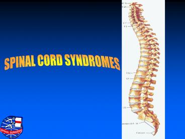
SPINAL CORD SYNDROMES
Description:
INCOMPLETE SPINAL CORD INJURY SYNDROMES The syndromes are named according to the presumed location of injury in the transverse plane of the spinal cord International ... – PowerPoint PPT presentation
Number of Views:2436
Avg rating:3.0/5.0
Title: SPINAL CORD SYNDROMES
1
SPINAL CORD SYNDROMES
2
INCOMPLETE SPINAL CORD INJURY SYNDROMES
- The syndromes are named according to the presumed
location of injury in the transverse plane of the
spinal cord - International standard classification is applied.
3
IMPORTANT TO CATEGORIZE ACCORDING TO LOCATION OF
INJURY
- Recognise types of injury
- Information helps to select treatment
- Each has different prognosis for recovery
4
CERVICO MEDULCARY SYNDROME(upper cervical cord
to medulla)
- Damage to upper cervical cord and medulla
- Upwards can extend upto pons
- Downwards upto C4.
5
CMS PRESENTATION
- Respiratory dysfunction
- Hypotension
- Tetraplegia
- Aneasthesia from C1 to C4
- Sensory loss on face Dejerine pattern or onion
skin pattern
6
CMS MECHANISM
- Traction injury
- Severe dislocation
- Antero posterior compression
- Protruded disc
- Past usually associated with death
- Present prompt first aid treatment, greater
number of survivors reach hospital
7
CMS EXAMINATION
- Face trigeminal nucleus pons
- Trigeminal tract- pons medulla and spinal cord
upto C4- descending spinal tract - Sensory loss around month lesion in medulla.
- Sensory loss forehead, chin, ear C3-C4
8
CMS LIMB WEAKNESS
- More weakness in arms
- Less weakness in legs
- (Mimics central cord syndrome)
- Mechanism Pyramidal arm fibers decussate at
this level antero medially and susceptible to
injury by odontoid and ant. rim of foramen
magnum. Selective bilateral arm paralysis is
possible cruciate paralysis of Bell
9
CMS INJURIES
- Atlanto occipital injury of Bell
- Atlanto axis injury dislocation
- Odontoid fracture
10
ACUTE CENTRAL CORD SRNDROME
- Acute compression
- Elderly people
- Hyperextension injury
- Dysproportionate greater motor loss in upper
extremities - Varying sensory loss
- Spontaneous recovery or improvement possible
11
CENTRAL SPINAL CORD SYNDROME
Cervical spondylosis, ant. and post. osteophytes.
Spinal cord is compressed. The central portion is
damaged
12
CSCS MECHANISM
- A - Hypertension injury
- Antero posterior compression
- Elderly people
- Central haematomyelia
- Surrounding oedema
- Mechanism- compression between bony spurs
ant. and ligamentum flavum post., central
necrosis, involves ant. horn cells.
13
CSCS MECHANISM
- B In absence of orteophytes
- Vascular aetiology
- Compromise of medullary artery perfusion
- Vertebral artery stretching
- Ant. spinal artery spasm / occlusion
- Venous infarcts
14
CSCS MECHANISM
- C - Acute traumatic prolapse of cervical disc
- D - Mechanical compression
15
CSCS v/s CMS
- Central cord Cruciate
- Syndrome Paralysis
- Site of lesions Mid-to lower cervical Lower
medulla and upper - cord cervical cord, anterior aspect
- Anterior horn cells Corticospinal arm fibers
- decussation
- Lateral corticospinal tract
- (medial part)
- Clinical manifestations Arms weaker than
legs, Arms weaker than legs, flaccid flaccid
arms acutely, legs arms acutely, legs normal or
normal or variably weak, variably weak,
upper motor - lower motor neuron neuron deficits in upper
limbs deficits in upper limbs develop - persists
- Trigeminal sensory deficit
- (onion skin , spinal tract of V)
- Cranial nerve dysfunction
- (IX, X, or XI)
- Prognosis for Variable Usually good
- neurological recovery
16
RESCENT EVIDENCEfor central cord syndrome
- Based on MRI and autopsy study
- No hemorrhage in cord
- No necrosis
- Only oedema
- Demyelination and myelin breakdown
- Mechanism- Direct mechanical
compression of cord
17
INDICATIONS FOR SURGERY
- Persistent compression
- Instability
- Neurological deterioration
18
ANT CORD SYNDROME
- Immediate complete paralysis in lower limbs
- Sparing of upper limbs
- Sparing of posterior column
- Hyperasthesia at the level of lesion
- Sparing of touch.
19
ANTERIOR CORD SYNDROME
A large prolapsed disc compresses the ant. spinal
cord post. column is intact
20
ACS MECHANISM
- Mechanical stress factors
- Cord is pulled between compression and dentate
ligament - Pyramided fibers bear the greatest stress
21
ACS PRESENTATION
- Spasticity
- Disturbance of gait
- Modified sensory changes
22
ACS TREATMENT
- Operative removal of lesion
- Substantial recovery
23
BROWN SEQUARD SYNDROME
- Not uncommon
- Lesion lat. half of spinal cord
- Ipsilateral motor and proprioceptive loss
- Contralateral pain and temp loss
24
BSS MECHANISM
Burst fracture with posterior displacement
causing unilateral compression
25
BSS MECHANISM
- Hyperextension injuries
- Flexion injuries
- Facet lock
- Associated with burst fracture
- CAUSE- spinal cord compression
26
BSS PRESENTATION
- Present from the beginning
- Gradual evolution within days possible
- Common in cervical spine.
- Sphincter may be spared
27
CONUS MEDULLARIS SYNDROME
- Anatomically all lumbar segments are opp. T12
vertebral body - All sacral segments are opp. L1 vertebral body
- Cord ends between L1 L2 disc space
28
CONUS MEDULLARIS SYNDROME
D12 burst fracture compress the conus. All lumbar
and sacral segments can be compressed
29
CMS PRESENTATION
- DL injuries common
- Lower motor neuron flaccid paralysis
- Flaccid sphincters
- Chronic spasticity
- Atrophy of muscles
- Perianal sensation may be preserved (sacral
sparing) - Low pressure high capacity neurogenic bladder
30
CAUDA EQUINA SYNDROME
- Injury to lumbar spine
- Roots of cauda equina involved
- Injury can be complete (Grade A)
- Or in varying degree of severity
- Motor fibers are always more susceptible than
sensory. - Some sensations are preserved
31
CAUDA EQUINA SYNDROME
Acute central disc prolapse L4/5. Medially placed
sacral roots sustain maximum compression
32
CES OUTCOME
- Prognosis for neurological recovery is much
better - Lower motor nerves have more resilience to trauma
- Fever secondary injury mechanisms
- Greater regeneration capability
33
SERIOUS CAUDA EQUINA SYNDROME
- Acute C4/C5 and L5/S1 disc prolapse
- Major damage to sacral roots
- Sparing of lumbar and S1 roots
- Complete bladder and bowel paralysis
- Perianal anaesthesia
- Sacral roots delicate
- - do not recover
34
ACUTE SPINAL CORD SYNDROME-SCIWORA
- Without radiological evidence of trauma (SCIWORA)
- Paediatric SCI
- Generally injury is less severe. Complete injury
possible. - Investigations do not include MRI. Only plain
x-ray tomography and CT. - In children there is laxity of ligaments
- Para spinal muscles weak.
35
ACUTE SPINAL CORD SYNDROME-SCIWORA
- MRI SCIWORA
- MRI detects ligamentous injury and haematoma in
soft tissues - Thus revealing damage to spine
36
ANT SPINAL ARTERY SYNDROME
- Ant. spinal artery supplies ant. 2/3 of cord when
occluded - Motor, pain and temperature sensations are lost
- Proprioception is preserved
- Rare in trauma
- Occurs in aortic disease, aortic surgery,
hypotension, spinal angioma - Pathology- occlusion of ant. spinal artery
37
CHRONIC POST TRAUMATIC SPINAL CORD SYNDROMES
- Develop late after trauma
- Months or years to develop
- Causes further sensory or motor loss and
involvement of sphincters - Post traumatic syringomyelia
- Microcystic myelomalacia (Marshy cord syndrome)
- Arachnoiditis
- Pain syndromes
38
CHRONIC POST TRAUMATIC SPINAL CORD SYNDROMES
- Pain syndromes
- Neurogenic Peripherial nerves.
- Mylogenic Spinal cord .
- Cephalogenic Brain.
39
REVERSIBLE OR TRANSIENT SYNDROME
- Spinal cord concussion
- transient loss of motor and sensory functions
with recovery within minutes. Clinical
examination is normal. - Cause Minor trauma.
- Mechanism Unknown , intracellular potassium leak
due to injury or vascular mechanism
40
BURNING HANDS SYNDROME
- Common in athlets and footballers.
- Transient paraesthesiae in both hands and upper
limbs - All such patients have radiological abnormalities
like - Ligamentous instability
- Disc disease
- Spinal stenosis
41
BURNING HANDS SYNDROME
- MRI shows posterior horn damage in intramedullary
injury - Always bilateral
- It unilateral then it is peripheral nerve root
injury.
42
THANK YOU

