
Middle ear - PowerPoint PPT Presentation
Title:
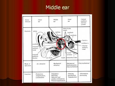
Middle ear
Description:
Middle ear Tensor tympani Enters middle ear through anterior wall of tympanum. Connected to manubrium/neck of the malleus Activated by the trigeminal nerve Muscle ... – PowerPoint PPT presentation
Number of Views:790
Avg rating:3.0/5.0
Title: Middle ear
1
Middle ear
2
(No Transcript)
3
Middle ear structures
- Middle ear cavity/tympanum
- Tympanic membrane
- Ossicles/Middle ear bones
4
Middle ear cavity
- 2-4 mm horizontal, 13 mm vertical, approximately
2 cm3 in volume - Comprised of
- Tympanic cavity between outer and inner ear
- Epitympanic recess above the TM
- Mastoid recess contained in the mastoid region of
the temporal bone
5
Tympanic cavity
- Similar to a box with six surfaces
- Lateral wall or membraneous wall Formed by TM
and squamous portion of TB. - Medial wall or labyrinthine wall Promontory
(outer wall of inner ear) - (oval and round windows, prominence of facial
nerve) - Anterior wall or carotid wall (opening of ET and
tendon of tensor tympani) - Posterior wall or mastoid wall (tympanic aditus,
fossa incudis, pyramidal prominence, facial nerve
through tympanic sulcus) - Inferior wall or jugular wall (tympanic plate of
TB) - Superior wall or tegmental wall (tegmen tympani,
continuing posteriorly to tympanic atrium)
6
Tympanic membrane
- Oval shaped lateral surface
- Thick periphery called annulus with the notch of
Rivinus - Located in a bony groove called tympanic sulcus
- 8-9 horizontal, 9-10 mm vertical axis, 0.1 mm
thick. - Held in place by fibers and cartilage
- Cone shaped, translucent
- 55 to 90 mm2 in area
http//www.ghorayeb.com/AuricleEACAnatomy2.html
7
- Three layers
- Outer Epidermis of EAM
- Inner Mucosal lining of middle ear space
- Middle Collagen fibers, support for TM.
- Middle layer composed of two sets of fibers
- Radiates outward from center
- Concentric rings of fibers
- Two regions in tympanic membrane
- Superior region Pars flaccida (defined by
anterior and posterior malleolar folds and
malleolar prominence) - Inferior region Pars tensa
8
Tympanic membrane, contd.
- Umbo Region of maximum concavity
- Position changes as EAM lengthens after birth
- Important landmarks visible
- Tympanic membrane attached to ossicles.
http//hyperphysics.phy-astr.gsu.edu/hbase/sound/e
ar.html
9
Ossicles
- Malleus Hammer
- Incus Anvil
- Stapes Stirrup http//www.ghorayeb.com/StapesPics
.html - Head of malleus and body of incus located in the
epitympanic recess - http//oto.wustl.edu/bbears/ossicle.htm
http//audilab.bmed.mcgill.ca/daren/3Dear/mid1.ht
ml
10
(No Transcript)
11
Malleus
- 9 mm long, 23-37 mg weight
- Head with articular facet
- Neck
- Anterior and lateral processes
- Handle or Manubrium
12
Incus
- About 23-32 mg weight
- Body
- Short crus/process in epitympanic recess, around
5 mm - Long crus/process with lenticular process, around
7 mm - (Plural of crus Crura)
13
Stapes
- 2-5 to 3.8 mm tall, 2.1 to 4.3 mg weight.
Footplate area around 3.2 mm2 - Head, with spine for attachment of stapedial
tendon - Neck
- Two bony crura, anterior and posterior
- Flat oval bone called footplate
- Medial surface of footplate fastened to wall of
oval window by annular ligament. - Unique connection allows for rocking, rather than
piston-like, movement of stapes.
14
(No Transcript)
15
Ossicular connections
- Malleus Manubrium of malleus connects to umbo of
TM. - Incus Head of malleus connects to body of incus
(incudo-mallear joint) - Stapes Inferior process of incus connects to
form lenticular process, which connects to the
stapes (incudo-stapedial joint) - Footplate of the stapes connects to the oval
window
16
Ossicular connections, contd.
- Suspended in the middle ear cavity by axial
ligaments and muscle tendons - Middle ear muscles
- Tensor tympani and stapedius
17
Tensor tympani
- Enters middle ear through anterior wall of
tympanum. - Connected to manubrium/neck of the malleus
- Activated by the trigeminal nerve
- Muscle runs parallel and superior to osseous
foundation of the ET, separated by septum canalis
musculotubarii) - Pulls malleus in anterior and posterior direction
18
Stapedius muscle
- Tendon enters the middle ear space through
opening in the posterior wall of the tympanum
(pyramidal eminence) - Attached to the head of the stapes
- Activated by the facial nerve
- Pulls stapes in the posterior direction