
Head and Neck Cancer - PowerPoint PPT Presentation
1 / 81
Title:
Head and Neck Cancer
Description:
Head and Neck Cancer Radiation Therapy 4412 Management of Head and Neck Cancer Through multidisciplinary treatment we try to: 1. decrease deformity 2. maintain the ... – PowerPoint PPT presentation
Number of Views:2445
Avg rating:3.0/5.0
Title: Head and Neck Cancer
1
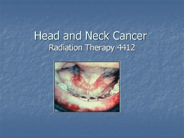
Head and Neck Cancer
- Radiation Therapy 4412
2
Management of Head and Neck Cancer
- Through multidisciplinary treatment we try to
- 1. decrease deformity
- 2. maintain the reduction of the tumor
- 3. restore function
- 4. preserve the structure and esthetics
- 5. cure the cancer
3
1. Compare and contrast the epidemiologic factors
prevalent in head and neck cancers
- 1/3 of patients that are treated have early stage
- 2/3 of patients will have locally advanced stages
- Lungs are the most common site for mets
- The nerve routes are important in treatment
planning, tumors can spread this way
4
- Almost half of all squamous cell ca occur in the
oral cavity - Head and neck cancer involves the upper
aerodigestive tract. - Oral cavity
- Pharynx
- Paranasal sinuses
- Larynx
- Thyroid gland
- Salivary glands
5
- Men- usually 50-60 years old
- Can occur in people younger than 40 years of age
- More women are smoking
- Smokeless tobacco
- Recurrences- usually within first 2 years
- Rarely after 4 years
- Most 5 year survivors will be alive at 10 years
6
2. List and describe the etiologic factors
associated with head and neck cancers
- Smokeless tobacco- squamous cell of cheek and gum
- Previous radiation exposure- thyroid /salivary
glands - Poor oral hygiene
- Ill fitting dentures/irritation to tissues
7
- Wood mill workers- nasal cavity/paranasal sinuses
- Lip cancer- UV exposure, unfiltered cigarettes
- Viruses
- Epstein Barr virus
- Herpes simplex (cold sores)
- HPV- oral/larynx
- Chronic abuse of marijuana- degree of risk
unknown - Diet
- Vitamin A and E deficiency
- Plummer-Vinson syndrome- iron deficiency anemia
8
- Alcohol
- Pharyngeal and laryngeal cancer
- Liver damage
- Secondary nutritional deficiencies
- Alcohol damages mucosa and makes it more
permeable - Impurities in the alcoholic beverages
9
- Smoking
- Head and neck cancers occur 6x more frequently
than non-smokers - Unfiltered cigarettes
- Cigar smoking is a risk
- Laryngeal cancer mortality increases as the
number of cigarettes smoked increases
10
Smoking, tobacco, alcohol a deadly combination!
- Alcohol is synergistic to tobacco- cooperate
together to produce a total effect greater than
the sum of the individual elements - Tars
- Aromatic hydrocarbons
- Ethanol suppresses the efficiency of DNA repair
- Nitrosamines most noncombustible product in snuff
and chewing tobacco
11
- Pre-cancerous signs
- Leukoplakia is a precancerous, slowly developing
change in the mucous membrane. They are
characterized by thickened, white, firmly
attached patches that are slightly raised. - Erythroplasia- A premalignant lesion that is
shiny, velvety and reddish in color - These are severe dysplastic changes and should be
taken seriously
12
Leukoplakia
13
3. Identify the prognostic indicators in head and
neck cancers
- Prognosis decreases as
- The affected area progresses backward from the
lips to the hypopharynx (excludes larynx) - Lesions that cross the midline
- Exhibits endophytic growth- invades within the
lamina propria and submucosa - Have cranial nerve involvement
- Fixed nodes
14
- Fixed lesion in the anatomic compartments
- Are poorly differentiated
- Nonsquamous cell
15
5. Compare and contrast endophytic and exophytic
tumor features of head and neck cancers
- Endophytic growth- growth pattern that invades
the lamina propria and submucosa - -more aggressive and harder to control locally
- Exophytic- a noninvasive neoplasm that projects
out from an epithelial surface - -characterized by raised, elevated borders
- Most head and neck cancers are infiltrative
lesions found in the epithelial lining
16
Staging
- Lymphatics of the head and neck are in direct
correlation to the prognosis - 1/3 of the bodys lymphatics are in the head and
neck area - Staging depends on
- Site of primary disease
- Extent of primary disease
- Size of primary tumor
17
- Staging contd
- Cell type and differentiation
- Lymphatic vascular space invasion of the tumor
- The nodal status
18
6. List and describe the different types of head
and neck cancers
- Most head and neck cancers will infiltrate into
the epithelial lining of the upper digestive
tract - 80 of all head and neck cancers will be squamous
cell
19
7. Compare and describe the different types of
head and neck cancers.8. Describe the different
treatmentconsiderations for the different types
of head and neck cancers.
20
Oral Cavity
- Oral cavity extends from the skin vermilion
junction of the lip to the posterior border of
the hard palate superiorly - And the circumvallate papillae inferiorly
- Anterior 2/3 of the tongue lips, buccal mucosa,
lower alveolar ridge, upper alveolar ridge,
retromolar trigone, floor of the mouth, and hard
palate - Page 694 Washington/Leaver
21
- Oral cavity cancers
- The most common aerodigestive tract cancers
- Occur mostly in men- 55 to 65 years old
- Alcohol and tobacco are synergistic
- Patients usually have poor oral and dental
hygiene - Plummer-Vinson syndrome is important etiologic
factor
22
- General practitioner or dentist will find the
cancer - Early diagnosis is important
- Leukoplakia and erythroplasia are serious
- Most oral cavity cancers will be nonhealing
ulcers with little pain - Localized pain is an advanced disease
23
- The cancer is usually raised, centrally
ulcerated, indurated edges and the base is
infiltrating - Mandatory biopsy
- Squamous cell carcinoma makes up 90-95
- Well or moderately well differentiated
- Has the lowest incidence (except glottic) of
nodal mets - Cervical node involvementadvanced disease
24
Lips and Gum
- Lip cancer is treated with radiation the same way
as skin cancer - Usually involves the lower lip and spreads by
direct invasion - Carcinoma in-situ and early lesions of the lip
may be surgically removed
25
- Radiation Therapy
- Portal should include primary lesion with a 2 cm
- A shield (stent) of lead and bolus material (to
absorb backscatter) is placed under the lip - This blocks the alveolar process and gums
- Treated with external beam, interstitial implant
or both - 100 SSD, 100 isodose line
26
Lip Cancer
27
Floor of Mouth
- Floor of the mouth lesions usually arise on the
anterior surface on either side of the midline. - They can spread to bone and tongue
- Approx 30 of these cancers will involve the
submaxillary and subdigastric nodes - Opposed lateral fields are used
- The tip of the tongue can be elevated out of the
portal with a cork or a bite block and tongue
depressor
28
- Bite blocks can also spare the roof of the mouth
from incidental irradiation - If the lesion has grown into the tongue, the
tongue is flattened to reduce the superior border
of the portal - Radiation therapy supraclavicular and bilateral
neck fields, followed with a boost of intraoral
cone, needle implants, or small external photon
beams
29
(No Transcript)
30
(No Transcript)
31
Tongue
- The anterior 2/3 of the tongue is included in the
oral cavity - The base of the tongue is considered oropharynx
- Small tumors in the anterior 2/3 of the oral
tongue are usually resected - Radiation therapy is used for inoperable patients
32
- Post-op radiation therapy
- Treats the primary site
- Treats the cervical lymph nodes
- And margins positive
- for tumor,
- extensive primary tumor with bone or skin
invasion, - and multiple positive nodes
33
- The anterior tongue drains into the
- Submandibular lymph nodes
- The posterior portion of the tongue drains into
the - Jugulodigastric
- Posterior pharyngeal
- Upper cervical lymph nodes
- Lesions of the tongue usually appear on the
lateral borders near the middle and posterior
third section - A limited number of tongue cancers can be excised
- Most are controlled with external beam and
interstitial boost fields
34
- Lesions at the base and posterior 1/3 of the
tongue invade - The floor of the mouth
- Tonsils
- or the muscles
- Are advanced
- Have a higher incidence of nodal mets
35
- Hemiglossectomy- surgical removal of half the
tongue. It is used for treatment of an early
stage lesion of the tongue - Radiation therapy- three field technique
- Utilizes external beam, electron beam
- Possibly an iridium implant and neck dissection
- Isocentric lateral opposed fields
- Lower anterior neck field
- Fields include subdigastric and submaxillary
nodes - Upper cervical nodes
36
T1 Squamous cell of tongue
37
Buccal Mucosa
- Buccal mucosa is the mucous membrane lining the
inner surface of the cheeks and lips - Most lesions arise on the lateral walls
- Have a history of leukoplakia
- Are raised, exophytic growths
- Lesion invades the skin and bone
- First sign is a bump on the tip of the tongue
- No pain associated at first until the nerves to
the tongue or ear become involved - Advanced lesions will bleed
38
- Stensens duct (parotid duct) can become
obstructed - The parotid gland becomes enlarged
- Small lesions are surgically removed
- Large lesions are treated with surgery and
radiation therapy or - Radiation therapy alone
- Complications- fibrosis of the cheek and trismus
39
Hard Palate
- Located between the upper alveolar ridge and
mucous membrane covering the palatine process of
the maxillary palatine bones - Mostly adenocarcinomas and rare
- Spread to the bone, invade the maxillary antrum
- Treatment- surgical resection, post-op radiation
therapy - History of ill fitting dentures or trauma
40
Retromolar Trigone
- Triangular space behind the last molar tooth
- Rare carcinomas
- Symptoms- tongue, ear canal pain, trismus
- Usually moderately differentiated squamous cell
carcinoma - Lymphatic spread to the submaxillary
subdigastric nodes - Treated with radiation therapy
41
PHARYNX
- Subdivided into three anatomic divisions
- Oropharynx
- Nasopharynx
- hypopharynx
- Common symptoms
- Persistant sore throat
- Painful swallowing
- Referred otalgia
- Cervical node enlargement
- Fetor oris, dyspnea, dysphasia, hoarseness,
dysarthria, hypersalivation indicates advanced
disease
42
(No Transcript)
43
- Diagnosis- indirect mirror exam, palpation,
biopsy, CT, MRI - Histopathology- squamous cell carcinomas
- Staging- AJCC Classification
- Mets- cervical lymph nodes (bilateral),
retropharyngeal nodes, lung
44
Oropharynx
- Consists of the base of the tongue, the tonsils
(fossa and pillars), soft palate, oropharyngeal
walls - The oropharynx is located between the axis and C3
vertebral bodies - Soft tissue regions- anterior tonsillary pillars,
soft palate, uvula, base of the tongue and the
lateral-posterior pharyngeal walls
45
- Tonsils are the most common site for disease
- Symptoms- sore throat and pain during swallowing
- Upper spinal accessory nodes are involved
bilaterally in 50 to 70 of the patients - Radiation therapy is treatment of choice
46
(No Transcript)
47
Cancer of tongue
48
Hypopharynx
- Pyriform sinuses, postcricoid, and lower
posterior pharyngeal walls below the base of the
tongue - It is situated between C3 to C6
- The cricoid cartilage is the inferior border
- Epiglottis is the superior border
- Hypopharyngeal cancer is advanced
- High rate of nodal mets
49
- Tumor is highly infiltrative
- The highest area for incidence is the pyriform
sinus - Radical surgery and radiation therapy is the
treatment of choice - Rouvieres (lateral retropharyngeal) lymph nodes
at the base of the skull are included with other
nodal groups in treatment (page 706, Washington)
50
- Tonsillar, pharyngeal wall and posterior cricoid
are treated using radiation therapy - (page 709, figure 30-28, Washington)
51
Unresectible T4 pyriform sinus tumor, surrounding
carotid artery
52
Nasopharynx
- Posterosuperior pharyngeal wall and lateral
pharyngeal wall, the eustachian tube orifice and
adenoids - The nasopharynx is a cuboidal structure lying on
a line from the zygomatic arch to the external
auditory meatus (EAM), extending inferiorly to
the mastoid tip - The nasopharynx lies behind the nasal cavities
and above the level of the soft palate
53
- The nasal cavity drains into the nsopharynx via
the two posterior nares - Two eustachian tubes are on the lateral walls
which connect to the middle ear - Nasopharyngeal disease can mimic an inflammatory
process - Can cause considerable respiratory or auditory
dysfunction
54
- The cranial nerve is frequently involved
- The ninth to the twelfth cranial nerves can be
affected - Enlargement of the retropharyngeal nodes
- Can affect the external carotid artery
- A lesion can invade directly into the third
cranial nerve - Commonly involves the sixth cranial nerve
55
- When cranial nerves are involved, this means the
disease is advanced and widespread - Histology- squamous cell
- Nasopharyngeal cancer is usually poorly
differentiated and shows an unusual growth
pattern - This disease is not associated with tobacco
consumption
56
- NPC is associated with the Epstein Barr virus
- Can occur in adolescence and young adults
- Occurs again between 50 and 70 years of age
- Uncommon in white populations
- Found mostly in southern China and the Middle East
57
- Positive cervical nodes in 75 to 85 of NPC
patients - About half of all cases will have bilateral or
contralateral disease - Radiation ports are large
- The lateral retropharyngeal (node of Rouviere)
which cannot be surgically removed, and
jugulodigastric nodes are almost always treated
58
- Primary lesion is small but the nodal disease is
extensive - Bone and lung common mets sites
- NPC spreads to adjacent sites and has a high
recurrence rate - Aggressive, large volume curative radiation
therapy is given
59
Larynx
- The larynx is contiguous with the lower portion
of the pharynx above and is connected with the
trachea below. - It extends from the tip of the epiglottis at the
level of the lower border of the C3 vertebra to
the lower border of the cricoid cartilage at the
level of C6
60
- There are 3 main parts to the larynx. These
parts are - The supraglottis - the area above the vocal cords
that contains the epiglottis cartilage - The glottis - the area around the vocal cords
- The subglottis - the part below the vocal
cords, containing the cricoid cartilage. It
continues down into the windpipe
61
(No Transcript)
62
- Glottic cancer- 65
- Supraglottic cancer- 25 to 33
- Subglottic- make up the rest of the cases
- Most common cancer in the aerodigestive tract is
the larynx - Male dominated disease
- 50-60 years of age
- Smoking high risk factor
63
- Extensive use of voice in occupation is risk
factor (singers, auctioneer) for laryngeal cancer - Alcohol high risk factor for supraglottic cancer
- Cancer of the glottis (true vocal cord) is not
life threatening - Choice of treatment is based on the preservation
of speech and airway
64
- Laryngeal cancer shows a mutation of the p53 gene
- Classic Symptoms- persistent sore throat and
hoarseness - Cervical lymph nodes involvement is associated
with supraglottic lesions - Carcinoma in situ is common on the vocal cords
65
- Glottic lesions are well to moderately
differentiated - Supraglottic lesions are less differentiated and
more aggressive - Glottic lesions will appear of the anterior 2/3
of one cord (approx 65-75) - Cord mobility is a factor in the classification
of lesions
66
- Treatment- radiation therapy is the treatment of
choice for nonfixed surface glottic lesions that
have not invaded muscle, bone or cartilage - Glottic cancer is treated with lateral opposing
fields 5X5cm or 6X6 cm - Large, fixed lesions will require aggressive
treatment - Radiation therapy offers the best voice
preservation
67
- Supraglottic lesions are usually large and bulky
- They do not usually invade the inferior false
cord or the ventricles - These lesions usually spread superiorly to the
epiglottis - Lymph nodes are usually involved in 40-50 of
the patients
68
- Subglottic lesions are treated with total
laryngectomy with - Post-op radiation therapy
69
Larynx- squamous cell, Rt anterior vocal fold
70
Salivary Glands
- Salivary glands are made up of
- Parotid-largest gland, located superficial to and
partly behind the ramus of the mandible, and
covers the masseter muscle - It fills the space between the ramus of the
mandible and the anterior border of the
sternocleidomastoid muscle - Contains extensive lymphatic capillary plexus
many aggregates of lymphocytic cells - Numerous intraglandular lymph nodes in the
superficial lobe
71
- Submandibular glands
- Sublingual glands
- Tumors of the salivary gland are rare
- The parotid is the most common site for tumors
- Nearly 2/3 of these tumors will be benign
- Low-dose ionizing radiation in childhood may have
been a risk factor - Dental x-rays have been implicated for both
benign and malignant tumors
72
- Most major and minor salivary gland cancers are
of unknown origin - Adenoid cystic, mucoepidermoid, and
adenocarcinoma are the most common cell types - Symptoms- asymptomatic parotid mass lasting 4-8
months before the tumor arises - Presenting symptoms- localized swelling and pain,
facial palsy, rapid growth - Facial nerve involvement suggests malignancy
- Diagnosis is done through lobectomy
73
- Treatment- Although most tumors are benign, local
recurrence is high - Total resection with margins sparing facial
nerves - Radiation therapy- post-op for residual,
recurrent or inoperable tumors - Accelerated fractionation- provides similar dose
levels of radiation therapy in a shorter amount
of overall time. This counteracts quick cellular
proliferation of aggressive tumors by giving more
dose in a shorter period of time.
74
Maxillary Sinus
- Maxillary sinus is a pyramid shaped cavity lined
by ciliated epithelium and bound by thin bone or
membranous partitions. - Carcinomas arising from the ciliated epithelium
or mucous glands perforate the bony walls almost
from the beginning - Tumors will also involve the superior portion of
the sinus and extend into the floor of the orbit
75
- Maxillary sinus cancers- 80 of all sinus cancers
- Long history of sinusitis, nasal obstructions and
bloody discharge - Squamous cell carcinomas
- Invade the floor of the orbit, ethmoid sinuses,
hard palate zygomatic arch - Displacement of the eye is common
76
- Nasal cavity and paranasal sinus tumors are often
associated with cranial nerve palsies- trigeminal
branches - CT and MRI are the most useful studies
- Submandibular node will be the first involved,
although cervical node spread is uncommon
77
- Treatment- Surgery is the treatment of choice
- Primary radiation therapy has a chance of optic
nerve damage from the high dose required for
tumor control - Surgery and radiation therapy used in most cases
- Lateral and anterior ports are used
- When the orbit is involved, eye blocking will not
be used - Care should be taken to miss the cord and
contralateral lens
78
- Angling the anterior beam a few degrees off the
vertical spares brain tissue - Nasal cavity risk- Bolus material will be
inserted to improve dose homogeneity - Angling the lateral port a few degrees off the
horizontal plane spares the contralateral optic
nerve and lens
79
Management of the Head and Neck Patient
80
- Washington, Page 718, Table 30-3, dose-tissue
response schedule - Page 719, Box 30-11, recommended skin care
program - Care of the head and neck patient
- Peridontal disease and caries
- Nutrition
- Mucositis/stomatitis
81
- Xerostomia
- Cataract formation
- Lacrimal glands
- Taste changes
- Skin reactions
Recommended
CrystalGraphics Presentations


