
ACTIN STAINING OF FIBROBLASTS - PowerPoint PPT Presentation
1 / 35
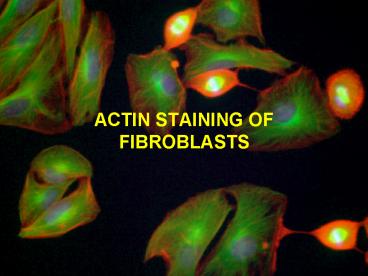
Title: ACTIN STAINING OF FIBROBLASTS
1
ACTIN STAINING OF FIBROBLASTS
2
Housekeeping
- Be vigilant about spills
- Wipe up as soon as possible without compromising
sterility or activities - Watch for splatters on back of hood
3
Lab Objectives
- Understand the cellular biology ( size, structure
and functions) of - Microtubulues, Intermediate Filaments
- G-Actin and F-actin
- Stress Fibers, Focal Contact/Adhesion, Adhesion
Belt, Contractile Ring - Become familiar with fluorescent microscopy
concepts and reagents used this week - Specific reagents such as formaldehyde, glycine,
Triton-X 100, PBS and Rhodamine phalloidin - Fluorescent microscope itself and components, be
able to diagram all parts and the light path
(show diagram) - Alternatives to organic dyes
4
Cytoskeleton
- Gives the cell mechanical strength, controls its
shape, drives and guides its movement - Four classes
- Muscle thick filament
- Microfilaments
- Intermediate filaments
- Microtubules
5
Appearance of cytoskeletal components in cultured
fibroblast cell
Transmission Electron Microscopy TEM
6
Comparison of cytoskeletal components
Rope-like bars Long, hollow cylinders
Helical polymers of actin about 10 nm made
from tubulin flexible, concentrated in
about 25nm the cortex of the
cell about 7nm
7
Microtubules
- Most rigid of cytoskeletal elements
- May occur singly or in groups
- Responsible for movement of materials within cell
- Gives cells shape
- Form cilia and flagella
- Helps cells divide
Negative stain
8
Placement of tubulin in non-metaphase and
metaphase cell
- Microtubules help with intracellular traffic in
non-dividing cell - Aid in splitting apart sister chromatids during
cell division
Fluorescently labeled antibody against tubulin
9
Intermediate Filaments
- Strengthen cell against mechanical stress
- Keratin is found in epithelial cells
- Vimentin is found in connective tissue, muscle
and support cells of the nervous system - Neurofilaments are found in nerve cells
10
Microfilaments
- Are the thinnest at 7nm in diameter
- Also called actin
- Ubiquitous, multifunctional
- Responsible for cell crawling
- Needed to produce contractile forces
- Interacts with myosin for muscle contraction
- Actin filaments are thin and flexible
- Usually found in bundles rather than individually
- Usually associated with the plasma membrane
11
Placement of actin in cells
Contractile ring In dividing cells
Microvilli cores And terminal web
Leading edge of Motile cell
Stress fibers And cell cortex
12
Functions of Actin in Non-Muscle Cells
- Contractile ring
- important in cell division
13
- Adhesion belts
- Intercellular anchoring junctions found in
epithelia
- Stress fibers
- Important for a cells structure and movement
- Are involved in forming adhesion plaques (points
of cell adhesion to the ECM)
14
- Actin bundles
- Bundles of actin, parallel in microvilli on many
columnar epithelial cells - Important for the structure and movement of the
microvilli, to help keep them clean
- Cell cortex
- Actin fibers connected to the plasma membrane
- Play a role in providing mechanical strength to
the surface, allowing for movement.
15
Lamellipodia, filipodia and blebs
Pinocytotic cup
- These membrane protrusions are under the control
of actin - Can view these as cells move across a surface
lamellipodia
Bridge still connecting recently divided cells
filipodia
bleb
16
Actin (microfilaments)
- The most abundant protein in eukaryotic cells
- 2 forms
- G-actin
- globular, a protein monomer associated with ATP
- ATP needed to bind units together to form a
filament. - F-actin
- filamentous, a polymerized chain of actin
monomers - Polymerization occurs on the positive end of the
filament
17
Actin Assembly/Polymerization
Direction of Growth
18
Solutions and Equipment
- PBS
- Used to wash the cells
- 4 Formaldehyde
- Cross-links and fixes proteins in cells, prevents
degradation - Glycine
- Blocks background (noise) by binding ketones
- 0.4 Triton X-100
- A detergent that permeabilizes cell membrane for
dye penetration
19
- Mounting Media
- Specially formulated to not cause interference in
the fluorescent image - Rhodamine Phalloidin
- Phalloidin
- From the poisonous mushroom Amanita phalloides
(Death Cap) - Binds specifically to f-actin
- Rhodamine
- Fluorescent marker conjugated to phalloidin
- Excited by green light l 520-560 nm
- Emits red light l 650-720nm
20
Fluorescence Microscopy
- Green light photons promote rhodamine electron
to a higher energy level. When they return to
original level, they emit a lower energy photon.
- What you can expect to see
- Black background, with red stress fibers visible
- Keep sample protected from light after rhodamine
phalloidin is added. - If a fluorescent sample is left exposed too long
to light, the fluorescence will fade. This is
called quenching or photobleaching
21
Fluorescence Microscope Diagram
22
A formula for those who like them
The fluorescence intensity in a stained object
can be related to a series of factors to which it
is directly proportional as elucidated in the
following formula
Equation 1 F fluorescence intensity f(q) is
a geometric factor g(l) is the quantum
efficiency of the detector and I0 denotes the
intensity of the excitation light. FF denotes the
quantum efficiency of the fluorescence dye, e is
the extinction coefficient of the dye, b the
optical path length (thickness with dye) and c
the concentration of the dye. All the factors in
the M bracket are to do with the imaging and
illumination side, the C bracket contains those
factors concerned with dye concentration sample
thickness, etc..
23
Other Fluorescent Dyes and Techniques
- Other chemicals with specificity for particular
parts of the cell - DAPI, Hoechst, or ethidium bromide for DNA
- Fluorescently tagged antibodies
- Used anywhere you have a protein you have an
antibody for. - Anti-tubulin antibody conjugated to fluorescein
- (excited by blue and glows green)
- GFP
- Must be expressed by the cells.
- Can localize a particular protein or entire cell
24
Quantum dots
- Tiny light emitting particles on the nanometer
scale - Inorganic semiconductors
- Cadmium selenide
- 2-8nm (200-1000atoms)
- Size determines the color
- Synthesized to emit l between 400-2000nm by
changing composition and size
25
QD advantages over organic dyes
- Same size regime as biological macromolecules
- Resistant to photobleaching
- Improved brightness
- Multicolor light emission from single light
source - Ultrasensitive chemical analysis and cellular
imaging - Can image within an organism
QD
Texas red
QD
Fluorescein -FITC
26
Multi colored fluorescence images require
changing excitation sources and layering the
images
- Blue DAPI (for DNA)
- Red Rhodamine phalloidin (actin)
- Green HPA lectin conjugated to Alexa Fluor 488
for golgi - Pink Alexa Fluor 680 conjugated to primary
antibody for mitochondrial protein
27
Disadvantages of QD
- Expensive
- Not refined yet
- Can be toxic
- Resistance to change on the part of scientists
28
Fluorescence tutorials from Molecular Probes, Inc.
- TAs will show these in class
- http//probes.invitrogen.com/resources/education/t
utorials/1Introduction/player.html - http//probes.invitrogen.com/resources/education/t
utorials/3Light_Sources_Filters/player.html
29
Application to Tissue Engineering
- Cytoskeletal changes are seen in
- Cell migration (chemotaxis)
- Physiological functions
- Organogenesis and embryonic development
- Wound healing and angiogenesis
- Understanding tissue development and cellular
changes helps the tissue engineer create new
tissues.
30
Cellular response to topography
- SEM images A 13nm islands, B 35nm, C 95nm
islands - Cells more spread out on 13nm islands compared to
flat polystyrene, No difference on 35nm islands,
less spread on 95mm islands - Distinct interactions between cell filipodia and
islands. Interactions increased with island size - Larger islands produced more amoeboid-like
pseudopodia
31
Cytoskeletal response to topography
- Actin red (rhodamine phalloidin), tubulingreen
(Fluorescein anti tubulin), nucleusblue (DAPI) - 13nm islands (a) supported fibroblasts with well
defined actin and tubulin skeletons - 35nm islands (b) less distinct cytoskeleton
- 95nm islands (c) had poorly organized
cytoskeleton - Time studies show that cells adhere and spread
better initially on the large islands but by 24
hours of culture the cells take on the amoeboid
morphology and proliferate very slowly.
32
Cell Morphology on Scaffolds
Lamellopodia Filopodia
Images courtesy of Aylin Sendemir
33
On Chitosan On Chitosan/BCP
Images courtesy of Aylin Sendemir
34
By The Way...
- If no times are given for washes, just after
solution is put on, take it off. - Incubation temp is RT
- Keep cells moist at all times during prep.
- Step 10 Put coverslip and parafilm in moist,
dark box, then tell TA. TA will distribute
phalloidin due to toxicity. - When adding warmed mounting media, you may want
to cut end of yellow pipette tip to have a larger
opening for easier dispensing.
35
Other exercises this week
- Groups 4, 5, 6 complete exercises B and C before
starting actin staining exercise - B-Add induction media to all three wells of 6
well plate - Remove mediaGENTLY
- No rinsing!
- Add induction media from TA--GENTLY
- C-Create at least one stock plate of 3T3-L1
- calculate cells for 10cm dish