
A Qualitative Review of Two Cortical Surface Modeling Packages: - PowerPoint PPT Presentation
1 / 1
Title:
A Qualitative Review of Two Cortical Surface Modeling Packages:
Description:
surf/?h.inflated - mri/orig - mri/T1 - mri/brain - mri/wm - mri/filled - surf/?h.orig ... surf/?h.sphere - surf/?h. sphere.reg - surf/?h.white - surf/?h.pial ... – PowerPoint PPT presentation
Number of Views:40
Avg rating:3.0/5.0
Title: A Qualitative Review of Two Cortical Surface Modeling Packages:
1
A Qualitative Review of Two Cortical Surface
Modeling Packages FreeSurfer and SureFit
Peggy Christidis, Shruti Japee, Ziad S. Saad and
Robert W. Cox National Institute of Mental
Health, National Institutes of Health, Department
of Health and Human Services
PROCESSING SEQUENCE
INTRODUCTION
Figure 4. Sample Volume and Surface from the
SureFit and FreeSurfer GUIs
SureFit
FreeSurfer
Traditionally, functional brain imaging data is
analyzed by projecting activation data from a
sequence of slices onto a standardized
3-dimensional anatomical space. However, the
cerebral cortex is better modeled by a
2-dimensional sheet that is highly folded and
curved. As such, a 3D space may underestimate
the neural distance between two points,
particularly if the points lie on opposite sides
of a sulcus. This anomaly has lead to the use of
computer-based tools that create 2D cortical
surfaces, which can be inflated, flattened, and
overlaid with functional activation data. This
review provides a discussion of two freely
available cortical surface modeling packages that
have gained wide use in the field of
neuroimaging FreeSurfer 1, 2, and SureFit 3.
Although in-depth descriptions of these tools
have been provided by their respective authors,
there has been to date no systematic qualitative
or quantitative comparison between these tools.
We provide here a qualitative comparison of these
two packages. The evaluation of FreeSurfer
(October, 2001 release) and SureFit (version
4.38, 2002) was based on a number of qualitative
criteria, including ease of installation, manual
editing procedure, quality of the documentation
and tutorials that accompany each package,
graphical user interfaces, and overall ease of
use.
- The intensity normalized, volume registered, and
averaged image served as input to both software
packages. FreeSurfer required the dataset to be
converted to its own specific format (COR files).
The FreeSurfer program mri_convert was used for
this purpose. This program reslices the input
volume to a 256 x 256 x 256 coronal volume with 1
mm3 voxels. - In the case of SureFit, AFNI was used to reslice
and reorient the volume to conform to SureFit
specifications. Since SureFit reads minc files,
the AFNI dataset (i.e., BRIK file) was converted
to minc format using the AFNI program
3dAFNItoMINC. - Parameter Specifications
- SureFit requires the user specify the gray and
white matter intensity peaks based on visual
assessment using an intensity histogram. The
subjectivity of this task introduces a potential
user bias that could influence the quality of
segmentation. With FreeSurfer, the gray and
white matter intensity peaks are automatically
determined by the program. - Ease of Input
- FreeSurfers file input and conversion procedure
was more straightforward compared to that of
SureFit. The latter required separate reslicing
and reorientation of the AFNI BRIK to conform to
the SureFit input file format. - Ease of Performing the Processing Sequence
- FreeSurfer has the added advantage of performing
the processing of the input volume to create
surfaces via command line. This scripting option
makes the processing of volumes fairly automated
and streamlined, eliminating constant user
intervention and supervision. Scripting is not
an option in SureFit, which is only usable
through the graphical user interface (GUI). - Ease and Quality of Surface Reconstruction
- Both software packages have decent and comparable
inflation procedures. In our experience, there
was no systematic difference between the final
surfaces created using either of the packages. - A first pass surface creation in both packages
takes approximately the same amount of time
(20-30 minutes). - An advantage of FreeSurfer is that it can process
both hemispheres simultaneously, while SureFit
does only one hemisphere at a time. - The quality of the first pass surface is
comparable between the two packages. - Figures 2 and 3 show flowcharts from the
FreeSurfer and SureFit manuals, which demonstrate
the processing steps and resulting volumes and
surfaces for each package
- Manual Editing Tools
- FreeSurfers manual editing GUI is far superior
to the editing GUI in SureFit. The latter
required meticulous voxel-by-voxel editing, or
editing using an erosion/dilation filter in two-
or three-dimensions. FreeSurfer allows
voxel-by-voxel editing as well as freehand brush
drawing with flexibilities of brush size and
shape. Both programs allow only one level of
undo. The manual editing GUIs for SureFit and
FreeSurfer are displayed in Figure 5. - The manual editing for both programs is done on
the segmented white matter volume. The original
view should also be loaded to assist with the
editing process.
INSTALLATION
- FreeSurfer is freely available upon registration
from the NMR group at Massachusetts General
Hospital (http//www.freesurfer.mgh.edu). - SureFit is freely available upon registration
from the David Van Essen laboratory at Washington
University at St. Louis (http//brainmap.wustl.edu
). - We downloaded the two packages and followed the
installation instructions accompanying each
package. Installation of these software packages
proceeded fairly smoothly. - However, FreeSurfer required considerable
alterations to the user environment. Once
installed, several environment variables needed
to be initialized and set, including for example,
the subject and functional directories. - Our assessment is that FreeSurfer is rather rigid
and awkward in its data input and directory
structure, requiring a bit of manipulation to
initialize and setup the correct access paths. - SureFit, on the other hand, presented only a
minor GL library glitch that was well documented
on their website and easily fixed.
Figure 5. Manual editing GUIs for SureFit and
FreeSurfer
SureFit
FreeSurfer
CONCLUSION
INPUT PREPROCESSING
Despite sharing similar underlying principles,
the packages discussed here differ widely in
their graphical user interfaces, editing tools,
and general ease of use (see Table 1). Although
SureFit received better marks for its GUIs and
easy installation, FreeSurfer had far superior
editing tools, a convenient command line option,
and excellent documentation, giving it a higher
rating overall. Nonetheless, it is up to the
user to consider our comments and determine which
package is better suited to their particular
application. Future work on this project will
include a method to make quantitative comparisons
between surfaces obtained from different surface
modeling packages.
Figure 2. Processing Sequence and Resulting
Output Volumes and Surfaces for FreeSurfer
Figure 3. Processing Sequence and Resulting
Output Volumes and Surfaces for SureFit
- The dataset we used was acquired as follows
- 4 consecutive MPRAGE scans acquired on a 3 Tesla
magnet - 124 slices acquired axially
- In plane resolution of 0.78 mm2 and slice
thickness of 1.2 mm - FOV 20 cm
- Using AFNI tools 4 (freely downloaded from the
AFNI website http//afni.nimh.nih.gov),
preprocessing of the data was performed as
follows - Each of the four volumes was first intensity
normalized to correct inhomogeneity artifacts.
Normalization can be done with either an AFNI
program called 3dUniformize, or a tool offered by
the Montreal Neurological Institute called
nu_correct, which performs a nonparametric
nonuniform intensity normalization (N3). - Three of the four intensity-normalized images
were then registered to the fourth image. - Finally, an average of the four
intensity-normalized and volume-registered scans
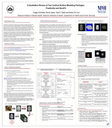
was created. - Figure 1 illustrates the preprocessing sequence
and AFNI tools used at each step. - Intensity normalization is a critical
preprocessing step since it makes the gray and
white matter intensity distribution more uniform,
thereby increasing the gray and white matter
contrast, while averaging increases the
signal-to-noise ratio. Intensity normalization
is essential, since both software packages
perform an intensity-based segmentation to
determine the gray/white matter boundary. - Although FreeSurfer performs intensity
normalization as part of its processing sequence,
we and others 5 have observed that the N3
normalization method does a better job in
correcting the nonuniformity effects, thereby
resulting in a better gray/white matter
segmentation.
FREESURFER STEPS
PROCESS VOLUMES SURFACES Raw image
intensity Orient Volume Define Volume of
interest(VOI) Resample (optional)
Oriented, cropped intensity
volume Set parameters Generate probabilistic
volumes Composite inner Composite
boundary outer
boundary Radial position
map Segment volume Initial
cortical segmentation Generate Surface
Initial surface reconstruction Correct
Errors Correct cortical segmentation Generate
fiducial surface Fiducial surface
reconstruction Map fMRI data (optional)
Functional activation maps
Input Files
Output Files
CONVERT/AVERAGE Convert/Motion Correct/ Average
MRI data in native scanner format
- mri/orig
I.
PROCESS VOLUME Normalize Intensity Strip
Skull Segment White Matter
- mri/orig - mri/T1 - mri/brain
- mri/T1 - mri/brain - mri/wm
Table 1. Comparison of various features of
FreeSurfer and SureFit
CREATE SURFACE Cutting Planes Filling Tesselate Sm
ooth Inflate
- mri/filled - surf/?h.orig - surf/?h.
smoothwm - surf/?h.curv - surf/?h.sulc -
surf/?h.inflated
- mri/wm - mri/filled - surf/?h.orig - surf/?h.
smoothwm
II.
MANUALLY EDIT DEFECTS Then return to Create
Surface repeat if required until no more large
topological defects remain.
- mri/wm
- mri/wm
III.
FIX SURFACE TOPOLOGY Fix Surface
Topology Smooth Inflate Sphere
- surf/?h.orig - surf/?h. smoothwm -
surf/?h.curv - surf/?h.sulc - surf/?h.
inflated - surf/?h.sphere
- surf/?h.orig - surf/?h.orig - surf/?h.
smoothwm - surf/?h.inflated
IV.
V.
REGISTER Register to Cortical Atlas
- surf/?h. sphere.reg
- surf/?h.sphere
VI.
- surf/?h.white - surf/?h.pial -surf/?h.thickness
- surf/?h.orig
MAKE FINAL SURFACES Final Surface Deformation
GRAPHICAL USER INTERFACE
Figure 1. Preprocessing of dataset for input into
FreeSurfer and SureFit
- Volume and Surface GUIs
- FreeSurfers volume and surface interfaces are
less user-friendly and flexible than those of
SureFit. - While SureFit allows rapid and free zoom,
translation, rotation, browse, etc., of the image
volume or surface, FreeSurfers GUIs are slower
and less smooth, especially the surface GUI. - FreeSurfers Surface loading and redrawing
functions are very slow, even with the best
graphics card. SureFit, on the other hand,
allows real-time control of the volume and
surface windows. - SureFit allows loading of three surfaces at a
time, while FreeSurfer allows only a single
surface view at a time. - Figure 4 shows an example of a volume and surface
as they appear in the SureFit and FreeSurfer GUIs
3dUniformize (or nu_correct)
to3d
REFERENCES
BRIK
I. files
- Dale, A.M., Fischl, B., et al. (1999). Cortical
surface-based analysis. I. Segmentation and
surface reconstruction. Neuroimage, 9(2)
179-194. - Fischl, B., Sereno, M.I., et al. (1999).
Cortical surface-based analysis. II. Inflation,
flattening, and a surface-based coordinate
system. Neuroimage, 9(2) 195-207. - Van Essen, D.C., Drury, H.A., et al. (2001). An
integrated software suite for surface-based
analyses of cerebral cortex. J Am Med Inform
Assoc, 8(5) 443-459. - Cox. R.W. (1996). AFNI Software for analysis
and visualization of functional magnetic
resonance neuroimages. Computers and Biomedical
Research, 29162-173. - Arnold, J.B., Liow, J.S., et al. (2001).
Qualitative and quantitative evaluation of six
algorithms for correcting intensity nonuniformity
effects. Neuroimage, 13(5) 931-943.
3dvolreg
3dMean
Final Result Intensity normalized, volume
registered, and averaged dataset.
Recommended
CrystalGraphics Presentations


