
Identification of Gram Positive Cocci: Staphylococcus - PowerPoint PPT Presentation
1 / 7
Title:
Identification of Gram Positive Cocci: Staphylococcus
Description:
Do not produce endospores, but are resistant to drying (desiccation) ... The antibiotic in the disc diffuses into the surrounding agar. ... – PowerPoint PPT presentation
Number of Views:808
Avg rating:3.0/5.0
Title: Identification of Gram Positive Cocci: Staphylococcus
1
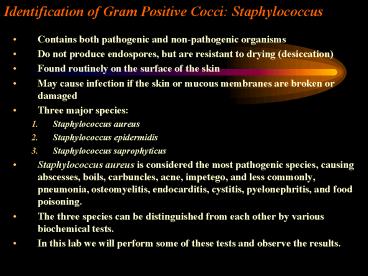
Identification of Gram Positive Cocci
Staphylococcus
- Contains both pathogenic and non-pathogenic
organisms - Do not produce endospores, but are resistant to
drying (desiccation) - Found routinely on the surface of the skin
- May cause infection if the skin or mucous
membranes are broken or damaged - Three major species
- Staphylococcus aureus
- Staphylococcus epidermidis
- Staphylococcus saprophyticus
- Staphylococcus aureus is considered the most
pathogenic species, causing abscesses, boils,
carbuncles, acne, impetego, and less commonly,
pneumonia, osteomyelitis, endocarditis, cystitis,
pyelonephritis, and food poisoning. - The three species can be distinguished from each
other by various biochemical tests. - In this lab we will perform some of these tests
and observe the results.
2
Chemical and Biochemical Tests
- The identification of organisms is based on
cellular, cultural, and biochemical
characteristics - All species of Staphylococcus are Gram Positive
Cocci (GPC) - On nutrient agar they tend to be white (or cream
colored), circular, entire, convex colonies. - On Sheep Blood Agar Staphylococcus aureus may
exhibit hemolysis of the agar in the area around
the colonies. - Tests to be performed
- Catalase test
- Coagulase test
- Growth and fermentation on Mannitol Salt Agar
- Susceptibility to the antibiotic Novobiocin
3
Catalase Test
- The Catalase test determines if the organism
produces the enzyme Catalase, which breaks down
hydrogen peroxide (H2O2) to water and oxygen
(O2). -
Catalase - 2 H2O2 2 H2O O2 (g)
- Catalase allows organisms to break down harmful
metabolites of aerobic respiration and may be
seen in aerobic and facultatively anaerobic
organisms. There are other enzymes produced by
some organisms to handle other toxic end-products
of metabolism, such as superoxide dismutase. Not
all organisms produce catalase. - Coagulase Test
- Pathogenic organisms require mechanisms to help
them overcome host defense systems. One mechanism
involves coating the bacterial cells in a body
substance, such as fibrin, to hide the
bacterial cells from the immune system. This
coating will not trigger an immune response by
the host cells. The enzyme coagulase causes
fibrin to be deposited on bacterial cells helping
them to become invisible to the host immune
system.
4
High Salt Tolerance
- Some organisms cannot tolerate a high salt
concentrations. - Media containing higher than normal salt
concentrations will inhibit the growth of these
non-salt tolerant organisms. - Mannitol salt agar contains a high salt
concentration so only salt tolerant organisms
will grow on it. - Also, Mannitol salt agar contains the sugar
Mannitol. - Some organisms can utilize this sugar as a food
source and will produce acidic by-products from
this metabolism. - The addition of acid to the medium by the
fermentation of Mannitol changes the pH. - If a pH indicator is present in the medium (such
as Phenol red) a color change will occur
dependant upon the pH of the medium (agar or
broth). - Mannitol Salt Agar contains the pH indicator
Phenol Red - This pH indicator is red at neutral pH (around
7.0), but turns yellow under acidic conditions. - Antibiotic Susceptibility/Resistance
- Antibiotic susceptibility is another test that
can be used to identify bacteria. - A paper disc impregnated with the antibiotic, in
this case Novobiocin, is placed on a lawn of
bacteria following inoculation. - The antibiotic in the disc diffuses into the
surrounding agar. - If the bacterial species is susceptible to the
antibiotic there is a circle of no-growth
around the disc where bacterial growth is
inhibited by the antibiotic. - If the bacteria is resistant to the antibiotic
the cells grow right up the the antibiotic disc. - The bacterial species or strain is reported as
being resistant to the antibiotic (R) or
susceptible to the antibiotic (S) depending on
the observations made. - The diameter of the area of no-growth around
the disc may determine the susceptibility or
resistance of the organism to the antibiotic.
5
Interpretation of Results
- Catalase
- Bubbling indicates a positive test for the
presence of the catalase enzyme. - Coagulase
- Agglutination of the Test latex with no
agglutination of the Control latex is
considered a positive () test for the presence
of this enzyme. All reactions occurring after 20
seconds should be ignored. - Agglutination of the Test latex with no
agglutination of the Control latex is
considered a positive () test for the presence
of this enzyme.
6
Mannitol Salt Agar
- Two different characteristics of the organism are
determined with this agar. The first is the
organisms ability to tolerate a high salt
environment. Evidence of growth on the slant
indicates the organism can grow in a high salt
environment. - Organisms that can ferment the sugar Mannitol
produce an acid end-product that changes the red
pH indicator (Phenol red) in the media to yellow. - Any yellow in the media is considered a positive
test for Mannitol fermentation. - It is possible to have growth, but no Mannitol
fermentation.
7
Novobiocin Susceptibility
- A zone of growth inhibition 17 mm or less in
diameter indicates resistance (R) to Novobiocin. - If the zone is greater than 11 mm the organism is
susceptible (S) to Novobiocin.
Recommended
CrystalGraphics Presentations


